Review
. 2022 Mar 19;11(6):1704.
doi: 10.3390/jcm11061704.

Different Methods to Improve the Monitoring of Noninvasive Respiratory Support of Patients with Severe Pneumonia/ARDS Due to COVID-19: An Update

Affiliations
Review

Different Methods to Improve the Monitoring of Noninvasive Respiratory Support of Patients with Severe Pneumonia/ARDS Due to COVID-19: An Update

Paolo Pelosi et al. J Clin Med. .

Abstract

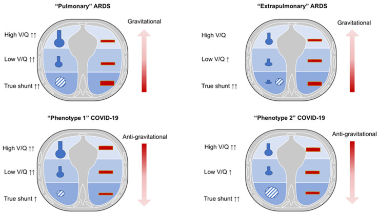
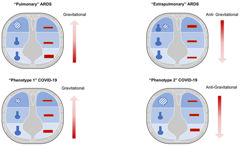
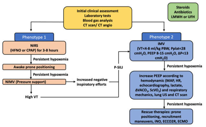
The latest guidelines for the hospital care of patients affected by coronavirus disease 2019 (COVID-19)-related acute respiratory failure have moved towards the widely accepted use of noninvasive respiratory support (NIRS) as opposed to early intubation at the pandemic onset. The establishment of severe COVID-19 pneumonia goes through different pathophysiological phases that partially resemble typical acute respiratory distress syndrome (ARDS) and have been categorized into different clinical-radiological phenotypes. These can variably benefit on the application of external positive end-expiratory pressure (PEEP) during noninvasive mechanical ventilation, mainly due to variable levels of lung recruitment ability and lung compliance during different phases of the disease. A growing body of evidence suggests that intense respiratory effort producing excessive negative pleural pressure swings (Ppl) plays a critical role in the onset and progression of lung and diaphragm damage in patients treated with noninvasive respiratory support. Routine respiratory monitoring is mandatory to avoid the nasty continuation of NIRS in patients who are at higher risk for respiratory deterioration and could benefit from early initiation of invasive mechanical ventilation instead. Here we propose different monitoring methods both in the clinical and experimental settings adapted for this purpose, although further research is required to allow their extensive application in clinical practice. We reviewed the needs and available tools for clinical-physiological monitoring that aims at optimizing the ventilatory management of patients affected by acute respiratory distress syndrome due to severe acute respiratory syndrome coronavirus-2 (SARS-CoV-2) infection.

Keywords: ARDS; COVID-19; coronavirus disease; noninvasive respiratory support (NIRS); respiratory failure.


Conflict of interest statement

The authors declare no conflict of interest.

Figures

Figure 1
Figure 1
Ventilation/perfusion characteristics in non-COVID-19 and COVID-19 ARDS in supine position. Legend. Pulmonary ARDS: in supine position, low ventilation/perfusion ratio (V/Q) is prevalent. Higher regional perfusion and true shunt are mostly diverted in the dependent lung regions. Extrapulmonary ARDS: in supine position, true shunt is prevalent with possible alveolar collapse. The pulmonary blood flow and shunt mainly distribute toward the dependent lung regions. COVID-19 phenotype 1: in supine position, a low V/Q is prevalent with anti-gravitational distribution of pulmonary blood flow. COVID-19 phenotype 2: in supine position, true shunt is increased, while the pulmonary blood flow is anti-gravitational. V/Q, ventilation/perfusion; COVID-19, coronavirus disease 2019; ARDS, acute respiratory distress syndrome.
Figure 2
Figure 2
Ventilation/perfusion characteristics in non-COVID-19 and COVID-19 ARDS in prone position. Legend. Pulmonary ARDS: in prone position, oxygenation may improve due to partial redistribution of pulmonary blood flow toward ventral regions, and not by effective alveolar recruitment. This might be associated with decreased carbon dioxide washout. Extrapulmonary ARDS: in prone position, oxygenation can improve because of possible alveolar recruitment of collapsed alveoli, while maintaining perfusion higher toward the dorsal lung regions. COVID-19 phenotype 1: in prone position, oxygenation may improve, partially redistributing pulmonary blood flow that remains anti-gravitational. COVID-19 phenotype 2: in prone position, oxygenation may improve, partially redistributing pulmonary blood flow from dorsal to ventral lung regions, and not by effective alveolar recruitment. This might be associated with decreased carbon dioxide washout. V/Q, ventilation/perfusion; COVID-19, coronavirus disease 201; ARDS, acute respiratory distress syndrome.
Figure 3
Figure 3
COVID-19 radiological phenotypes at computed tomography and follow-up. Legend. Axial chest HRCT showing the main features of COVID-19 phenotypes. (A) Phenotype 1: diffuse bilateral ground glass opacities and consolidation. (B) Phenotype 2: patchy, dependent, bilateral areas of parenchymal consolidations. (C) Phenotype 3: diffuse interlobular septal thickening, bronchial dilatation/distortion, and perilobular fibrosis. (D) Persistence of thin subpleural reticular and curvilinear opacities after 9 months from ARDS resolution. HRCT, high resolution computed tomography scan; COVID-19, coronavirus disease 2019; ARDS, acute respiratory distress syndrome.
Figure 4
Figure 4
Correlation between oscillatory compliance and lung aeration. Legend. Upper panel. CT scans of an animal model of induced lung injury; from left to right: baseline, during left lung atelectasis induced by 10 min of single-lung ventilation at 100% oxygen, after a RM, post broncho-alveolar lavage and after RM after broncho-alveolar lavage. Lower panel: corresponding VtissNA% (closed symbols, solid line) and Cx5 (open symbols, dotted line) showing that oscillatory compliance and lung derecruitment show a specular trend being inversely correlated. (With permission, courtesy of Prof. Dellaca [60]). Cx5, 5Hz oscillatory compliance; VtissNA%, percentage of non-aerated tissue volume; CT, computed tomography; RM recruitment maneuvers.
Figure 5
Figure 5
Suggested algorithm for the ventilatory management of COVID-19 ARDS. Legend. Algorithm for the ventilatory management of COVID-19 ARDS. NIRS, non-invasive respiratory support; HFNO, high-flow nasal oxygen; CPAP, continuous positive airway pressure; NIMV, non-invasive mechanical ventilation; IMV, invasive mechanical ventilation; VT, tidal volume; PBW, predicted body weight; PEEP, positive end-expiratory pressure; P, driving pressure; p-SILI, patient self-inflicted lung injury (patients on NIPPV who are generating massive negative pressures and high tidal volumes in the range of 10–12 cc/kg IBW); MAP, mean arterial pressure; HR, heart rate; ΔVACO2, venous–arterial carbon dioxide tension difference; ScVO2, central venous oxygen saturation; US, ultrasound; CT, computed tomography; iNO, inhaled nitric oxide; ECCO2R, extracorporeal carbon dioxide removal; ECMO, extracorporeal membrane oxygenation; LMWH, low-molecular weight heparin; UFH, unfractioned heparin.
Figure 6
Figure 6
Correlation between PEEP and dynamic compliance and PaO2/FiO2 ratio in COVID-19 and non-COVID-19 ARDS. Legend. Correlation between PEEP values and change in both dynamic compliance and PaO2/FiO2 ratio in COVID-19 pneumonia (panel A and C, respectively) and ARDS (panel B and D, respectively) patients. PEEP, positive end-expiratory pressure; COVID-19, coronavirus disease 2019; ARDS, acute respiratory distress syndrome.
Figure 7
Figure 7
Practical flow-chart of respiratory assistance for patients with COVID-19 acute respiratory failure based on inspiratory effort assessment. Legend. HFNC trial might be started with close monitoring of inspiratory effort. If low values of ΔPes are detected, HFNC should be kept with close monitoring of esophageal pressure swings and gas exchange. In case of high inspiratory effort, non-invasive respiratory assistance should be upgraded to NIV. If ΔPes is reduced by positive pressure application, NIV might be continued with continuous monitoring of inspiratory effort. In case NIV fails to reduce inspiratory effort, a rapid switch to IMV should be considered. HFNO, high-flow nasal oxygen; NIV, non-invasive mechanical ventilation; MV, invasive mechanical ventilation; ΔPes, esophageal pressure.

References

    1. WHO WHO Coronavirus (COVID-19) Dashboard. 2021. [(accessed on 16 February 2022)]. Available online: https://covid19.who.int.
    1. Hermann M., Laxar D., Krall C., Hafner C., Herzog O., Kimberger O. Duration of invasive mechanical ventilation prior to extracorporeal membrane oxygenation is not associated with survival in acute respiratory distress syndrome caused by coronavirus disease 2019. Ann. Intensive Care. 2022;12:6. doi: 10.1186/s13613-022-00980-3. - DOI - PMC - PubMed
    1. Ball L., Barisione E., Mastracci L., Campora M., Costa D., Robba C., Battaglini D., Micali M., Costantino F., Cittadini G., et al. Extension of Collagen Deposition in COVID-19 Post Mortem Lung Samples and Computed Tomography Analysis Findings. Int. J. Mol. Sci. 2021;22:7498. doi: 10.3390/ijms22147498. - DOI - PMC - PubMed
    1. Baratella E., Ruaro B., Marrocchio C., Starvaggi N., Salton F., Giudici F., Quaia E., Confalonieri M., Cova M.A. Interstitial Lung Disease at High Resolution CT after SARS-CoV-2-Related Acute Respiratory Distress Syndrome According to Pulmonary Segmental Anatomy. J. Clin. Med. 2021;10:3985. doi: 10.3390/jcm10173985. - DOI - PMC - PubMed
    1. Radermacher P., Maggiore S.M., Mercat A. Fifty Years of Research in ARDS. Gas Exchange in Acute Respiratory Distress Syndrome. Am. J. Respir. Crit. Care Med. 2017;196:964–984. doi: 10.1164/rccm.201610-2156SO. - DOI - PubMed

LinkOut - more resources