. 2022 Feb 23;12(3):332.
doi: 10.3390/life12030332.

Effect of C60 Fullerene on Recovery of Muscle Soleus in Rats after Atrophy Induced by Achillotenotomy

Affiliations

Effect of C60 Fullerene on Recovery of Muscle Soleus in Rats after Atrophy Induced by Achillotenotomy

Dmytro Nozdrenko et al. Life (Basel). .

Abstract

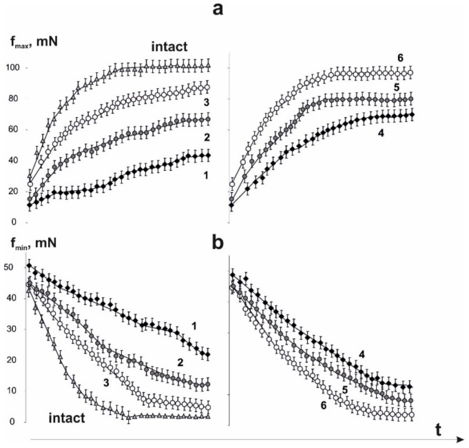
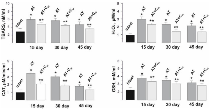
Biomechanical and biochemical changes in the muscle soleus of rats during imitation of hind limbs unuse were studied in the model of the Achilles tendon rupture (Achillotenotomy). Oral administration of water-soluble C60 fullerene at a dose of 1 mg/kg was used as a therapeutic agent throughout the experiment. Changes in the force of contraction and the integrated power of the muscle, the time to reach the maximum force response, the mechanics of fatigue processes development, in particular, the transition from dentate to smooth tetanus, as well as the levels of pro- and antioxidant balance in the blood of rats on days 15, 30 and 45 after injury were described. The obtained results indicate a promising prospect for C60 fullerene use as a powerful antioxidant for reducing and correcting pathological conditions of the muscular system arising from skeletal muscle atrophy.

Keywords: C60 fullerene; achillotenotomy; atrophy; biomechanical and biochemical parameters of skeletal muscle contraction; muscle soleus of rat.


Conflict of interest statement

The authors have no conflict of interest to declare.

Figures

Figure 1
Figure 1
The force of contraction of the muscle soleus after AT in rats, caused by 10 consecutive 6 s non-relaxation pools of stimulation: without C60 fullerenes administration (a); with C60 fullerenes administration at a dose of 1 mg/kg (b). Native muscle–intact, t1–time of the maximum strength response development, t2–recovery time of strength parameters to their initial values, S–integrated power of muscle contraction, calculated as the total area under the corresponding strength curve.
Figure 2
Figure 2
Biomechanical parameters of muscle soleus after AT in rats at 10 consecutive 6 s non-relaxation contractions: change in the maximum force response as a percentage of the values in the intact group (a); integrated muscle power as a percentage of values in the intact group (b); time of development of the maximum force response (c); recovery time of force parameters to their original values (d). Native muscle–intact; 1,2,3–the values of the corresponding parameters on 15th, 30th and 45th days after AT, respectively, without administration of C60 fullerenes (*p < 0.05 compare to the intact group at all 1, 2,…10 consecutive contractions); 4,5,6–the values of the corresponding parameters on 15th, 30th and 45th days after AT, respectively, after using C60 fullerenes at a dose of 1 mg/kg (**p < 0.05 compared to the group without the use of C60 fullerene at all 1, 2,…10 consecutive contractions).
Figure 3
Figure 3
Biomechanical parameters of muscle soleus after AT in rat at 1 Hz stimulation for 1800 s: without C60 fullerenes administration (a); with the use of C60 fullerenes at a dose of 1 mg/kg (b); integrated muscle power (S), presented as a percentage of values in the intact group (c); time reduction of the force response by 50% from the initial values (t50) (d). Native muscle–intact; 1,2–the corresponding values of the parameters without and with C60 fullerenes use, respectively. *p < 0.05 compare to the intact group; **p < 0.05 compare to the group without the use of C60 fullerene.
Figure 4
Figure 4
Mechanograms of the transition of muscle soleus after AT in rats from dentate to smooth tetanus with the use of increasing stimulation with a maximum frequency of 30 Hz for 6 s: without C60 fullerenes administration (a); with C60 fullerenes administration in a daily dose of 1 mg/kg (b). Native muscle–intact; fmax is the maximum force of a single contraction, fmin is the minimum value of the force response in one tooth of the dentate tetanus; 1,2,3–the values of the corresponding parameters on 15, 30 and 45 days after AT, respectively, without C60 fullerenes administration; 4,5,6–the values of the corresponding parameters on 15, 30 and 45 days after AT, respectively, with the use of C60 fullerenes at a dose of 1 mg/kg.
Figure 5
Figure 5
Changes in fmax (a) and fmin (b) parameters of muscle soleus after AT for each of the single contractions during the transition of the force response to smooth tetanus using an increasing stimulation signal with a maximum frequency of 30 Hz for 6 s: 1,2,3–parameter values on days 15, 20 and 45 after AT, respectively, without C60 fullerenes administration; 4,5,6–the values of the parameters on days 15, 20 and 45 after AT, respectively, with the use of C60 fullerenes at a dose of 1 mg/kg.
Figure 6
Figure 6
Indicators of pro- and antioxidant balance (TBARS, H2O2, CAT and GSH) in the blood of rats after 1 Hz stimulation of muscle soleus for 1800 s on 15, 20 and 45 days after AT. * p < 0.05 compare to the intact group; ** p < 0.05 compare to the group without C60 fullerenes administration.

References

    1. Ohira Y., Yoshinaga T., Nomura T., Kawano F., Ishihara A., Nonaka I., Roy R.R., Edgerton V.R. Gravitational unloading effects on muscle fiber size, phenotype and myonuclear number. Adv. Space Res. 2002;30:777–781. doi: 10.1016/S0273-1177(02)00395-2. - DOI - PubMed
    1. Nozdrenko D.N., Shut A.N., Prylutskyy Y.I. The possible molecular mechanism of the nonlinearity muscle contraction and its experimental substantiation. Biopolym. Cell. 2005;21:80–83. doi: 10.7124/bc.0006E0. - DOI
    1. Goldberg A.L., Dupont-Versteegden Etlinger J.D., Goldspink D.F., Jablecki C. Mechanism of work-induced hypertrophy of skeletal muscle. Med. Sci. Sports. 1975;7:185–198. - PubMed
    1. Fluckey J.D., Dupont-Versteegden E.E., Knox M., Gaddy D., Tesch P.A., Peterson C.A. Insulin facilitation of muscle protein synthesis following resistance exercise in hindlimb-suspended rats is independent of a rapamycin-sensitive pathway. Am. J. Physiol. Endocrinol. Metab. 2004;287:E1070–E1075. doi: 10.1152/ajpendo.00329.2004. - DOI - PubMed
    1. Paddon-Jones D., Sheffield-Moore M., Cree M.G., Hewlings S.J., Aarsland A., Wolfe R.R., Ferrando A.A. Atrophy and impaired muscle protein synthesis during prolonged inactivity and stress. J. Clin. Endocrinol. Metab. 2006;91:4836–4841. doi: 10.1210/jc.2006-0651. - DOI - PubMed

LinkOut - more resources