. 2022 Feb 28;8(3):248.
doi: 10.3390/jof8030248.

Multigene Phylogeny Reveals Endophytic Xylariales Novelties from Dendrobium Species from Southwestern China and Northern Thailand

Affiliations

Multigene Phylogeny Reveals Endophytic Xylariales Novelties from Dendrobium Species from Southwestern China and Northern Thailand

Xiaoya Ma et al. J Fungi (Basel). .

Abstract

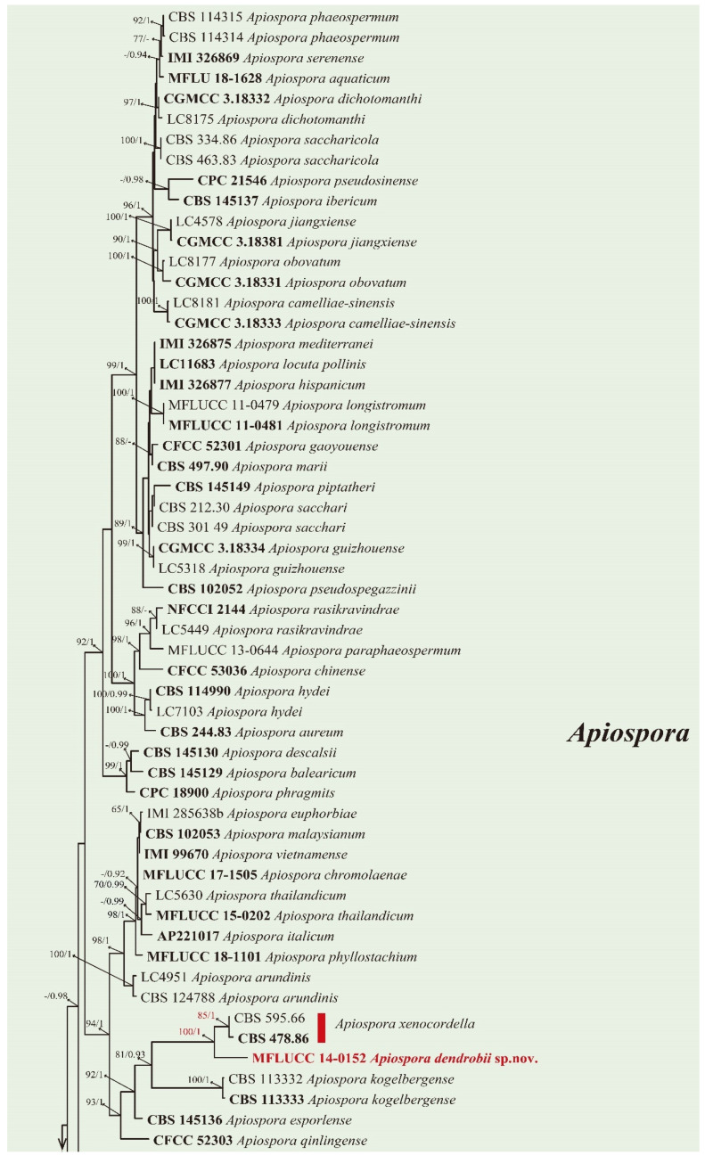
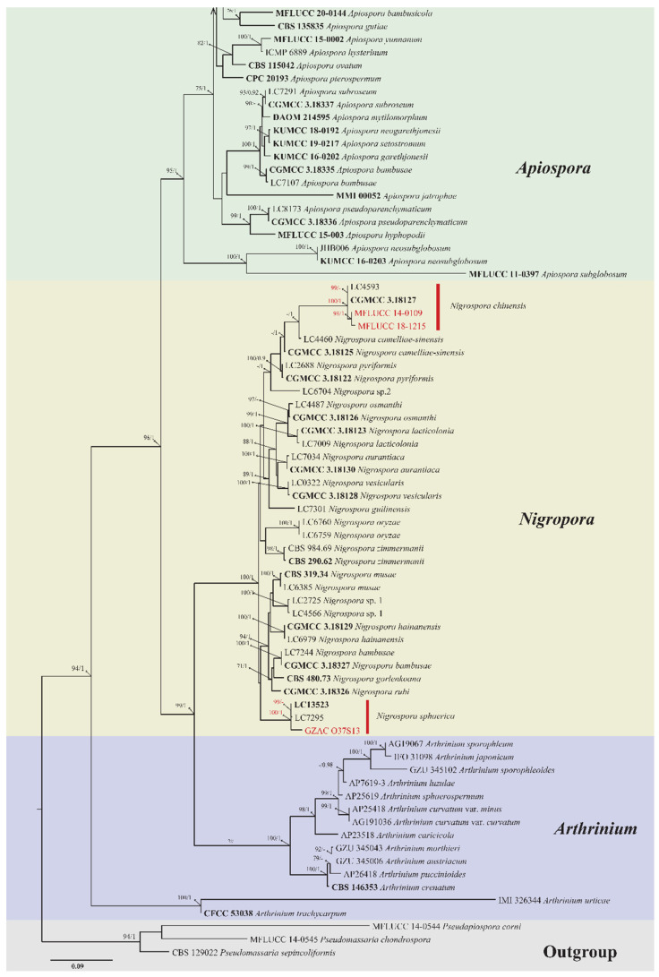
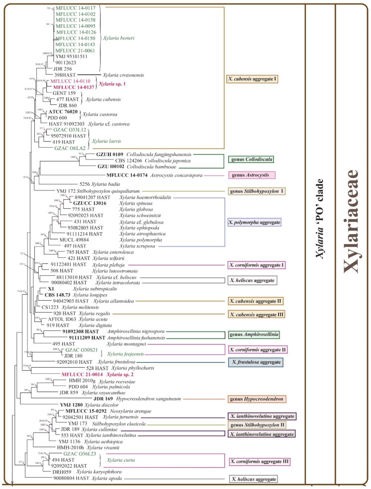
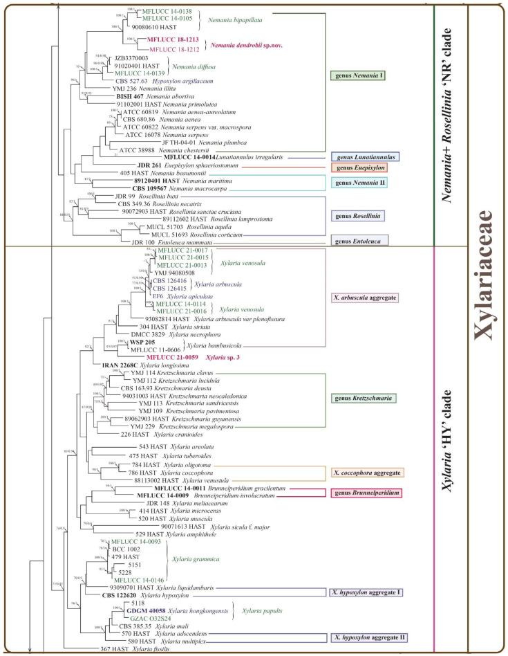
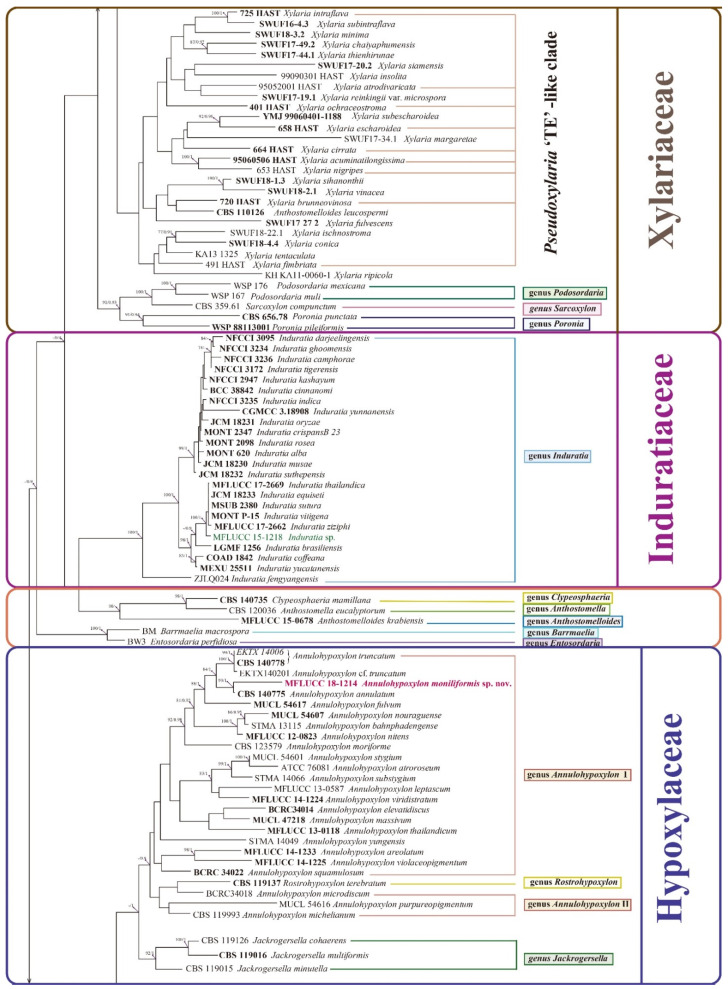
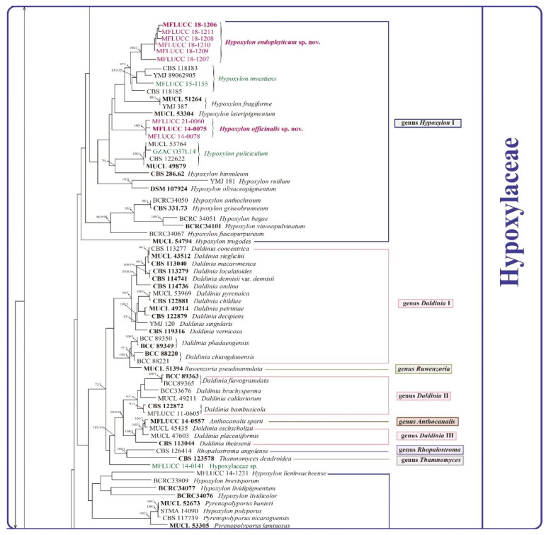
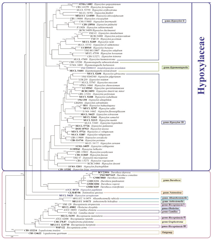
Xylariales are common endophytes of Dendrobium. However, xylarialean species resolution remains difficult without sequence data and poor sporulation on artificial media and asexual descriptions for only several species and old type material. The surface-sterilized and morph-molecular methods were used for fungal isolation and identification. A total of forty-seven strains were identified as twenty-three species belonging to Apiosporaceae, Hypoxylaceae, Induratiaceae, and Xylariaceae. Five new species-Annulohypoxylon moniliformis, Apiospora dendrobii, Hypoxylon endophyticum, H. officinalis and Nemania dendrobii were discovered. Three tentative new species were speculated in Xylaria. Thirteen known fungal species from Hypoxylon, Nemania, Nigrospora, and Xylaria were also identified. Another two strains were only identified at the genus and family level (Induratia sp., Hypoxylaceae sp.). This study recorded 12 new hosts for xylarialean endophytes. This is the first report of Xylariales species as endophytes from Dendrobium aurantiacum var. denneanum, D. cariniferum, D. harveyanum, D. hercoglossum, D. moniliforme, and D. moschatum. Dendrobium is associated with abundant xylarialean taxa, especially species of Hypoxylon and Xylaria. We recommend the use of oat agar with low concentrations to induce sporulation of Xylaria strains.

Keywords: Xylariomycetidae; endophytes; multi-locus phylogeny; oat media; orchids.


Conflict of interest statement

The authors declare no conflict of interest.

Figures

Figure 1
Figure 1
A multi-locus phylogenetic tree based on the combined ITS-LSU-TUB2-TEF-1α sequences of Apiosporaceae species resulting from a maximum likelihood analysis. Pseudomassaria chondrospora (MFLUCC 14-0545), Pseudomassaria sepincoliformis (CBS 129022), and Pseudopiospora corni (MFLUCC 14-0544) are selected as outgroup. The isolates from this study are in red. The ex-type isolates are in bold. Maximum likelihood values (ML) equal or greater than 70 and/or 0.9 Bayesian posterior probabilities (BPP) are labelled at the end of nodes. Dashes are indicated values lower than 70% ML and/or 0.9BPP. Scale bar corresponds to 0.09 substitutions per site.
Figure 1
Figure 1
A multi-locus phylogenetic tree based on the combined ITS-LSU-TUB2-TEF-1α sequences of Apiosporaceae species resulting from a maximum likelihood analysis. Pseudomassaria chondrospora (MFLUCC 14-0545), Pseudomassaria sepincoliformis (CBS 129022), and Pseudopiospora corni (MFLUCC 14-0544) are selected as outgroup. The isolates from this study are in red. The ex-type isolates are in bold. Maximum likelihood values (ML) equal or greater than 70 and/or 0.9 Bayesian posterior probabilities (BPP) are labelled at the end of nodes. Dashes are indicated values lower than 70% ML and/or 0.9BPP. Scale bar corresponds to 0.09 substitutions per site.
Figure 2
Figure 2
A multi-locus phylogenetic tree based on the combined ITS-LSU-TUB2-RPB2 sequences of related Xylariales species resulting from a maximum likelihood phylogenetic analysis. The tree is rooted at the outgroup Lopadostoma gastrinum (CBS 134632) and Lopadostoma insulare (CBS 133214). The isolates from this study are in red (tentative new and new species) and green (known species). Strains needing reevaluation are in purple. Different aggregate partition is based on their various stromata, mainly represented by several Xylaria species following Hsieh et al. 2010. The ex-type isolates are in bold. Equal or greater than 70% maximum likelihood values (ML) and/or 0.9 Bayesian posterior probabilities (BPP) are labelled at the end of nodes. Dashes are indicated values lower than 70% ML and/or 0.9BPP. Scale bar corresponds to 0.09 substitutions per site.
Figure 2
Figure 2
A multi-locus phylogenetic tree based on the combined ITS-LSU-TUB2-RPB2 sequences of related Xylariales species resulting from a maximum likelihood phylogenetic analysis. The tree is rooted at the outgroup Lopadostoma gastrinum (CBS 134632) and Lopadostoma insulare (CBS 133214). The isolates from this study are in red (tentative new and new species) and green (known species). Strains needing reevaluation are in purple. Different aggregate partition is based on their various stromata, mainly represented by several Xylaria species following Hsieh et al. 2010. The ex-type isolates are in bold. Equal or greater than 70% maximum likelihood values (ML) and/or 0.9 Bayesian posterior probabilities (BPP) are labelled at the end of nodes. Dashes are indicated values lower than 70% ML and/or 0.9BPP. Scale bar corresponds to 0.09 substitutions per site.
Figure 3
Figure 3
Apiospora dendrobii (MFLU 21-0153). (a) Colony on PDA (left-front view, right-reverse view). (bd) Conidiomatal mass. (eg) Conidiophores with conidia. (hj) Chlamydospores. (k) Conidia. Notes: (bk) on 4% OA. Scale bars: (b) = 100 μm, (ek) = 10 μm.
Figure 4
Figure 4
Nigrospora chinensis (MFLUCC 14–0109). (a) Colony on PDA (left-front view, right-reverse view). (b) Colony on MEA. (c,d) Conidiomatal masses. (e) Mycelia. (f) Conidia. Notes: (cf) on 2% MEA.
Figure 5
Figure 5
Annulohypoxylon moniliformis (MFLU-O35L22) (a) Colony on PDA (left-front view, right-reverse view). (b) Ascomata on WA (slide cultre). (cg) Asci with ascospores on the robber band (for supporting slide) in slide culture. Scale bars: (b) = 200 μm, (cg) = 10 μm.
Figure 6
Figure 6
Hypoxylon endophyticum (MFLU 21-0154) (a) Colony on PDA (left-front view, right-reverse view). (b) Conidiomata. (c,d) Conidiophores with conidia. (eh) Conidia. Notes: (bg) on WA, slide culture, (h) on PDA. Scale bars: (b) = 200 μm, (c,d) = 10 μm, (eg) = 5 μm, (h) = 10 μm.
Figure 7
Figure 7
Hypoxylon officinalis (MFLU 21-0152) (a) Colony on PDA (MFLUCC 14-0075). (b) Colony on PDA (MFLUCC 14-0078). (c) Colony on PDA (MFLUCC 21-0060). (d) Conidiomata. (e) Mycelia. (fh) Conidiophores with conidia. (ik) Conidia. Notes: (d,fg) on MEA, (e) on WA, (hk) on 4% OA. Scale bars: (d) = 500 μm, (ek) = 5 μm.
Figure 8
Figure 8
Hypoxylon investiens (MFLUCC 15-1155) (a) Colony on PDA (left-front view, right-reverse view). (b) Aggregated conidiomata. (cf) Conidia. (gk) Ascospores. Notes: (b) on PDA, (cf) on WA (slide culture), (gk) on MEA. Scale bars: (b) = 1 cm, (cf) = 5 μm, (gk) = 10 μm.
Figure 9
Figure 9
Front and reverse views of the isolates on PDA, isolated Dendrobium species; (A) Hypoxylon pulicicidum (GZAC O37L14). (B) Induratia sp. (MFLUCC 15-1218). (C) Nigrospora sphaerica (GZAC O37S13). (D) Xylaria papulis (GZAC O32S24). (E) Xylaria feejeensis (GZACA O30S21). (F) Xylaria laevis (GZACA O33L12). (G) Xylaria curta (GZAC O36L23). (H) Xylaria berteri (MFLUCC 14-0142).
Figure 10
Figure 10
Hypoxylaceae sp. (a) Colony on PDA (MFLUCC 14-0141) (left-front view, right-reverse view). (b,c) Mycelia. (d,e) Conidiophore-like structures. (f) β conidia-like structures Notes: (bf) on 4% OA.
Figure 11
Figure 11
Nemania dendrobii (GZAC O49S1A) (a) Colony on PDA (MFLUCC 18-1213) (left-front view, right-reverse view). (b) Mycelia. (c) Conidiophore with conidia. Notes: (b,c) on WA.
Figure 12
Figure 12
Nemania bipapillata (MFLUCC 14-0105). (a) Colony on PDA (MFLUCC 14-0105) (left-front view, right-reverse view). (b) Colony on PDA (MFLUCC 14-0138) (left-front view, right-reverse view). (c,d) Conidiomata. (e,f), (hj) Conidiophores with conidia. (g) Stromatic hypha with protuberances. (k) Chlamydospores. (l) Conidia. (m,n) Conidiomatal masses. (oq) Conidia. Notes: (cl) on PDA, (mq) on WA. Scale bars: (c,d) = 200 μm, (el) = 5 μm, (m,n) = 500 μm, (o) = 10 μm, (p,q) = 5 μm.
Figure 13
Figure 13
Nemania diffusa (MFLUCC 14-0139) (a) Colony on PDA (left-front view, right-reverse view). (b,c) Conidiophores with conidia-like structures.
Figure 14
Figure 14
Xylaria sp. 1 (a) Colony on PDA (MFLUCC 14–0137) (left-front view, right-reverse view). (b,c) Conidiomata. (dh) Conidiophores with conidia. (im) Conidia. n Mature mycelia. Notes: (bn) on 4% OA. Scale bars: (b,c) = 500 μm, (dn) = 5 μm.
Figure 15
Figure 15
Xylaria sp.2. (a) Colony on 2% MEA (MFLUCC 21-0014) (left-front view, right-reverse view). (b) Conidiophore with conidium. (c,d) Mycelia. (e) Conidium. Scale bars: (be) = 5 μm. Notes: (be) on 4% OA.
Figure 16
Figure 16
Xylaria sp. 3 (MFLUCC 21-0059). (a) Colony on 4% OA (MFLUCC 21-0059) (left-front view, right-reverse view). (b,c) Conidia with conidiophores. (d,e) Conidia. Notes: (be) on 4% OA. Scale bars: (be) = 5 μm.
Figure 17
Figure 17
Xylaria berteri. (a) Conidiomata. (bd) Conidiophores with conidia. (eg) Conidia. (h) Conidiomata. (im) Conidiophore-like structures. (np) Conidia-like structures. Notes: (ag) on WA, slide culture, (hp) on PDA. Scale bars: (a) = 200 μm, (bg) = 10 μm, (h) = 500 μm, (ip) = 10 μm.
Figure 18
Figure 18
Xylaria grammica on PDA and WA (slide culture). (a) MFLUCC 14-0093 colony on PDA (left-front view, right-reverse view). (b) MFLUCC 14-0146 colony on PDA (left-front view, right-reverse view). (ci) Conidiophores with conidia-like structures. (jo) Conidia-like structures. Scale bars: (ci) = 5 μm, (jo) = 10 μm. Note: (co) on WA of slide culture.
Figure 19
Figure 19
Xylaria venosula. (a) Coloy on PDA (MFLUCC 14-0114) (left-front view, right-reverse view). (b) Stroma on 2% MEA. (cg) Conidiophores with conidia. (hj) Conidia. (k) Appressoria-like structure. Notes: (ck) on 4% OA. Scale bars: (hj) = 5 μm.

Similar articles

Cited by

References

    1. Moudi M., Go R. Morphological study of four sections of genus Dendrobium SW.(Orchidaceae) in Peninsular Malaysia. Pak. J. Bot. 2017;49:569–577.
    1. Wu M., Shu Y., Song L., Liu B., Zhang L., Wang L., Liu Y., Bi J., Xiong C., Cao Z., et al. Prenatal exposure to thallium is associated with decreased mitochondrial DNA copy number in newborns: Evidence from a birth cohort study. Environ. Int. 2019;129:470–477. doi: 10.1016/j.envint.2019.05.053. - DOI - PubMed
    1. Kabir M., Rahman M., Jamal A., Rahman M., Khalekuzzaman M. Multiple shoot regeneration in Dendrobium fimbriatum Hook an ornamental orchid. J. Anim. Plant Sci. 2013;23:1140–1145.
    1. Arnold A.E., Mejia L.C., Kyllo D., Rojas E.I., Maynard Z., Robbins N., Herre E.A. Fungal endophytes limit pathogen damage in a tropical tree. Proc. Natl. Acad. Sci. USA. 2003;100:15649–15654. doi: 10.1073/pnas.2533483100. - DOI - PMC - PubMed
    1. Fracchia S., Aranda-Rickert A., Rothen C., Sede S. Associated fungi, symbiotic germination and in vitro seedling development of the rare Andean terrestrial orchid Chloraea riojana. Flora. 2016;224:106–111. doi: 10.1016/j.flora.2016.07.008. - DOI

LinkOut - more resources