

Circulating Endothelial Cell Levels Correlate with Treatment Outcomes of Splanchnic Vein Thrombosis in Patients with Chronic Myeloproliferative Neoplasms
- PMID: 35330364
- PMCID: PMC8954048
- DOI: 10.3390/jpm12030364
Circulating Endothelial Cell Levels Correlate with Treatment Outcomes of Splanchnic Vein Thrombosis in Patients with Chronic Myeloproliferative Neoplasms
Abstract
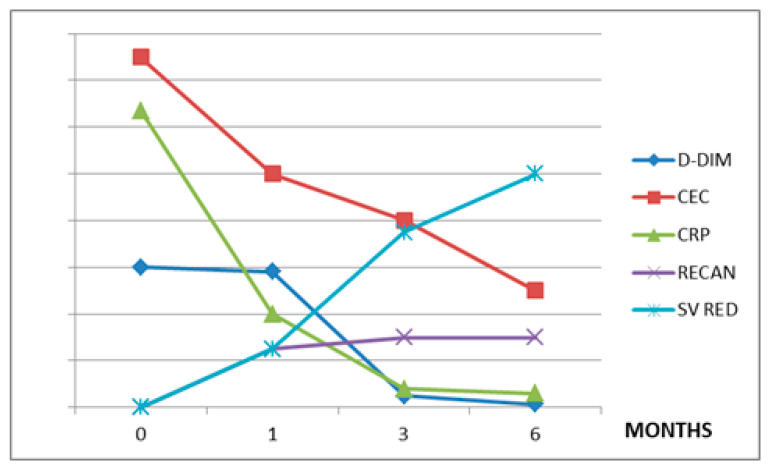
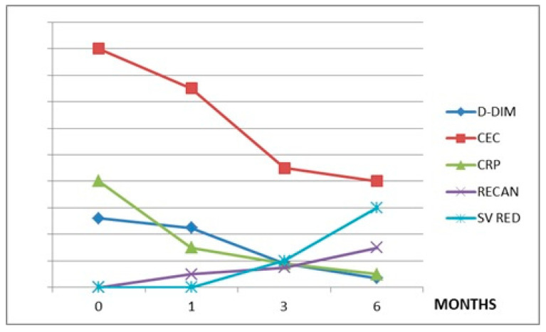
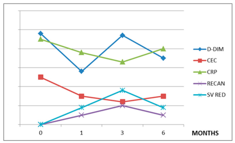
Circulating endothelial cells (CECs) are viable, apoptotic or necrotic cells, identified by CD 146 surface antigen expression, considered a biomarker of thrombotic risk, given their active role in inflammatory, procoagulant and immune processes of the vascular compartment. Growing evidence establishes that CECs are also involved in the pathogenesis of several hematological and solid malignancies. The primary aim of this study was to verify if CEC levels could predict both the course and treatment responses of splanchnic vein thrombosis (SVT), either in patients affected by myeloproliferative neoplasms (MPNs) or liver disease. Thus, a retrospective multicenter study was performed; fifteen patients receiving anticoagulant oral treatment with vitamin k antagonists (VKA) for SVT were evaluated. Nine patients were affected by MPN, and all of them received cytoreduction in addition to anticoagulant therapy; four of these patients had primary myelofibrosis (PMF) and were treated with ruxolitinib (RUX), and one patient with primary myelofibrosis, two patients with essential thrombocythemia (ET), and two patients with polycythemia vera (PV) were treated with hydroxyurea (HU). Six patients affected by liver diseases (three with liver cirrhosis and three with hepatocellular carcinoma) were included as the control group. CECs were assayed by flow cytometry on peripheral blood at specific time points, for up to six months after enrollment. The CEC levels were related to C-reactive protein (CRP) levels, splenic volume reduction, and thrombus recanalization, mainly in MPN patients. In patients with liver cirrhosis (LC) and hepatocellular carcinoma (HCC), for which the mechanism of SVT development is quite different, the relationship between CEC and SV reduction was absent. In conclusion, the CEC levels showed a significant correlation with the extent of venous thrombosis and endothelial cell damage in myeloproliferative neoplasm patients with splanchnic vein thrombosis. Although preliminary, these results show how monitoring CEC levels during cytoreductive and anticoagulant treatments may be useful to improve SVT outcome in MPN patients.
Keywords: circulating endothelial cells; myeloproliferative neoplasms; portal vein thrombosis; recanalization.
Conflict of interest statement
The authors have no conflicts of interest to declare.
Figures
Similar articles
-
Splanchnic Vein Thrombosis in Myelofibrosis-An Underappreciated Hallmark of Disease Phenotype.Int J Mol Sci. 2023 Oct 29;24(21):15717. doi: 10.3390/ijms242115717. Int J Mol Sci. 2023. PMID: 37958701 Free PMC article. Review.
-
Splanchnic vein thrombosis in myeloproliferative neoplasms: treatment algorithm 2018.Blood Cancer J. 2018 Jun 26;8(7):64. doi: 10.1038/s41408-018-0100-9. Blood Cancer J. 2018. PMID: 29946154 Free PMC article. Review.
-
Splanchnic vein thrombosis in myeloproliferative neoplasms: pathophysiology and molecular mechanisms of disease.Ther Adv Hematol. 2017 Mar;8(3):107-118. doi: 10.1177/2040620716680333. Epub 2016 Dec 8. Ther Adv Hematol. 2017. PMID: 28246554 Free PMC article. Review.
-
ADAMTS13, von Willebrand Factor, Platelet Microparticles, Factor VIII, and Impact of Somatic Mutations in the Pathogenesis of Splanchnic Vein Thrombosis Associated with BCR-ABL-Negative Myeloproliferative Neoplasms.Life (Basel). 2024 Apr 9;14(4):486. doi: 10.3390/life14040486. Life (Basel). 2024. PMID: 38672756 Free PMC article.
-
Natural history of polycythemia vera and essential thrombocythemia presenting with splanchnic vein thrombosis.Ann Hematol. 2020 Apr;99(4):791-798. doi: 10.1007/s00277-020-03965-z. Epub 2020 Feb 22. Ann Hematol. 2020. PMID: 32086587
Cited by
-
Are circulating endothelial cells the next target for transcriptome-level pathway analysis in ARDS?Am J Physiol Lung Cell Mol Physiol. 2023 Apr 1;324(4):L393-L399. doi: 10.1152/ajplung.00353.2022. Epub 2023 Feb 7. Am J Physiol Lung Cell Mol Physiol. 2023. PMID: 36749906 Free PMC article.
-
The role and mechanisms of microvascular damage in the ischemic myocardium.Cell Mol Life Sci. 2023 Oct 29;80(11):341. doi: 10.1007/s00018-023-04998-z. Cell Mol Life Sci. 2023. PMID: 37898977 Free PMC article. Review.
-
A phase I study of docetaxel plus synthetic lycopene in metastatic prostate cancer patients.Clin Transl Med. 2024 Mar;14(3):e1627. doi: 10.1002/ctm2.1627. Clin Transl Med. 2024. PMID: 38515274 Free PMC article. Clinical Trial.
-
Splanchnic Vein Thrombosis in Myelofibrosis-An Underappreciated Hallmark of Disease Phenotype.Int J Mol Sci. 2023 Oct 29;24(21):15717. doi: 10.3390/ijms242115717. Int J Mol Sci. 2023. PMID: 37958701 Free PMC article. Review.
-
Pathological and genomic features of myeloproliferative neoplasms associated with splanchnic vein thrombosis in a single-center cohort.Ann Hematol. 2023 Jun;102(6):1409-1420. doi: 10.1007/s00277-023-05217-2. Epub 2023 Apr 20. Ann Hematol. 2023. PMID: 37079068 Review.
References
-
- Monestiroli S., Mancuso P., Burlini A., Pruneri G., Dell’Agnola C., Gobbi A., Martinelli G., Bertolini F. Kinetics and viability of circulating endothelial cells as surrogate angiogenesis marker in an animal model of human lymphoma. Cancer Res. 2001;61:4341–4344. - PubMed
LinkOut - more resources
Full Text Sources
Research Materials
Miscellaneous
