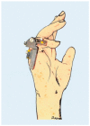
. 2022 Mar 1;12(3):378.
doi: 10.3390/jpm12030378.

A Personalized Approach to Treat Advanced Stage Severely Contracted Joints in Dupuytren's Disease with a Unique Skeletal Distraction Device-Utilizing Modern Imaging Tools to Enhance Safety for the Patient

Affiliations

A Personalized Approach to Treat Advanced Stage Severely Contracted Joints in Dupuytren's Disease with a Unique Skeletal Distraction Device-Utilizing Modern Imaging Tools to Enhance Safety for the Patient

Wibke Müller-Seubert et al. J Pers Med. .

Abstract

Background: While surgical therapy for Dupuytren's disease is a well-established standard procedure, severe joint flexion deformities in advanced Dupuytren's disease remain challenging to treat. Skeletal distraction has proven to be an additional treatment option.

Methods: We analyzed the surgical treatment algorithm, including the application of a skeletal distraction device, in patients with a flexion deformity due to Dupuytren's disease, Iselin stage III or IV, who were operated on from 2003 to 2020 in our department.

Results: From a total of 724 patients, we included the outcome of 55 patients' fingers in this study, who had undergone additional skeletal joint distraction with our Erlangen device. Additional fasciotomy or fasciectomy, in a one- or two-staged procedure, was performed in all patients, according to the individual findings and necessities. The range of motion of the PIP joint improved from 12° to 53°. A number of complications, in all steps of the treatment, were noted in a total of 36.4% of patients, including the development of fractures (16.4%), followed by vessel injury, pin infections, and complex regional pain syndrome (5%).

Conclusions: Additional skeletal distraction improves the range of motion of severely contracted joints in Dupuytren's disease. Nevertheless, careful patient selection is necessary, due to the moderate rate of complications.

Keywords: Dupuytren contracture; distracted driving; joint dislocation.


Conflict of interest statement

The authors declare no conflict of interest.

Figures
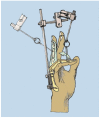
Figure 1
Figure 1
Schematic depiction of Messina’s skeletal distraction device, modified from Brenner P, Ray Ghazi M. (ed.) Morbus Dupuytren: Ein chirurgisches Therapiekonzept, Springer Verlag Berlin-Heidelberg-New York, 2003, ISBN 978-3-7091-6723-6 (copyright R E Horch).
Figure 2
Figure 2
Erlangen miniaturized distraction device with turning worm-screw mechanism (copyright R E Horch 2020).
Figure 3
Figure 3
Hyperspectral imaging showing malperfusion (yellow parts) after extension subfigure (B) in comparison to the non-extended finger subfigure (A). Red color represents well-perfused skin, as measured with the no-contact hyperspectral imaging device, consistently distributed over the visible palm of the hand. A change in optimal oxygen saturation (red = 100% oxygen saturation) is shown as yellow, representing decreasing amounts of oxygen saturation. Under distraction, more yellow can be observed in the right depiction (B). Corresponding clinical images of the non-extended (C) and extended finger (D).
Figure 4
Figure 4
Individualized treatment regimen of DD.
Figure 5
Figure 5
Patient with stage IV DD of the little finger.
Figure 6
Figure 6
Preoperative X-ray of stage IV flexion deformity and schematic drawing.
Figure 7
Figure 7
Intraoperative application of the skeletal distraction device in combination with fasciectomy.
Figure 8
Figure 8
Completely extended PIP joint of the same patient shown in Figure 5, Figure 6 and Figure 7 at 6 weeks of distraction showing complete release of the PIP joint.
Figure 9
Figure 9
Presented patient 8 weeks after fasciectomy.

References

    1. Loos B., Puschkin V., Horch R.E. 50 years experience with Dupuytren’s contracture in the Erlangen University Hospital—A retrospective analysis of 2919 operated hands from 1956 to 2006. BMC Musculoskelet. Disord. 2007;8:60. doi: 10.1186/1471-2474-8-60. - DOI - PMC - PubMed
    1. Karabeg R., Jakirlic M., Arslanagic S., Dujso V., Obradovic G., Zeco A. Results of surgery treatment of Dupuytren’s contracture in 115 patients. Med. Arch. 2012;66:329–331. doi: 10.5455/medarh.2012.66.329-331. - DOI - PubMed
    1. Evans R.B., Dell P.C., Fiolkowski P. A clinical report of the effect of mechanical stress on functional results after fasciectomy for dupuytren’s contracture. J. Hand Ther. 2002;15:331–339. doi: 10.1016/S0894-1130(02)80004-7. - DOI - PubMed
    1. Jones N.F., Huang J.I. Emergency microsurgical revascularization for critical ischemia during surgery for Dupuytren’s contracture: A case report. J. Hand Surg. Am. 2001;26:1125–1128. doi: 10.1053/jhsu.2001.28428. - DOI - PubMed
    1. Tubiana R., Fahrer M., McCullough C.J. Recurrence and other complications in surgery of Dupuytren’s contracture. Clin. Plast. Surg. 1981;8:45–50. doi: 10.1016/S0094-1298(20)30472-7. - DOI - PubMed

LinkOut - more resources