

Reducing the Excess Activin Signaling Rescues Muscle Degeneration in Myotonic Dystrophy Type 2 Drosophila Model
- PMID: 35330385
- PMCID: PMC8948895
- DOI: 10.3390/jpm12030385
Reducing the Excess Activin Signaling Rescues Muscle Degeneration in Myotonic Dystrophy Type 2 Drosophila Model
Abstract
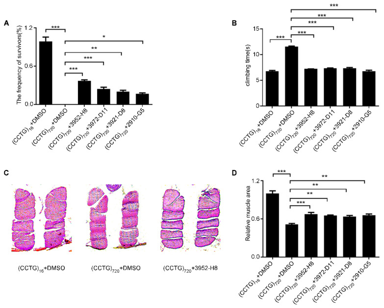
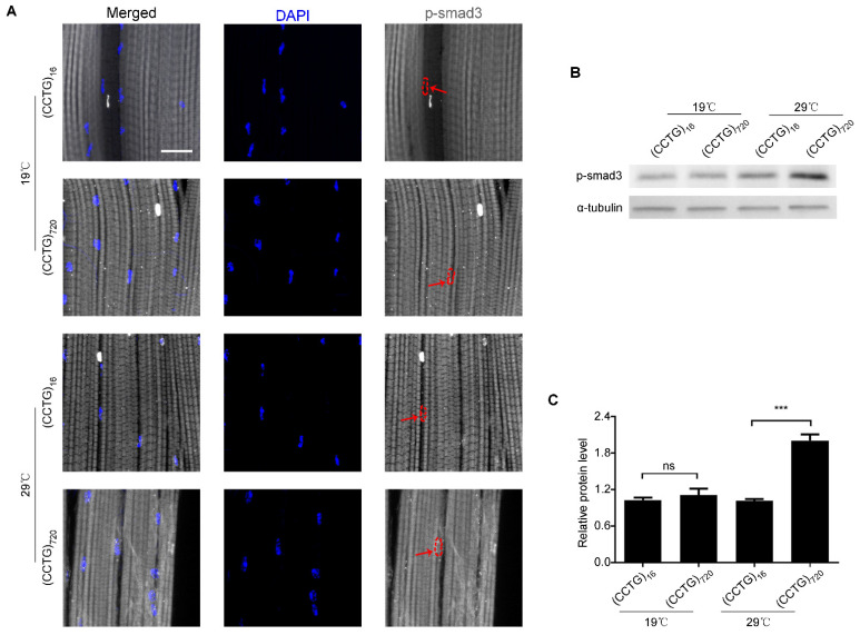
Expanded non-coding RNA repeats of CCUG are the underlying genetic causes for myotonic dystrophy type 2 (DM2). There is an urgent need for effective medications and potential drug targets that may alleviate the progression of the disease. In this study, 3140 small-molecule drugs from FDA-approved libraries were screened through lethality and locomotion phenotypes using a DM2 Drosophila model expressing 720 CCTG repeats in the muscle. We identified ten effective drugs that improved survival and locomotor activity of DM2 flies, including four that share the same predicted targets in the TGF-β pathway. The pathway comprises two major branches, the Activin and BMP pathways, which play critical and complex roles in skeletal development, maintenance of homeostasis, and regeneration. The Drosophila model recapitulates pathological features of muscle degeneration in DM2, displaying shortened lifespan, a decline in climbing ability, and progressive muscle degeneration. Increased levels of p-smad3 in response to activin signaling were observed in DM2 flies. Decreased levels of activin signaling using additional specific inhibitors or genetic method ameliorated climbing defects, crushed thoraxes, structure, and organization of muscle fibers. Our results demonstrate that a decrease in activin signaling is sufficient to rescue muscle degeneration and is, therefore, a potential therapeutic target for DM2.
Keywords: Drosophila; TGF-β pathway; chemical screen; myotonic dystrophy type 2.
Conflict of interest statement
The authors declare no conflict of interest.
Figures


Similar articles
-
Expanded CCUG repeat RNA expression in Drosophila heart and muscle trigger Myotonic Dystrophy type 1-like phenotypes and activate autophagocytosis genes.Sci Rep. 2017 Jun 6;7(1):2843. doi: 10.1038/s41598-017-02829-3. Sci Rep. 2017. PMID: 28588248 Free PMC article.
-
Modulating CCTG repeat expansion toxicity in DM2 Drosophila model through TDP1 inhibition.EMBO Mol Med. 2025 May;17(5):967-992. doi: 10.1038/s44321-025-00217-3. Epub 2025 Mar 25. EMBO Mol Med. 2025. PMID: 40133672 Free PMC article.
-
Myotonic dystrophy types 1 and 2.Handb Clin Neurol. 2011;101:193-237. doi: 10.1016/B978-0-08-045031-5.00015-3. Handb Clin Neurol. 2011. PMID: 21496635 Review.
-
Molecular mechanisms of muscle atrophy in myotonic dystrophies.Int J Biochem Cell Biol. 2013 Oct;45(10):2280-7. doi: 10.1016/j.biocel.2013.06.010. Epub 2013 Jun 21. Int J Biochem Cell Biol. 2013. PMID: 23796888 Free PMC article. Review.
-
Immortalized human myotonic dystrophy muscle cell lines to assess therapeutic compounds.Dis Model Mech. 2017 Apr 1;10(4):487-497. doi: 10.1242/dmm.027367. Epub 2017 Feb 10. Dis Model Mech. 2017. PMID: 28188264 Free PMC article.
Cited by
-
Development of Therapeutic Approaches for Myotonic Dystrophies Type 1 and Type 2.Int J Mol Sci. 2022 Sep 10;23(18):10491. doi: 10.3390/ijms231810491. Int J Mol Sci. 2022. PMID: 36142405 Free PMC article. Review.
-
Aligning with the 3Rs: alternative models for research into muscle development and inherited myopathies.BMC Vet Res. 2024 Oct 18;20(1):477. doi: 10.1186/s12917-024-04309-z. BMC Vet Res. 2024. PMID: 39425123 Free PMC article. Review.
-
Modeling Myotonic Dystrophy Type 2 Using Drosophila melanogaster.Int J Mol Sci. 2023 Sep 16;24(18):14182. doi: 10.3390/ijms241814182. Int J Mol Sci. 2023. PMID: 37762484 Free PMC article. Review.
-
Decoding Nucleotide Repeat Expansion Diseases: Novel Insights from Drosophila melanogaster Studies.Int J Mol Sci. 2024 Nov 2;25(21):11794. doi: 10.3390/ijms252111794. Int J Mol Sci. 2024. PMID: 39519345 Free PMC article. Review.
-
Drosophila Models Reveal Properties of Mutant Lamins That Give Rise to Distinct Diseases.Cells. 2023 Apr 12;12(8):1142. doi: 10.3390/cells12081142. Cells. 2023. PMID: 37190051 Free PMC article.
References
-
- Brook J.D., McCurrach M.E., Harley H.G., Buckler A.J., Church D., Aburatani H., Hunter K., Stanton V.P., Thirion J.P., Hudson T., et al. Molecular basis of myotonic dystrophy: Expansion of a trinucleotide (CTG) repeat at the 3′ end of a transcript encoding a protein kinase family member. Cell. 1992;68:799–808. doi: 10.1016/0092-8674(92)90154-5. - DOI - PubMed
Grants and funding
- 81571253/National Natural Science Foundation of China
- 81771385/National Natural Science Foundation of China
- 2019SK1010/Hunan Science and Technology major project of Birth Defect cooperative control
- 82071273/National Natural Science Foundation of China
- 82101958/National Natural Science Foundation of China
LinkOut - more resources
Full Text Sources
Molecular Biology Databases
Miscellaneous
