Physiology of Astroglial Excitability
- PMID: 35330636
- PMCID: PMC8788756
- DOI: 10.1093/function/zqaa016
Physiology of Astroglial Excitability
Abstract
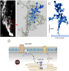
Classic physiology divides all neural cells into excitable neurons and nonexcitable neuroglia. Neuroglial cells, chiefly responsible for homeostasis and defense of the nervous tissue, coordinate their complex homeostatic responses with neuronal activity. This coordination reflects a specific form of glial excitability mediated by complex changes in intracellular concentration of ions and second messengers organized in both space and time. Astrocytes are equipped with multiple molecular cascades, which are central for regulating homeostasis of neurotransmitters, ionostasis, synaptic connectivity, and metabolic support of the central nervous system. Astrocytes are further provisioned with multiple receptors for neurotransmitters and neurohormones, which upon activation trigger intracellular signals mediated by Ca2+, Na+, and cyclic AMP. Calcium signals have distinct organization and underlying mechanisms in different astrocytic compartments thus allowing complex spatiotemporal signaling. Signals mediated by fluctuations in cytosolic Na+ are instrumental for coordination of Na+ dependent astrocytic transporters with tissue state and homeostatic demands. Astroglial ionic excitability may also involve K+, H+, and Cl-. The cyclic AMP signalling system is, in comparison to ions, much slower in targeting astroglial effector mechanisms. This evidence review summarizes the concept of astroglial intracellular excitability.
Keywords: astrocyte; astrocytic processes; astroglial excitability; calcium signaling; ionic signaling; sodium signaling.
© The Author(s) 2020. Published by Oxford University Press on behalf of American Physiological Society.
Figures


Similar articles
-
Ionic signalling in astroglia beyond calcium.J Physiol. 2020 May;598(9):1655-1670. doi: 10.1113/JP277478. Epub 2019 Feb 28. J Physiol. 2020. PMID: 30734296 Review.
-
Physiology of neuroglia of the central nervous system.Handb Clin Neurol. 2025;209:69-91. doi: 10.1016/B978-0-443-19104-6.00005-X. Handb Clin Neurol. 2025. PMID: 40122632 Review.
-
Sodium homeostasis and signalling: The core and the hub of astrocyte function.Cell Calcium. 2024 Jan;117:102817. doi: 10.1016/j.ceca.2023.102817. Epub 2023 Nov 4. Cell Calcium. 2024. PMID: 37979342
-
Na+-dependent transporters: The backbone of astroglial homeostatic function.Cell Calcium. 2020 Jan;85:102136. doi: 10.1016/j.ceca.2019.102136. Epub 2019 Dec 3. Cell Calcium. 2020. PMID: 31835178 Review.
-
Astroglial calcium signalling in Alzheimer's disease.Biochem Biophys Res Commun. 2017 Feb 19;483(4):1005-1012. doi: 10.1016/j.bbrc.2016.08.088. Epub 2016 Aug 18. Biochem Biophys Res Commun. 2017. PMID: 27545605 Review.
Cited by
-
Principles of Astrogliopathology.Adv Neurobiol. 2021;26:55-73. doi: 10.1007/978-3-030-77375-5_3. Adv Neurobiol. 2021. PMID: 34888830 Free PMC article.
-
Role of Microglia and Astrocytes in Alzheimer's Disease: From Neuroinflammation to Ca2+ Homeostasis Dysregulation.Cells. 2022 Sep 1;11(17):2728. doi: 10.3390/cells11172728. Cells. 2022. PMID: 36078138 Free PMC article. Review.
-
The Association Between Antidepressant Effect of SSRIs and Astrocytes: Conceptual Overview and Meta-analysis of the Literature.Neurochem Res. 2021 Oct;46(10):2731-2745. doi: 10.1007/s11064-020-03225-6. Epub 2021 Feb 1. Neurochem Res. 2021. PMID: 33527219
-
Transgenic Mouse Overexpressing Spermine Oxidase in Cerebrocortical Neurons: Astrocyte Dysfunction and Susceptibility to Epileptic Seizures.Biomolecules. 2022 Jan 25;12(2):204. doi: 10.3390/biom12020204. Biomolecules. 2022. PMID: 35204705 Free PMC article. Review.
-
Astroglial Serotonin Receptors as the Central Target of Classic Antidepressants.Adv Neurobiol. 2021;26:317-347. doi: 10.1007/978-3-030-77375-5_13. Adv Neurobiol. 2021. PMID: 34888840 Free PMC article.
References
-
- von Haller A. Commentarii Societatis Regiae Scientiarum Gottingensis. Gottingen: Vadenhoek, 1753.
-
- Hooke R. Micrographia. UK: Folio Society, 2017:1655.
-
- Malpighi M. In de Viscerum Structura Exercitatio Anatomica. Bologna: Typographia Iacobi Montij, 1666.
-
- Shapiro S. Antony van Leeuwenhoek; a review of his life and work. J Biol Photogr Assoc 1955;23(2–3):49–57. - PubMed
-
- Swedenborg E. The Brain, Considered Anatomically, Physiologically and Phylosopically (translated and edited by Tafel R. L.) in 4 volumes. London: James Speirs, 1882.
Publication types
MeSH terms
Substances
LinkOut - more resources
Full Text Sources
Miscellaneous
