Case Reports
. 2022 Mar 21;17(5):1646-1655.
doi: 10.1016/j.radcr.2022.02.075. eCollection 2022 May.

Pulmonary lymphangioleiomyomatosis (LAM): A literature overview and case report

Affiliations
Case Reports

Pulmonary lymphangioleiomyomatosis (LAM): A literature overview and case report

Cung-Van Cong et al. Radiol Case Rep. .

Abstract

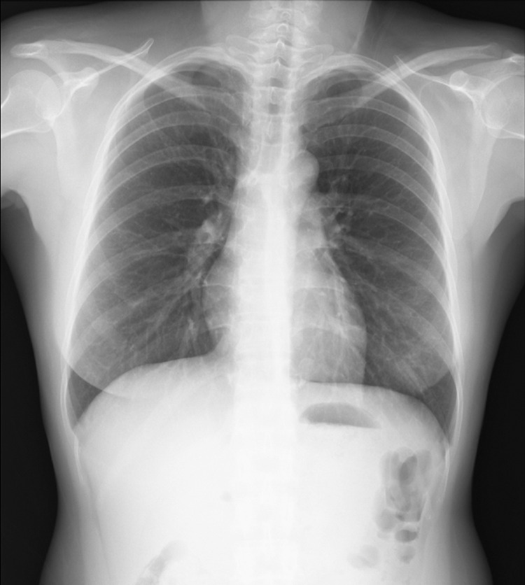
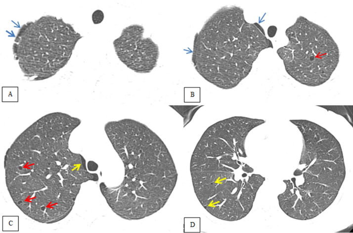
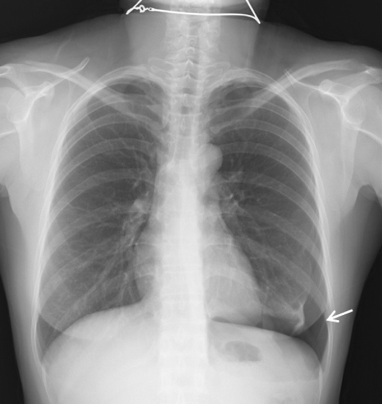
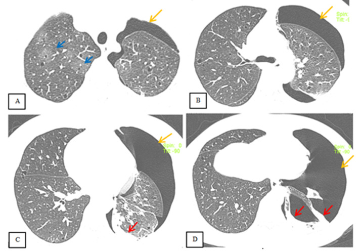
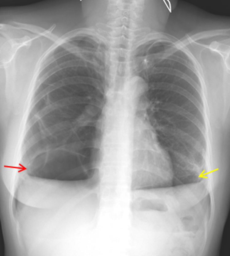
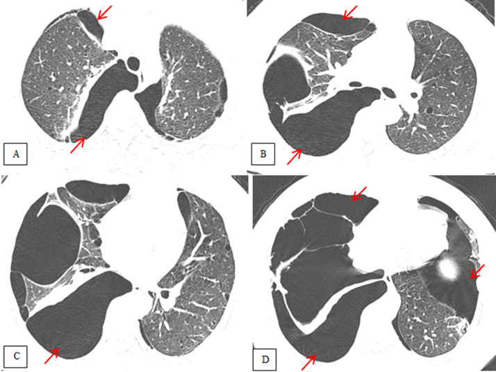
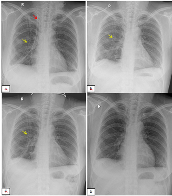
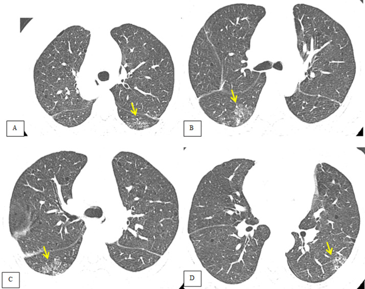
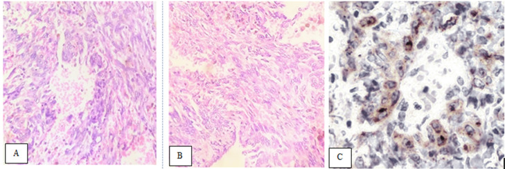
Lymphangioleiomyomatosis is a rare multisystem disease associated with genetic mutations. The disease usually occurs in women of childbearing age and is characterized by infiltration of immature smooth muscle cells into the lungs, airways, and axial lymphatic systems of the chest and abdomen. The disease often destroys lung parenchyma and produces air cysts. Lymphangioleiomyomatosis cell infiltration of the lymphatic axis can affect hilar lymph nodes, mediastinal ganglia, and extrathoracic lymph nodes. The disease can cause lymphatic dilation in the lungs and thoracic ducts, causing chylous effusion into the pleural or abdominal cavities. Invasion of cells into the walls of pulmonary veins can lead to venous obstruction and pulmonary venous hypertension with hemoptysis. Most patients present with cough, dyspnea, pneumothorax, hemoptysis, and abnormal lung function. Definitive diagnosis is usually based on histopathology and immunohistochemistry. We present a case of LAM in a 36-year-old female patient who was confirmed by specimens obtained from pneumothorax surgery and positive immunohistochemical staining with HMB-45.

Keywords: Computed tomography; LAM; Lung lymphangioleiomyomatosis; PEComa.


Figures

Fig 1 –
Fig. 1
Chest X-ray radiograph of the patient at the first examination at NLH (note that the patient underwent surgery for pneumothorax 1 year prior). The main sign is increased chest volume (hypertension of the lungs) and the parenchymal structure is similar in lung regions. In addition, no parenchymal abnormalities are seen.
Fig 2 –
Fig. 2
Chest CT image of the patient's lungs at 4 different levels. (A) The right-sided, low-grade pneumothorax appears, to be localized (green arrows). (B) Lower view shows right-sided pneumothorax (green arrows) and the presence of a 5-mm-diameter, thin-walled air cyst in the upper lobe of left lung. (C) A lower section shows pneumothorax (yellow arrow) and multiple small air cysts (red arrows). (D) Subsection of the carina shows no right-sided pneumothorax and few small air cysts (yellow arrows) (Color version of the figure is available online.)
Fig 3 –
Fig. 3
Chest X-ray taken 13 days after admission. X-ray shows increased chest volume and left-sided pneumothorax (white arrow). Note that the parenchymal structure is uniform in all regions.
Fig 4 –
Fig. 4
Chest CT taken 13 days after admission. (A) A left pneumothorax (yellow arrow) is visible, with no air in the right pleural space, and small air cysts in the parenchyma of the right upper lobe in the area of ground-glass opacity (GGO) (blue arrow). (B, C, D) Severe left pneumothorax (yellow arrow) is visible with passive atelectasis (red arrow). Many small air cysts remain scattered throughout the lung parenchyma (A, B, C, D) (Color version of the figure is available online.)
Fig 5 –
Fig. 5
Chest X-ray 3 months after laparoscopic surgery for left pneumothorax. Right pneumothorax (red arrow) and a suspected left pneumothorax (yellow arrow) are visible (Color version of the figure is available online.)
Fig 6 –
Fig. 6
Chest CT 3 months after laparoscopic surgery for left pneumothorax. (A–C) Bilateral pneumothorax; the right side is more severe than the left (red arrows) (Color version of the figure is available online.)
Fig 7 –
Fig. 7
A series of postoperative follow-up X-ray radiograph (for 13 days). (A) Two drainage tubes (red arrow) and the mediastinal pleural space and right posterior pleural space (yellow arrow) are visible. (B) The pleural-mediastinal drainage tube was withdrawn after 3 days. (C) The right pleural drainage tube remains in place, behind (yellow arrow). (D) On day 13 after surgery, the drainage tube has been removed, and no air is visible in the bilateral pleural spaces (Color version of the figure is available online.)
Fig 8 –
Fig. 8
Chest CT of the patient taken 1 month after discharge. A–D: Bilateral pneumothorax is no longer visible. Nodular infiltrates are seen in segment VI of both lungs (yellow arrow). Small air cocoons persist but are inconspicuous (Color version of the figure is available online.)
Fig 9 –
Fig. 9
Microscopic examination of lung lesion biopsies obtained through laparoscopic surgery. (A) Histopathological examination of the first surgical specimen. (B) Histopathological examination of the second surgical specimen. Hematoxylin and eosin (HE) staining shows proliferation of LAM cell nodules with central spindle characteristics and peripheral epithelial cells (HE x 200). Histopathological lesions show that the lung parenchyma has dilated alveoli, the interstitial tissue, the alveolar wall contains proliferated rhombus cells, the nuclei are oval-shaped and regular, benign, and without mitotic nuclei. The cells are arranged in clusters and scattered in the dilated alveolar regions. Histopathological diagnosis: lesions consistent with pulmonary lymphangioma (LAM). (C) Immunohistochemical staining with HMB-45 was positive in epithelial cells (IHCS x 400).

Similar articles

Cited by

References

    1. Richard Webb W., Higgins Charles B. Thoracic imaging: pulmonary and cardiovascular radilogy. Wolters Kluwer. 2017;3:649–653.
    1. Richard Webb W., Muller Nestor L., Naidich David P. High – resolution CT of the lung. Wolters Kluwer. 2015;3:501–510.
    1. McCormack FX, Gupta N, Finlay GR, Young LR, Taveira-DaSilva AM, Glasgow CG, et al. ATS/JRS committee on lymphangioleiomyomatosis. Official American thoracic Society/Japanese respiratory society clinical practice guidelines: lymphangioleiomyomatosis diagnosis and management. Am J Respir Crit Care Med. 2016;194(6):748–761. doi: 10.1164/rccm.201607-1384ST. - DOI - PMC - PubMed
    1. Sathirareuangchai S, Shimizu D, Vierkoetter KR. Pulmonary lymphangioleiomyomatosis: a case report and literature review. Hawaii J Health Soc Welf. 2020;79(7):224–229. - PMC - PubMed
    1. Sclafani A, VanderLaan P. Lymphangioleiomyomatosis. N Engl J Med. 2018;378(23):2224. doi: 10.1056/NEJMicm1712581. - DOI - PubMed

Publication types

LinkOut - more resources