. 2022 Mar 7:13:802577.
doi: 10.3389/fmicb.2022.802577. eCollection 2022.

Dissecting the Species-Specific Virome in Culicoides of Thrace

Affiliations

Dissecting the Species-Specific Virome in Culicoides of Thrace

Konstantinos Konstantinidis et al. Front Microbiol. .

Abstract

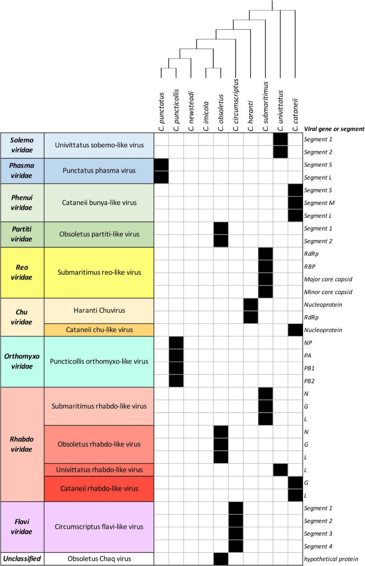
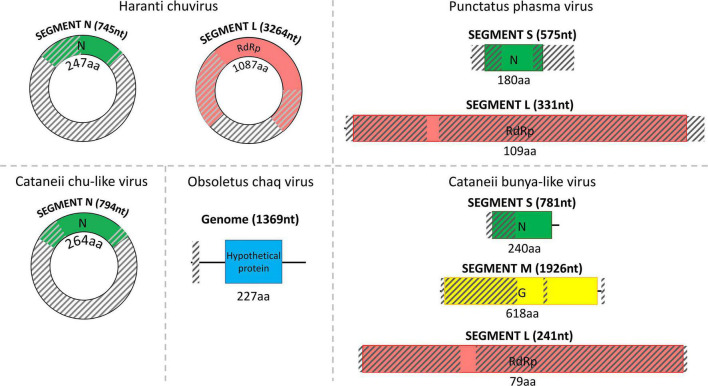
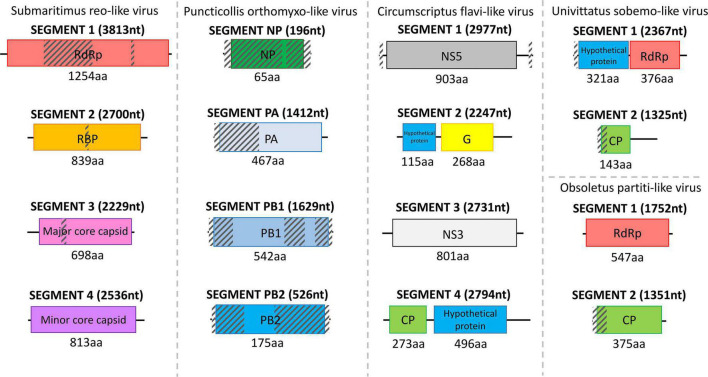
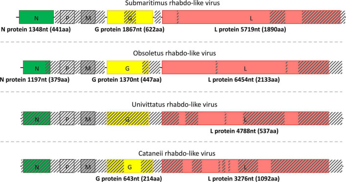
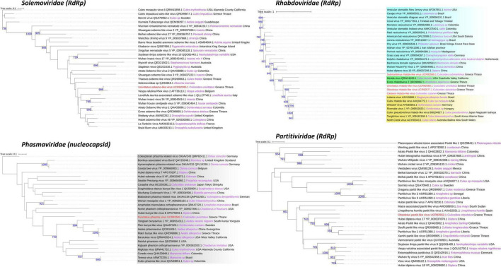
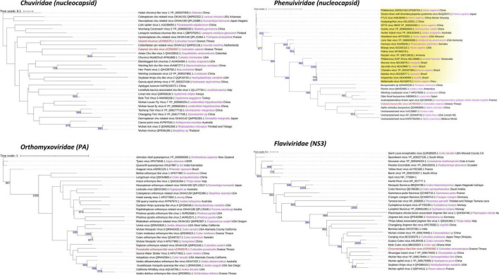
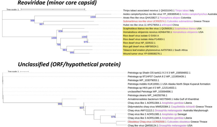
Biting midges (Culicoides) are vectors of arboviruses of both veterinary and medical importance. The surge of emerging and reemerging vector-borne diseases and their expansion in geographical areas affected by climate change has increased the importance of understanding their capacity to contribute to novel and emerging infectious diseases. The study of Culicoides virome is the first step in the assessment of this potential. In this study, we analyzed the RNA virome of 10 Culicoides species within the geographical area of Thrace in the southeastern part of Europe, a crossing point between Asia and Europe and important path of various arboviruses, utilizing the Ion Torrent next-generation sequencing (NGS) platform and a custom bioinformatics pipeline based on TRINITY assembler and alignment algorithms. The analysis of the RNA virome of 10 Culicoides species resulted in the identification of the genomic signatures of 14 novel RNA viruses, including three fully assembled viruses and four segmented viruses with at least one segment fully assembled, most of which were significantly divergent from previously identified related viruses from the Solemoviridae, Phasmaviridae, Phenuiviridae, Reoviridae, Chuviridae, Partitiviridae, Orthomyxoviridae, Rhabdoviridae, and Flaviviridae families. Each Culicoides species carried a species-specific set of viruses, some of which are related to viruses from other insect vectors in the same area, contributing to the idea of a virus-carrier web within the ecosystem. The identified viruses not only expand our current knowledge on the virome of Culicoides but also set the basis of the genetic diversity of such viruses in the area of southeastern Europe. Furthermore, our study highlights that such metagenomic approaches should include as many species as possible of the local virus-carrier web that interact and share the virome of a geographical area.

Keywords: Culicoides; Greece; Thrace; arboviruses; emerging infectious diseases; metagenomics; vectors; virome analysis.


Conflict of interest statement

PP was employed by the company Evrofarma S.A. AN was employed by the company Geotechno Ygeionomiki O.E. The remaining authors declare that the research was conducted in the absence of any commercial or financial relationships that could be construed as a potential conflict of interest.

Figures

FIGURE 1
FIGURE 1
List of detected viruses within Culicoides samples. Black filled squares indicate presence of the corresponding viral genes or segments.
FIGURE 2
FIGURE 2
Structure of the detected chu/chu-like viruses, phasma virus, bunya-like virus, and uncharacterized Chaq virus separated by dashed lines. The total length of each assembled viral sequence is indicated in nucleotides between parentheses (nt), whereas the corresponding encoded protein length is shown below each colorful rectangular region in amino acids (aa). Translation of all viral genomic sequences was done by ExPASy Translate online tool. The diagonally shaded regions upon each viral genomic sequence depict areas that could not be successfully assembled, and their lengths were estimated after MAFFt alignment against the most closely related viral nucleotide sequences.
FIGURE 3
FIGURE 3
Structure of the detected reo-like virus, orthomyxo-like virus, flavi-like virus, sobemo-like virus, and partiti-like virus separated by dashed lines. The length of each assembled viral sequence is indicated in nucleotides between parentheses (nt), whereas the corresponding encoded protein length is shown below each colorful rectangular region in amino acids (aa). Translation of all viral genomic sequences was done by ExPASy Translate online tool. The diagonally shaded regions upon each viral genomic sequence depict areas that could not be successfully assembled, and their lengths were estimated after MAFFt alignment against the most closely related viral nucleotide sequences.
FIGURE 4
FIGURE 4
Structure of the detected rhabdo-like viruses separated by dashed lines. The length of each assembled viral sequence is indicated in nucleotides between parentheses (nt), whereas the corresponding encoded protein length is shown below each colorful rectangular region in amino acids (aa). Translation of all viral genomic sequences was done by ExPASy Translate online tool. The diagonally shaded regions upon each viral genomic sequence depict areas that could not be successfully assembled, and their lengths were estimated after MAFFt alignment against the most closely related viral nucleotide sequences.
FIGURE 5
FIGURE 5
Solemoviridae, Phasmaviridae, Rhabdoviridae, and Partitiviridae family phylogenetic trees of the identified viruses in this study (red text). Phylogenetic analysis was performed according to the protein indicated between the parentheses after each viral family name using its amino acid sequence. FastME minimum evolution substitution model was utilized as part of the NGPhylogeny.fr methodology. All of the presented phylogenetic trees were rooted according to the outgroup rooting method. Bootstrap values (blue text) were obtained from 1,000 bootstrap replicates, and only those greater than 700 are displayed at the start of each node. Host and country origin information of homologous viruses was also extracted, if applicable, depicted here in purple and black text, respectively. Viruses with known genus taxonomy have also been highlighted accordingly. More specifically, Orthophasmavirus genus is colored gray in the Phasmaviridae family, whereas Sigmavirus, Ohlsrhavirus, and Merhavirus genera are displayed with cyan, yellow, and green gradients, respectively, in the Rhabdoviridae family.
FIGURE 6
FIGURE 6
Chuviridae, Orthomyxoviridae, Phenuiviridae, and Flaviviridae family phylogenetic trees of the identified viruses in this study (red text). Phylogenetic analysis was performed according to the protein indicated between the parentheses after each viral family name using its amino acid sequence. FastME minimum evolution substitution model was utilized as part of the NGPhylogeny.fr methodology. All of the presented phylogenetic trees were rooted according to the outgroup rooting method. Bootstrap values (blue text) were obtained from 1,000 bootstrap replicates, and only those greater than 700 are displayed at the start of each node. Host and country origin information of homologous viruses was also extracted, if applicable, depicted here in purple and black text, respectively. Viruses with known genus taxonomy have also been highlighted accordingly. More specifically, Phlebovirus genus is colored yellow in the Phenuiviridae family.
FIGURE 7
FIGURE 7
Reoviridae and unclassified family phylogenetic trees of the identified viruses in this study (red text). Phylogenetic analysis was performed according to the protein indicated between the parentheses after each viral family name using its amino acid sequence. FastME minimum evolution substitution model was utilized as part of the NGPhylogeny.fr methodology. All of the presented phylogenetic trees were rooted according to the outgroup rooting method. Bootstrap values (blue text) were obtained from 1,000 bootstrap replicates, and only those greater than 700 are displayed at the start of each node. Host and country origin information of homologous viruses was also extracted, if applicable, depicted here in purple and black text, respectively. Viruses with known genus taxonomy have also been highlighted accordingly. More specifically, Phytoreovirus genus is colored yellow in the Reoviridae family.

Similar articles

Cited by

References

    1. Aguiar E. R. G. R., Olmo R. P., Paro S., Ferreira F. V., de Faria I. J., da S., et al. (2015). Sequence-Independent Characterization of Viruses Based on the Pattern of Viral Small RNAs Produced by the Host. Nucleic Acids Res. 43 6191–6206. 10.1093/nar/gkv587 - DOI - PMC - PubMed
    1. Altschul S. F., Gish W., Miller W., Myers E. W., Lipman D. J. (1990). Basic Local Alignment Search Tool. J. Mol. Biol. 215 403–410. 10.1016/S0022-2836(05)80360-2 - DOI - PubMed
    1. Atoni E., Zhao L., Karungu S., Obanda V., Agwanda B., Xia H., et al. (2019). The Discovery and Global Distribution of Novel Mosquito-Associated Viruses in the Last Decade (2007-2017). Rev. Med. Virol. 29:e2079. 10.1002/rmv.2079 - DOI - PubMed
    1. Augot D., Mathieu B., Hadj-Henni L., Barriel V., Mena S. Z., Smolis S., et al. (2017). Molecular Phylogeny of 42 Species of Culicoides (Diptera, Ceratopogonidae) from Three Continents. Parasite 24:23. 10.1051/parasite/2017020 - DOI - PMC - PubMed
    1. Batson J., Dudas G., Haas-Stapleton E., Kistler A. L., Li L. M., Logan P., et al. (2021). Single Mosquito Metatranscriptomics Identifies Vectors, Emerging Pathogens and Reservoirs in One Assay. eLife 10:e68353. 10.7554/eLife.68353 - DOI - PMC - PubMed

LinkOut - more resources