

DELAYED DIAGNOSIS OF AVM PRESENTING WITH STROKE AND SEIZURES IN A YOUNG NIGERIAN: A CASE REPORT
- PMID: 35330890
- PMCID: PMC8935678
DELAYED DIAGNOSIS OF AVM PRESENTING WITH STROKE AND SEIZURES IN A YOUNG NIGERIAN: A CASE REPORT
Abstract
Background: Brain arteriovenous malformations (BAVM) are a cause of intracerebral haemorrhage (ICH) and seizures especially in young patients. ICH due to BAVMs seem to have relatively better neurologic outcomes compared to other causes of spontaneous ICH as patients often recover fully. In this report we highlight a case of delayed diagnosis of BAVM in a young man who presented with seizures and stroke.
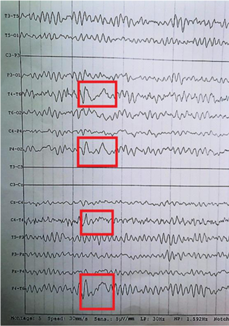
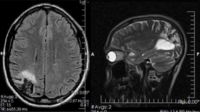
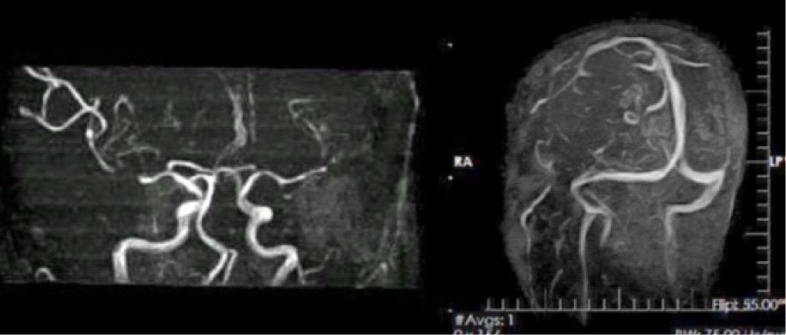
Case summary: A 36-year-old man was referred on account of focal, secondarily generalized tonic clonic convulsions. He had suffered a right ICH 3 years before the index presentation. His general physical and neurologic examination were normal. Electroencephalography revealed right sided focal epileptiform discharges and brain MRI revealed a right parieto-occipital AVM. The seizures were controlled with carbamazepine and he was referred for neurosurgical evaluation.
Conclusion: BAVMs are an important cause of intracerebral haemorrhage and attendant neurologic morbidity especially in young individuals. Neuroimaging plays a central role in BAVM diagnosis and MRI is of great value where facilities and expertise for conventional angiography do not exist. In some instances, delayed presentation of BAVM cases may be due to relatively better neurologic outcomes in BAVM-related ICH.
© Association of Resident Doctors, UCH, Ibadan.
Figures
References
-
- Friedlander RM. Arteriovenous malformations of the brain. N Engl J Med. 2007 Jun 28; 356(26):2704–2712. - PubMed
-
- Akiyama K, Minakawa T, Tsuji Y, Isayama K. Arteriovenous malformation associated with moyamoya disease: Case report. Surg Neurol. 1994 Jun;41(6):468–471. - PubMed
-
- Russin J, Spetzler RF. In: Surgical Management of Cranial and Spinal Arteriovenous Malformations. Stroke. 1158. Grotta JC, Albers GW, Broderick JP, Kasner SE, Lo EH, Mendelow AD, et al., editors. Elsevier; 2016. p. 1170.
-
- Abecassis IJ, Xu DS, Batjer HH, Bendok BR. Natural history of brain arteriovenous malformations: a systematic review. Neurosurg Focus. 2014;37(3):E7. - PubMed
Publication types
LinkOut - more resources
Full Text Sources