. 2022 Mar 24;14(1):13.
doi: 10.1186/s13099-022-00486-0.

Exosomal CagA from Helicobacter pylori aggravates intestinal epithelium barrier dysfunction in chronic colitis by facilitating Claudin-2 expression

Affiliations

Exosomal CagA from Helicobacter pylori aggravates intestinal epithelium barrier dysfunction in chronic colitis by facilitating Claudin-2 expression

Yinjie Guo et al. Gut Pathog. .

Abstract

Background: The chronic infection with Helicobacter pylori (H. pylori), especially cytotoxin-associated gene A-positive (CagA+) strains, has been associated with various extragastric disorders. Evaluating the potential impacts of virulence factor CagA on intestine may provide a better understanding of H. pylori pathogenesis such as colitis. The intestinal mucosal barrier is essential for maintaining its integrity and functions. However, how persistent CagA+ H. pylori colonization influences barrier disruption and thereby affects chronic colitis is not fully understood.

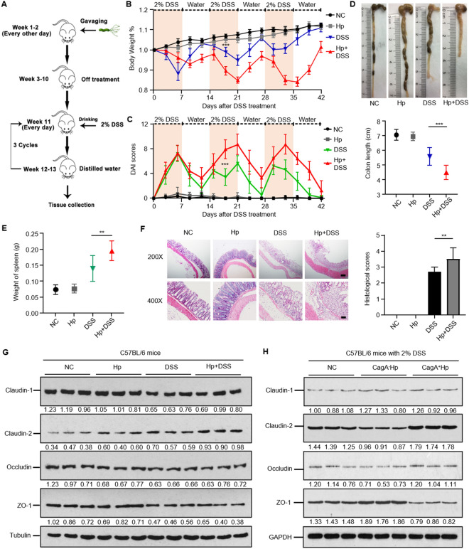
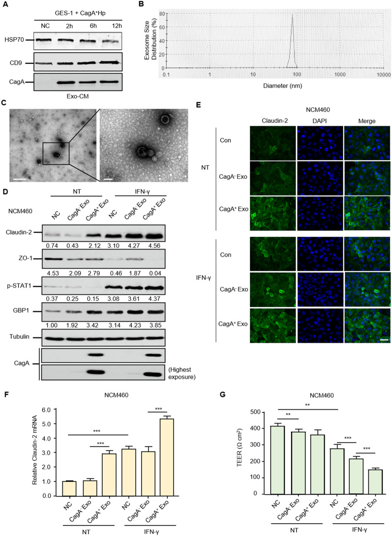
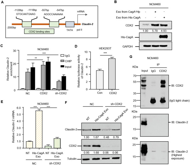
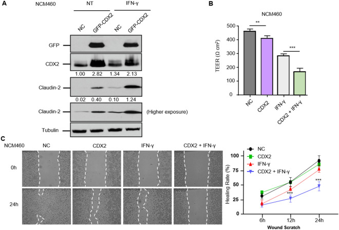
Results: Chronic colitis models of CagA+ H. pylori-colonized mice treated with 2% Dextran sulphate sodium (DSS) were established to assess the disease activity and pertinent expression of tight junction proteins closely related to mucosal integrity. The aggravating effect of CagA+ H. pylori infection on DSS-induced chronic colitis was confirmed in mouse models. In addition, augmented Claudin-2 expression was detected in CagA+ H. pylori infection conditions and selected for mechanistic analysis. Next, GES-1 human gastric epithelial cells were cultured with CagA+ H. pylori or a recombinant CagA protein, and exosomes isolated from conditioned media were then identified. We assessed the Claudin-2 levels after exposure to CagA+ exosomes, CagA- exosomes, and IFN-γ incubation, revealing that CagA+ H. pylori compromised the colonic mucosal barrier and facilitated IFN-γ-induced intestinal epithelial destruction through CagA-containing exosome-mediated mechanisms. Specifically, CagA upregulated Claudin-2 expression at the transcriptional level via a CDX2-dependent mechanism to slow the restoration of wounded mucosa in colitis in vitro.

Conclusions: These data suggest that exosomes containing CagA facilitate CDX2-dependent Claudin-2 maintenance. The exosome-dependent mechanisms of CagA+ H. pylori infection are indispensable for damaging the mucosal barrier integrity in chronic colitis, which may provide a new idea for inflammatory bowel disease (IBD) treatment.

Keywords: CDX2; CagA; Claudin-2; Colitis; Exosome; Tight junction.


Conflict of interest statement

The authors declare that they have no competing interests.

Figures

Fig. 1
Fig. 1
CagA+ H. pylori infection exacerbates DSS-induced chronic colitis in mice. A Schematic depiction of the experimental design for H. pylori (CagA+ strain) inoculation followed by chronic colitis models administrated 2% DSS. B, C Body weights (B) and disease activity index (DAI) changes (C) in different groups. Data from the second and third DSS-treatment cycles (7 day 2% DSS and 7 day diluted water) showed that H. pylori-infected mice had significantly less body weight loss and disease manifestation than control mice with no H. pylori infection in DSS-treated conditions. ***p < 0.001, compared to DSS group. Student’s t test for body weights, and χ2 test for DAI scores. D, E, F Colon lengths (D), spleen weights (E), microscopic appearance and H&E histological sections of the colon F in each group. Scale bars, 200 µm (200 ×) and 100 µm (400 ×). **p < 0.01, ***p < 0.001. Student’s t test was used for colon length and spleen weights, and the χ2 test was used for histological scores. G The relative expression of tight junction proteins (TJs) in the colon of H. pylori and chronic colitis mice. The augmented Claudin-2 was observed in the H. pylori + DSS group in comparison with the DSS group. H The TJs protein expression of chronic colitis mice subgroups without H. pylori, with CagA H. pylori infection, and with CagA+ H. pylori infection. Claudin-2 was significantly upregulated in CagA+ H. pylori-infected group compared with CagA H. pylori mice. All data were presented as means ± SD (n = 10)
Fig. 2
Fig. 2
Exosomes from GES-1 human gastric epithelial cells cultured with CagA+ H. pylori significantly disrupt the intestinal mucosal barrier meanwhile and upregulate Claudin-2 in vitro. A Western blotting analysis confirmed exosomes isolated from GES-1 and CagA+ H. pylori coculture media via characteristic biomarkers HSP70 and CD9. The presence of CagA within the exosome was also shown. Exo-CM, exosome derived from conditioned medium. B, C Features of exosomes in terms of size distribution (B) and morphology (C) on transmission electron microscopy. Scale bar, 0.5 µm and 200 nm. D Western blotting verified the augmentation of Claudin-2 proteins by CagA+ exosomes, while the CagA+ exosome group partially maintained Claudin-2 protein expression under IFN-γ conditions. p-STAT1 and GBP1 were employed as positive controls to show the inflammatory status in response to CagA+ exosomes, CagA exosomes, or IFN-γ. The p-STAT1 pathway was stringently dependent on IFN-γ, as expected, and the presence of a slight GBP1 band also showed a low degree of inflammation after the entry of CagA+ exosomes. The shrinkage of ZO-1, an acknowledged tight junction protein in both CagA+ exosomes and IFN-γ conditions, was observed as a positive control. E Cell immunostaining showed that CagA+ exosomes contributed to membrane Claudin-2 formation, whose increase was also observed in IFN-γ induced barrier function disorders. Scale bar, 20 µm. F RT–qPCR revealed the mRNA expression tendencies in different groups. G CagA+-containing exosomes accelerate the dysintegrity of the NCM460 cell monolayer under inflammatory factor IFN-γ conditions. The transepithelial electrical resistance (TEER) values of the cell monolayer were tested after CagA+ exosomes and IFN-γ 24 h. The data are expressed as the means ± SD of three independent experiments. ***p < 0.001, t test
Fig. 3
Fig. 3
CagA activates CDX2 to transcriptionally upregulate Claudin-2 expression. A Schematic binding prediction of the transcription factor CDX2 in the Claudin-2 gene promoter. The two positions and sequences with relatively high scores are indicated. B Western blotting confirmed the incremental CDX2 protein expression by CagA. In addition to exosomes from GES-1 gastric epithelial cells cultured with CagA+ H. pylori, recombinant His-CagA protein was utilized to perform CagA and GES-1 cell cocluture and subsequent exosome isolation. Both exosomes from CagA+ H. pylori infection and recombinant CagA protein incubation triggered CDX2 expression. The presence of CagA in exosomes was proven using an antibody against the His-tag. Notably, the more striking contrast of the CDX2 band was shown above at shorter exposure times due to its abundant endogenous expression in the colon. C Identification of the CDX2-Claudin-2 interaction using chromatin immunoprecipitation. CDX2 interferences were introduced into NCM460 cell. CDX2 protein was able to pull down Claudin-2 DNA fragments. IgG and Input were performed as controls. D The positive transcriptional regulation of Claudin-2 by CDX2 was confirmed by luciferase reporter assay. The ratio of firefly luciferase activity to Renilla activity was calculated to show the binding of CDX2 to gene promoter activities. Each experiment was performed in triplicate. E, F CDX2 depletion impeded CagA-associated Claudin-2 transcription and protein expression. Data are presented as the mean ± SD value from three biological replicates. **p < 0.01, ***p < 0.001, t test. G Coimmunoprecipitation precluded the protein interaction of CDX2 and Claudin-2. The equal CDX2 loading in the IP panel was preverified, and immunoblotting with Claudin-2 provided no binding of CDX2 and Claudin-2 even under the longest exposure
Fig. 4
Fig. 4
CDX2 is indispensable for Claudin-2 expression in mucosal barrier integrity and colonic epithelial restitution. A Overexpression of CDX2 upregulated Claudin-2 protein expression, and their abundances were detected even under IFN-γ incubation. The stable CDX2 overexpression model was employed using GFP-tagged vectors. B CDX2 partially impaired colonic mucosal barrier integrity in IFN-γ stimulation. The TEER values of the NCM460 cell monolayer under each condition were measured. **p < 0.01, ***p < 0.001, t test. C CDX2-associated Claudin-2 impeded colonic wound healing and partially strengthened the IFN-γ-induced mucosal deficiency. The wounds were recorded at 0 h, 6 h, 12 h, and ultimate 24 h. CDX2 + IFN-γ vs. IFN-γ, ***p < 0.001, t test

Similar articles

Cited by

References

    1. Liou JM, Malfertheiner P, Lee YC, et al. Screening and eradication of Helicobacter pylori for gastric cancer prevention: the Taipei global consensus. Gut. 2020;69:2093–2112. - PubMed
    1. Fox JG, Wang TC. Helicobacter pylori infection: pathogenesis. Curr Opin Gastroenterol. 2002;18:15–25. - PubMed
    1. Yong X, Tang B, Li BS, et al. Helicobacter pylori virulence factor CagA promotes tumorigenesis of gastric cancer via multiple signaling pathways. Cell Commun Signal. 2015;13:30. - PMC - PubMed
    1. Hatakeyama M. Helicobacter pylori CagA and gastric cancer: a paradigm for hit-and-run carcinogenesis. Cell Host Microbe. 2014;15:306–316. - PubMed
    1. Campuzano-Maya G. Hematologic manifestations of Helicobacter pylori infection. World J Gastroentero. 2014;20:12818–12838. - PMC - PubMed

LinkOut - more resources