. 2022 Oct 20;60(4):2102347.
doi: 10.1183/13993003.02347-2021. Print 2022 Oct.

Pirfenidone exacerbates Th2-driven vasculopathy in a mouse model of systemic sclerosis-associated interstitial lung disease

Affiliations

Pirfenidone exacerbates Th2-driven vasculopathy in a mouse model of systemic sclerosis-associated interstitial lung disease

Anna Birnhuber et al. Eur Respir J. .

Abstract

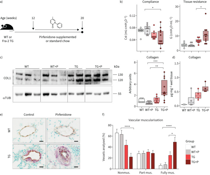
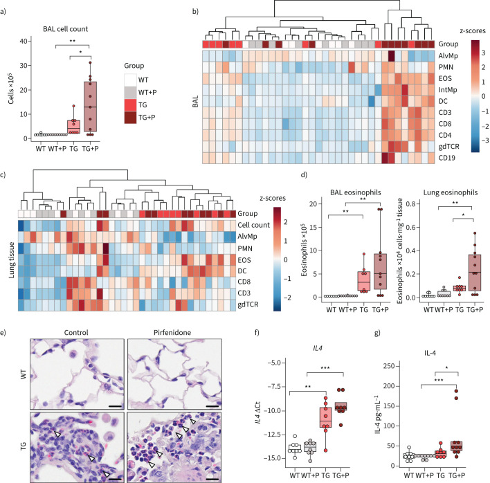
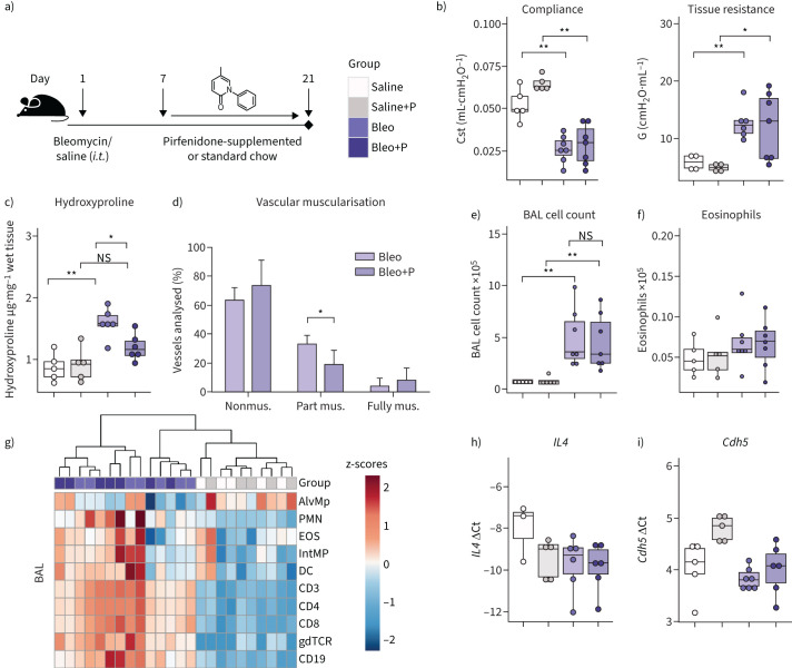
Background: Systemic sclerosis (SSc) is an autoimmune disease characterised by severe vasculopathy and fibrosis of various organs including the lung. Targeted treatment options for SSc-associated interstitial lung disease (SSc-ILD) are scarce. We assessed the effects of pirfenidone in a mouse model of SSc-ILD.

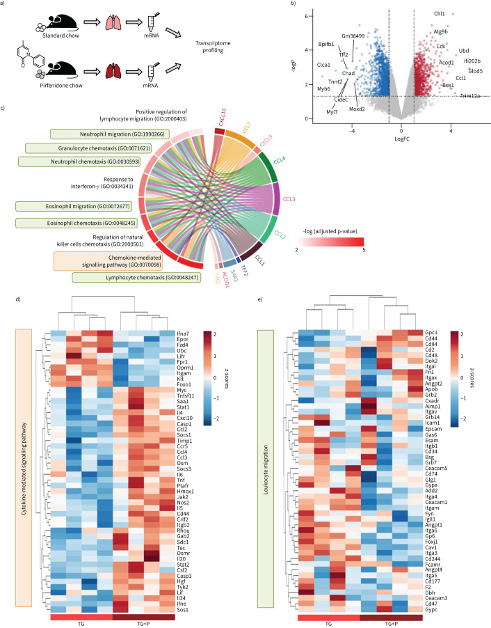
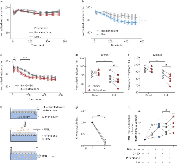
Methods: Pulmonary function, inflammation and collagen deposition in response to pirfenidone were assessed in Fra-2-overexpressing transgenic (Fra-2 TG) and bleomycin-treated mice. In Fra-2 TG mice, lung transcriptome was analysed after pirfenidone treatment. In vitro, pirfenidone effects on human eosinophil and endothelial cell function were analysed using flow cytometry-based assays and electric cell-substrate impedance measurements, respectively.

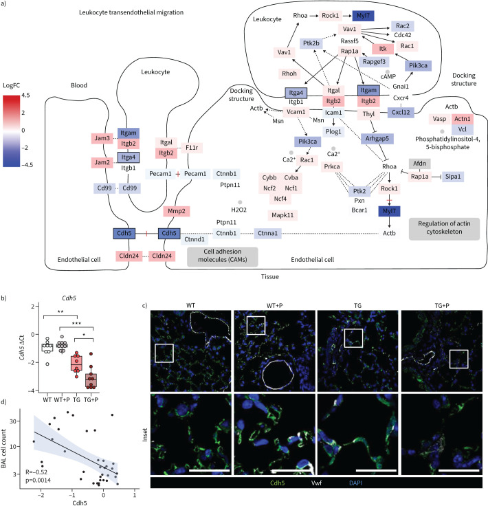
Results: Pirfenidone treatment attenuated pulmonary remodelling in the bleomycin model, but aggravated pulmonary inflammation, fibrosis and vascular remodelling in Fra-2 TG mice. Pirfenidone increased interleukin (IL)-4 levels and eosinophil numbers in lung tissue of Fra-2 TG mice without directly affecting eosinophil activation and migration in vitro. A pronounced immune response with high levels of cytokines/chemokines and disturbed endothelial integrity with low vascular endothelial (VE)-cadherin levels was observed in pirfenidone-treated Fra-2 TG mice. In contrast, eosinophil and VE-cadherin levels were unchanged in bleomycin-treated mice and not influenced by pirfenidone. In vitro, pirfenidone exacerbated the IL-4 induced reduction of endothelial barrier resistance, leading to higher leukocyte transmigration.

Conclusion: This study shows that antifibrotic properties of pirfenidone may be overruled by unwanted interactions with pre-injured endothelium in a setting of high T-helper type 2 inflammation in a model of SSc-ILD. Careful ILD patient phenotyping may be required to exploit benefits of pirfenidone while avoiding therapy failure and additional lung damage in some patients.


Conflict of interest statement

Conflict of interest: A. Heinemann has received remunerations from AstraZeneca for lecture activities; grants from Austrian Science Funds, FWF. H. Olschewski received consulting fees from Actelion, Bayer AG, Böhringer Ingelheim, Inventiva, Janssen Pharmaceutica, MSD (Merck Sharp & Dohme), payment for educational events from Böhringer Ingelheim and remunerations for participation on advisory boards from Bayer AG, Pfizer, MSD (Merck Sharp & Dohme) and IQVIA Biotech LLC, and is Deputy Director of the Ludwig Boltzmann Institute for Lung Vascular Research. G. Kwapiszewska is Director of LBI-LVR. All other authors have nothing to disclose.

Figures

FIGURE 1
FIGURE 1
Pulmonary remodelling is worsened following pirfenidone treatment in Fra-2-overexpressing transgenic (Fra-2 TG) mice. a) Schematic representation of pirfenidone (P) treatment in Fra-2 TG and wild-type (WT) mice. Lung function measurements and organ collection was performed in ∼20-week-old mice after 8 weeks of pirfenidone treatment. b) Lung function measurements showing quasi-static compliance (Cst) and tissue dampening (G). c) Western blot analysis and corresponding quantification of Collagen I (COL1) in lung homogenates of WT and TG mice with (+P) and without pirfenidone. α-tubulin (αTUB) served as loading control. One of two Western blots is shown. d) Hydroxyproline measurement of collagen in lung tissue. Data are indicated as boxplots with dot-plot overlays. Statistical analysis was performed using nonparametric Kruskal–Wallis testing with post-analysis to compare specific groups. e) Representative images of double immunohistochemical staining for von Willebrand factor (brown) and α-smooth muscle actin (purple). Scale bars=10 µm. f) Percentage of nonmuscularised (nonmus.), partially muscularised (part mus.) and fully muscularised (fully mus.) vessels <100 μm in diameter. n=4 (TG) or n=6 (WT, WT+P and TG+P). Data are shown as mean±sd. *: p<0.05, **: p<0.01, ***: p<0.001, ****: p<0.0001.
FIGURE 2
FIGURE 2
Pirfenidone increases inflammatory cell counts in the bronchoalveolar lavage (BAL) and eosinophilic infiltration into the lung tissue in Fra-2-overexpressing transgenic (Fra-2 TG) mice. a) Inflammatory cell count in the BAL of wild-type (WT) and Fra-2 TG mice with (+P) and without pirfenidone treatment. b and c) Heat map representations with hierarchical clustering of relative proportions of inflammatory cell populations in b) BAL and c) lung tissue of WT and TG mice with (+P) and without pirfenidone treatment. Data were normalised using sqrt(sqrt(cellcount)); z-scores are shown. d) Eosinophil cell count in BAL and lung tissue of WT and TG mice with (+P) and without pirfenidone treatment. e) Chromotrop 2R staining of eosinophil granules (arrowheads) in the lung tissue of WT and TG mice with and without pirfenidone treatment. Scale bars=10 µm. f) Quantitative real-time PCR analysis of interleukin-4 (IL4) gene expression. g) IL-4 protein content in lung tissue homogenates. Data are presented as boxplots with dot-plot overlays. AlvMp: alveolar macrophages; PMN: polymorphonuclear granulocytes/neutrophils; EOS: eosinophils; IntMp: interstitial macrophages; DC: dendritic cells; CD3: CD3+ T-cells; CD8: CD8+ cytotoxic T-cells; CD4: CD4+ T-helper cells; gdTCR: γδ T-cell receptor positive cells; CD19: CD19+ B-cells. Statistical analysis was performed using nonparametric Kruskal–Wallis testing with post-analysis to compare specific groups. *: p<0.05, **: p<0.01, ***: p<0.001.
FIGURE 3
FIGURE 3
Transcriptomic profiling highlights inflammatory pathways upregulated upon pirfenidone treatment in Fra-2-overexpressing transgenic (Fra-2 TG) mice. a) Schematic representation of the experimental setup. b) Volcano plot showing differential gene regulation in Fra-2 TG mouse lungs with (+P) and without pirfenidone. Top 10 regulated genes according to their log-fold-change (logFC) are labelled by name. c) Top 10 significantly regulated gene ontologies (GO biological process) and their significantly regulated genes within the dataset. d and e) Heatmap representation with hierarchical clustering of genes within the gene ontologies d) GO:0019221 cytokine-mediated signalling pathway and e) GO:0050900 leukocyte migration.
FIGURE 4
FIGURE 4
Pirfenidone leads to decreased vascular endothelial (VE)-cadherin expression in the lungs of Fra-2-overexpressing transgenic (Fra-2 TG) mice. a) Schematic of the Kyoto Encyclopedia of Genes and Genomes (KEGG) pathway “Leukocyte transendothelial migration”. The log fold change of gene expression is indicated by colour, significance of regulation is indicated by box borders. b) Quantitative real-time PCR analysis of VE-cadherin (Cdh5). Data are presented as boxplots with dot-plot overlays. Statistical analysis was performed using nonparametric Kruskal–Wallis testing with post-analysis to compare specific groups. *: p<0.05, **: p<0.01, ***: p<0.001. c) Low- and high-magnification immunofluorescence images of VE-cadherin (Cdh5) and von Willebrand factor (Vwf) staining in lung tissue from wild-type (WT) and Fra-2 TG mice with (+P) and without pirfenidone treatment. Nuclear counterstain was performed using 4′,6-diamidino-2-phenylindole (DAPI). Scale bars=10 µm. d) Spearman's rank correlation analysis of inflammatory cells in the bronchoalveolar lavage fluid (BAL cell count) and Cdh5 expression.
FIGURE 5
FIGURE 5
Pirfenidone ameliorates pulmonary remodelling without affecting lung function and inflammation in a bleomycin (Bleo)-induced mouse model of pulmonary fibrosis. a) Schematic representation of pirfenidone (P) treatment in the bleomycin-induced mouse model of pulmonary fibrosis. Lung function measurements and organ collection was performed 21 days after bleomycin and 14 days after pirfenidone treatment. b) Lung function measurements showing quasi-static compliance (Cst) and tissue dampening/resistance (G). c) Hydroxyproline measurement of collagen in lung tissue of saline and bleomycin-treated mice with (+P) and without pirfenidone. Data are presented as boxplots with dot-plot overlays. Statistical analysis was performed using nonparametric Kruskal–Wallis testing with post-analysis to compare specific groups. d) Percentage of nonmuscularised (nonmus.), partially muscularised (part mus.) and fully muscularised (fully mus.) vessels <100 μm in diameter. n=5 (Bleo) or n=7 (Bleo+P). Data are presented as mean±sd. e) Inflammatory cell and f) eosinophil counts in the bronchoalveolar lavage (BAL) of bleomycin and saline-treated mice with (+P) and without pirfenidone treatment. g) Heat map representation with hierarchical clustering of relative proportions of inflammatory cell populations in BAL of bleomycin and saline-treated mice with (+P) and without pirfenidone treatment. Data were normalised using sqrt(sqrt(cellcount)); z-scores are shown. h and i) Quantitative real-time PCR analysis of h) interleukin-4 (IL4) and i) vascular endothelial-cadherin (Cdh5) gene expression. Data are presented as boxplots with dot-plot overlays. i.t.: intratracheal; ns: nonsignificant. *: p<0.05, **: p<0.01.
FIGURE 6
FIGURE 6
Priming with interleukin (IL)-4 sensitises human lung microvascular endothelial cells (HMVECs) and leads to increased loss of barrier function and increased polymorphonuclear leukocyte (PMNL) transmigration upon pirfenidone treatment. a) Electric Cell-substrate Impedance Sensing (ECIS) measurement of HMVEC barrier resistance in response to pirfenidone. Dimethyl sulfoxide (DMSO) and basal medium served as vehicle and negative control, respectively. b) ECIS measurements of HMVEC barrier resistance in response to IL-4 alone compared to basal medium (vehicle control distilled water). c) ECIS measurements of HMVEC barrier resistance in response to IL-4 in combination with pirfenidone or DMSO vehicle control. d and e) Detailed analysis of b) and c) at d) 10 min and e) 120 min post-treatment. Statistical analysis was performed using two-way ANOVA with multiple comparison testing. IL-4 effect: *: p<0.05, **: p<0.01, ***: p<0.001; pirfenidone effect: #: p<0.05. f) Schematic representation of the transendothelial migration experimental setup. Endothelial cells (ECs) were cultured on 3 µm transmembrane inserts and PMNLs (consisting of eosinophils and neutrophils) were allowed to migrate to 0% or 10% fetal bovine serum, respectively, in the presence or absence of IL4 and/or pirfenidone. g) Transendothelial migration of PMNLs through 3 µm transwell insets with and without HMVEC monolayers in basal medium with 0% serum. h) Transendothelial migration of PMNLs through established HMVEC monolayers in the presence of IL-4 and/or pirfenidone and corresponding vehicle controls. Statistical analysis was performed by one-way ANOVA with Dunnett's post-test using 0% serum with DMSO as control. *: p<0.05, **: p<0.01; t-test was used to compare IL-4 with and without pirfenidone treatment. #: p<0.05.

Comment in

References

    1. Shima Y. Cytokines involved in the pathogenesis of SSc and problems in the development of anti-cytokine therapy. Cells 2021; 10: 1104. doi:10.3390/cells10051104 - DOI - PMC - PubMed
    1. Huang X-L, Wang Y-J, Yan J-W, et al. . Role of anti-inflammatory cytokines IL-4 and IL-13 in systemic sclerosis. Inflamm Res 2015; 64: 151–159. doi:10.1007/s00011-015-0806-0 - DOI - PubMed
    1. Mostmans Y, Cutolo M, Giddelo C, et al. . The role of endothelial cells in the vasculopathy of systemic sclerosis: a systematic review. Autoimmun Rev 2017; 16: 774–786. doi:10.1016/j.autrev.2017.05.024 - DOI - PubMed
    1. Matucci-Cerinic M, Kahaleh B, Wigley FM. Review: evidence that systemic sclerosis is a vascular disease. Arthritis Rheum 2013; 65: 1953–1962. doi:10.1002/art.37988 - DOI - PubMed
    1. Volkmann ER, Fischer A. Update on morbidity and mortality in systemic sclerosis-related interstitial lung disease. J Scleroderm Relat Disord 2021; 6: 11–20. doi:10.1177/2397198320915042 - DOI - PMC - PubMed

Publication types

MeSH terms