

Shunt-type plexiform lesions identified in the Sugen5416/hypoxia rat model of pulmonary arterial hypertension using synchrotron-based phase-contrast micro-CT
- PMID: 35332070
- PMCID: PMC9202485
- DOI: 10.1183/13993003.02802-2021
Shunt-type plexiform lesions identified in the Sugen5416/hypoxia rat model of pulmonary arterial hypertension using synchrotron-based phase-contrast micro-CT
Abstract
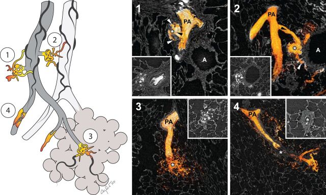
Human like plexiform lesions identified in the prolonged Sugen5416/hypoxia rat model, visualised by synchrotron tomography imaging https://bit.ly/3KQvDHg
Conflict of interest statement
Conflict of interest: C. Westöö declares a regional salary grant for medical resident researchers from ALF Forskningsutrymme för ST-läkare. T. Dreier declares an industrial PhD salary from Excillum AB, and payment to their institution from the Swedish Foundation for Strategic Research (SSF) in connection with the present manuscript. M.E. Kumar declares American Heart Association Scientist Development Grant AHA 16SDG30030006, Stanford Pediatrics startup funds 1246483-200-JHAJH, Stanford Spectrum Child Health Research Institute 1170805-100-GHEBG, Stanford Maternal and Child Health Research Institute Pilot Grant 1220318-108-JHACT, Vera Moulton Wall Center for Pulmonary Vascular Research grant 1144545-401-GHDCK and a Bravo Family Endowed Faculty Scholarship, in connection with the present manuscript. E. Spiekerkoetter declares that they have a research scholarship from Vera Moulton Wall Center for Pulmonary Vascular Disease, in connection with the present manuscript; and that they have received grants from the National Institutes of Health (National Heart, Lung and Blood Institute grants R01HL158868 and R01 HL128734) and Department of Defense (grants PR161256 and PR 181774) in the 36 months prior to manuscript submission; and that they receive royalties from Stanford University for Method of use patent “Use of FK506 for the treatment of Pulmonary Arterial Hypertension”; that they have a provisional US patent for “Enzastaurin and Fragile Histidine Trial (FHIT) Increasing Agents for the Treatment of Pulmonary Hypertension”; and that they are chair of the American Heart Association 3CPR Early Career Committee, and a member of the Stanford University Institutional Review Board. K. Tran-Lundmark declares funding from the Swedish Heart-Lung Foundation, the Crafoord Foundation, the Swedish Society of Medicine, the Knut and Alice Wallenberg Foundation and the Skåne County Council, in connection with the present manuscript; as well as roles as American Thoracic Society (ATS) Pulmonary Circulation Program Committee member (unpaid); American Heart Association (AHA) 3CPR Early Career Committee (unpaid) and Association for European Paediatric and Congenital Cardiology (AEPC) Councillor in the Working Group for Pulmonary Hypertension, Heart Failure and Transplantation (unpaid). All other authors declare no competing interests.
Figures
References
-
- Norvik C, Westoo CK, Peruzzi N, et al. Synchrotron-based phase-contrast micro-CT as a tool for understanding pulmonary vascular pathobiology and the 3-D microanatomy of alveolar capillary dysplasia. Am J Physiol Lung Cell Mol Physiol 2020; 318: L65–L75. doi: 10.1152/ajplung.00103.2019 - DOI - PubMed
Publication types
MeSH terms
Grants and funding
LinkOut - more resources
Full Text Sources