Review
. 2022 Mar 24;7(1):95.
doi: 10.1038/s41392-022-00934-y.

Notch signaling pathway: architecture, disease, and therapeutics

Affiliations
Review

Notch signaling pathway: architecture, disease, and therapeutics

Binghan Zhou et al. Signal Transduct Target Ther. .

Abstract

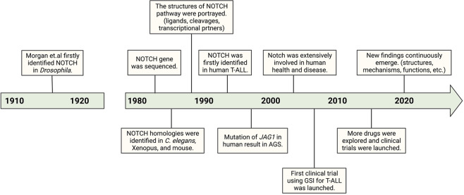
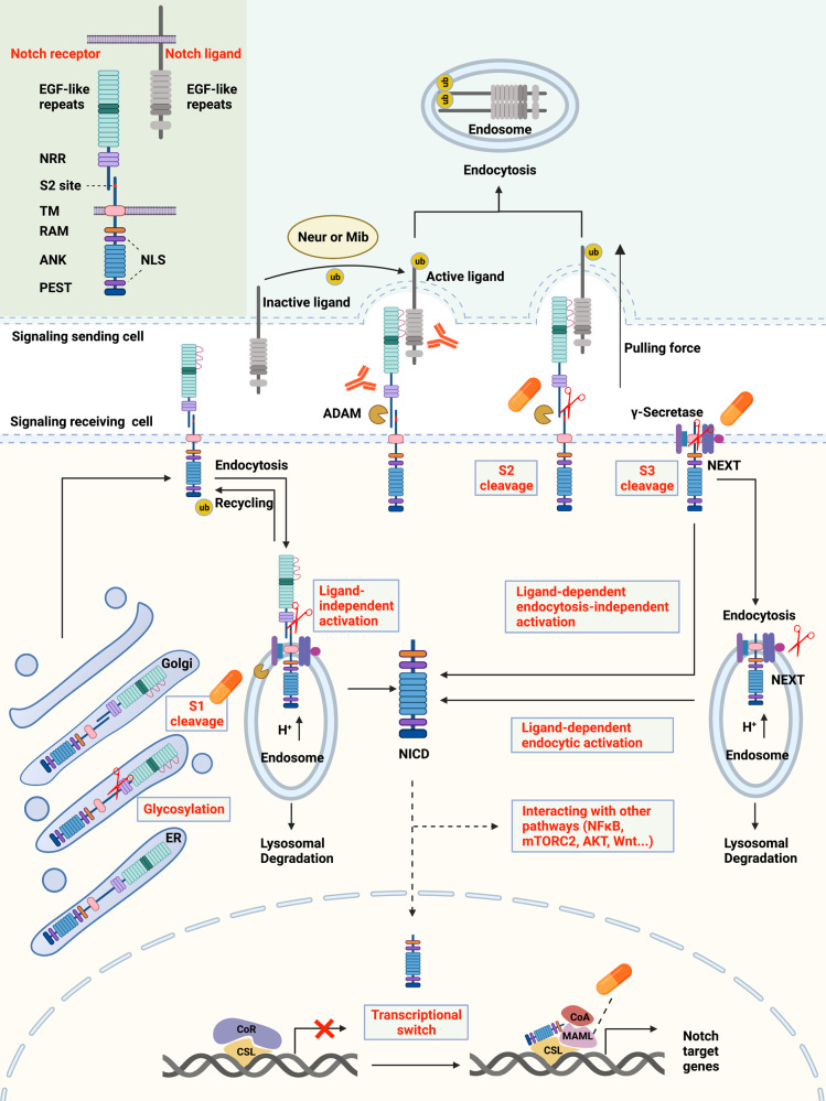
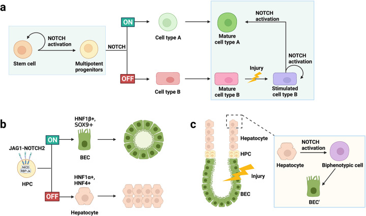
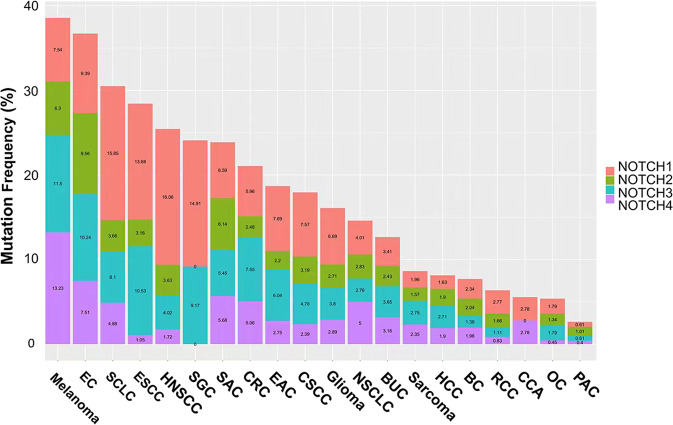
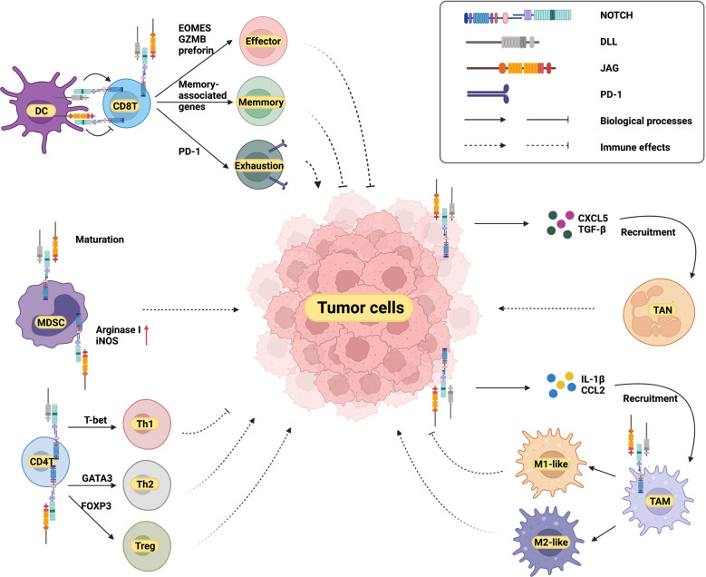
The NOTCH gene was identified approximately 110 years ago. Classical studies have revealed that NOTCH signaling is an evolutionarily conserved pathway. NOTCH receptors undergo three cleavages and translocate into the nucleus to regulate the transcription of target genes. NOTCH signaling deeply participates in the development and homeostasis of multiple tissues and organs, the aberration of which results in cancerous and noncancerous diseases. However, recent studies indicate that the outcomes of NOTCH signaling are changeable and highly dependent on context. In terms of cancers, NOTCH signaling can both promote and inhibit tumor development in various types of cancer. The overall performance of NOTCH-targeted therapies in clinical trials has failed to meet expectations. Additionally, NOTCH mutation has been proposed as a predictive biomarker for immune checkpoint blockade therapy in many cancers. Collectively, the NOTCH pathway needs to be integrally assessed with new perspectives to inspire discoveries and applications. In this review, we focus on both classical and the latest findings related to NOTCH signaling to illustrate the history, architecture, regulatory mechanisms, contributions to physiological development, related diseases, and therapeutic applications of the NOTCH pathway. The contributions of NOTCH signaling to the tumor immune microenvironment and cancer immunotherapy are also highlighted. We hope this review will help not only beginners but also experts to systematically and thoroughly understand the NOTCH signaling pathway.


Conflict of interest statement

The authors declare that the research was conducted in the absence of any commercial or financial relationships that could be construed as a potential conflict of interest. Figures were created with biorender.com.

Figures

Fig. 1
Fig. 1
A brief history of the NOTCH signaling pathway. T-ALL, T cell acute lymphoblastic leukemia; AGS, Alagille syndrome; GSI, γ-secretase inhibitor
Fig. 2
Fig. 2
Overview of the NOTCH signaling pathway and therapeutic targets. In signal-receiving cells, NOTCH receptors are first generated in the ER and then trafficked to the Golgi apparatus. During trafficking, NOTCH receptors are glycosylated at the EGF-like repeat domain (red curves). Then, in the Golgi apparatus, NOTCH receptors are cleaved into heterodimers (S1 cleavage) and transported to the cell membrane. With the help of ubiquitin ligases, some of the NOTCH receptors on the cell membrane are endocytosed into endosomes. Endosomes contain an acidic environment with ADAMs and γ-secretase. The NOTCH receptors in endosomes can be recycled to the cell membrane, cleaved into NICD, or transported into lysosomes for degradation. In signal-sending cells, NOTCH ligands are distributed on the cell membrane and can bind to NOTCH receptors on signal-receiving cells. However, the ligands are inactive before ubiquitylation by Neur or Mib. After ubiquitylation, ligands can be endocytosed, thus producing a pulling force for the binding receptors. Without the pulling force, the S2 site (red marks) of NOTCH receptors is hidden by the NRR domain, and thus, the NOTCH receptors are resistant to cleavage by ADAMs. With the pulling force, the NRR domain is extended, therefore exposing the S2 site for cleavage. ADAMs and the pulling force are both necessary for S2 cleavage. After S2 cleavage, the remaining part of the NOTCH receptor is called NEXT. NEXT can be further cleaved on the cell membrane by γ-secretase or endocytosed into endosomes. In the former mode, NICD is released on the cell membrane. In the latter mode, NEXT can be cleaved into NICD or transported into lysosomes for degradation. In total, there are three approaches to generate NICD, classified as ligand-independent activation, ligand-dependent endocytosis-independent activation, and ligand-dependent endocytic activation. NICD can be translocated into the nucleus or remain in the cytoplasm to crosstalk with other signaling pathways, such as NFκB, mTORC2, AKT, and Wnt. The classical model proposes that, in the absence of NICD, CSL binds with corepressors to inhibit the transcription of target genes. Once NICD enters the nucleus, it can bind with CSL and recruit MAMLs, releasing corepressors, recruiting coactivators, and thus promoting the transcription of NOTCH target genes. There are two main approaches to inhibit NOTCH signaling for therapy. One is designing inhibitors of the key components of the pathways, including the enzymes that participate in S1 cleavage, ADAMs, γ-secretase, and MAML. The other one is producing antibody-drug conjugates against NOTCH receptors and ligands. The protein structures of NOTCH ligands and receptors are shown in the top left corner. NICD, NOTCH intracellular domain; ADAM, a disintegrin and metalloproteinase domain-containing protein; Neur, Neuralized; Mib, Mindbomb; NRR, negative regulatory region; NEXT, NOTCH extracellular truncation; CSL, CBF-1/suppressor of hairless/Lag1; MAMLs, Mastermind-like proteins; TM, transmembrane domain; RAM, RBPJ association module; ANK, ankyrin repeats; PEST, proline/glutamic acid/serine/threonine-rich motifs; NLS, nuclear localization sequence; CoR, corepressor; CoA, coactivator; ub, ubiquitin
Fig. 3
Fig. 3
The role of NOTCH signaling in body development and damage repair. NOTCH signaling is involved in regulating the differentiation and function of stem cells, affecting organ production and damage repair. a NOTCH signaling promotes the self-renewal of stem cells, induces multipotent progenitors for lineage selection, and generates different terminal cells; when the organ is damaged, cell type A is damaged and destroyed, and the stimulated cell type B rapidly upregulates the expression of NOTCH signaling to promote their own proliferation, and is partially redifferentiated into cell type A. b Highly activated NOTCH induces the expression of bile duct cell-enriched transcription factors and promotes the differentiation of multipotent hepatocyte progenitors into bile duct epithelial cells. c In liver injury, BEC are damaged and destroyed. NOTCH signaling is highly expressed in hepatocytes, which are further transformed into biphenotypic cells, which manifests the biliary tract morphology, and finally generate new BEC (BEC’) to form small tubular structures. HPC, hematopoietic progenitor cell; BEC, bile duct epithelial cell; SOX9, SRY-related high-mobility group box 9; HNF, hepatocyte nuclear factor
Fig. 4
Fig. 4
Mutation frequencies of NOTCH receptors in different cancers. Data are obtained from cBioPortal (http://cbioportal.org). We included data from two studies: MSK-IMPACT Clinical Sequencing and TCGA PanCancer Atlas Studies, with a total of 21289 patients. And we only used samples with mutation information, including missense, truncating, inframe, splice, and structural variation/fusion. This figure shows the mutation frequency of the four receptors of NOTCH in different cancer types. EC, endometrial carcinoma; SCLC, small-cell lung cancer; ESCC, esophageal squamous cell carcinoma; HNSCC, head, and neck squamous cell carcinoma; SGC, salivary gland cancer; SAC, stomach adenocarcinoma; CRC, colorectal cancer; EAC, esophagogastric adenocarcinoma; CSCC, cervical squamous cell carcinoma; NSCLC, non-small-cell lung cancer; BUC, bladder urothelial carcinoma; HCC, hepatocellular carcinoma; BC, breast cancer; RCC, renal cell carcinoma; CCA, cholangiocarcinoma; OC, ovarian cancer; PAC, prostate adenocarcinoma
Fig. 5
Fig. 5
NOTCH signaling pathway in antitumor immunity. NOTCH signaling plays important roles in both tumor-suppressive and tumor-promoting immune cells. NOTCH signaling promotes the differentiation of many immune cells. DLL and JAG mediate both similar and distinct effects. DC, dendritic cell; CD8T, CD8+ T cell; MDSC, myeloid-derived suppressor cell; CD4T, CD4+ T cell; Th1, type1 T helper cell; Th2, type2 T helper cell; Treg, regulatory T cell; TAM, tumor-associated macrophage; TAN, tumor-associated neutrophil; PD-1, programmed death-1; EOMES, eomesodermin; GZMB, granzyme B; DLL, delta-like ligand; CCL2, C-C motif chemokine ligand 2

References

    1. Metz CW, Bridges CB. Incompatibility of mutant races in Drosophila. Proc. Natl Acad. Sci. USA. 1917;3:673–678. - PMC - PubMed
    1. Mohr OL. Character changes caused by mutation of an entire region of a chromosome in Drosophila. Genetics. 1919;4:275–282. - PMC - PubMed
    1. Bridges CB. Non-disjunction as proof of the chromosome theory of heredity (concluded) Genetics. 1916;1:107–163. - PMC - PubMed
    1. Yochem J, Weston K, Greenwald I. The Caenorhabditis elegans lin-12 gene encodes a transmembrane protein with overall similarity to Drosophila Notch. Nature. 1988;335:547–550. - PubMed
    1. Austin J, Kimble J. Transcript analysis of glp-1 and lin-12, homologous genes required for cell interactions during development of C. elegans. Cell. 1989;58:565–571. - PubMed

Publication types

Substances