. 2022 Mar 24;12(1):5102.
doi: 10.1038/s41598-022-08316-8.

HydroZitLa inhibits calcium oxalate stone formation in nephrolithic rats and promotes longevity in nematode Caenorhabditis elegans

Affiliations

HydroZitLa inhibits calcium oxalate stone formation in nephrolithic rats and promotes longevity in nematode Caenorhabditis elegans

Nalinthip Lordumrongkiat et al. Sci Rep. .

Abstract

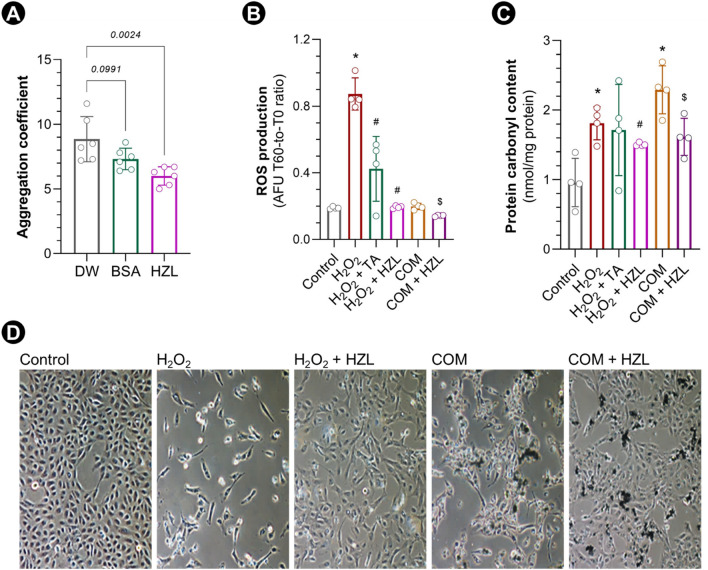
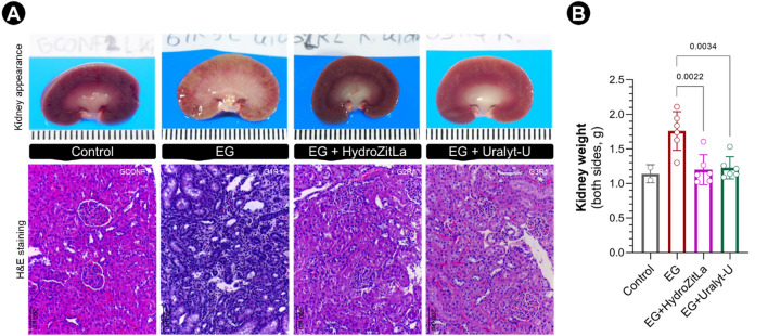
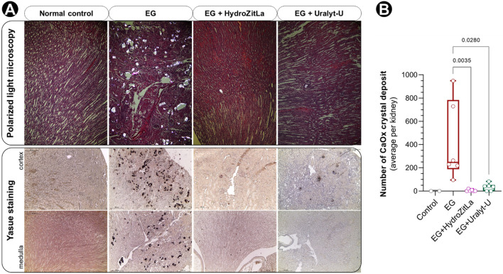
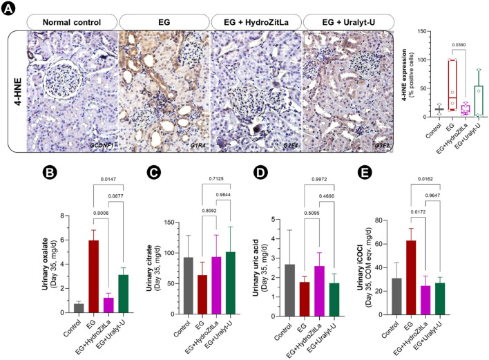
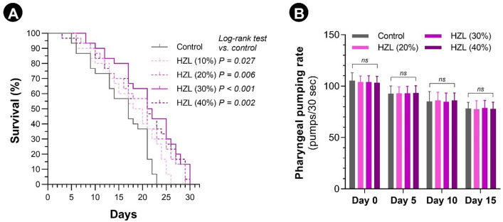
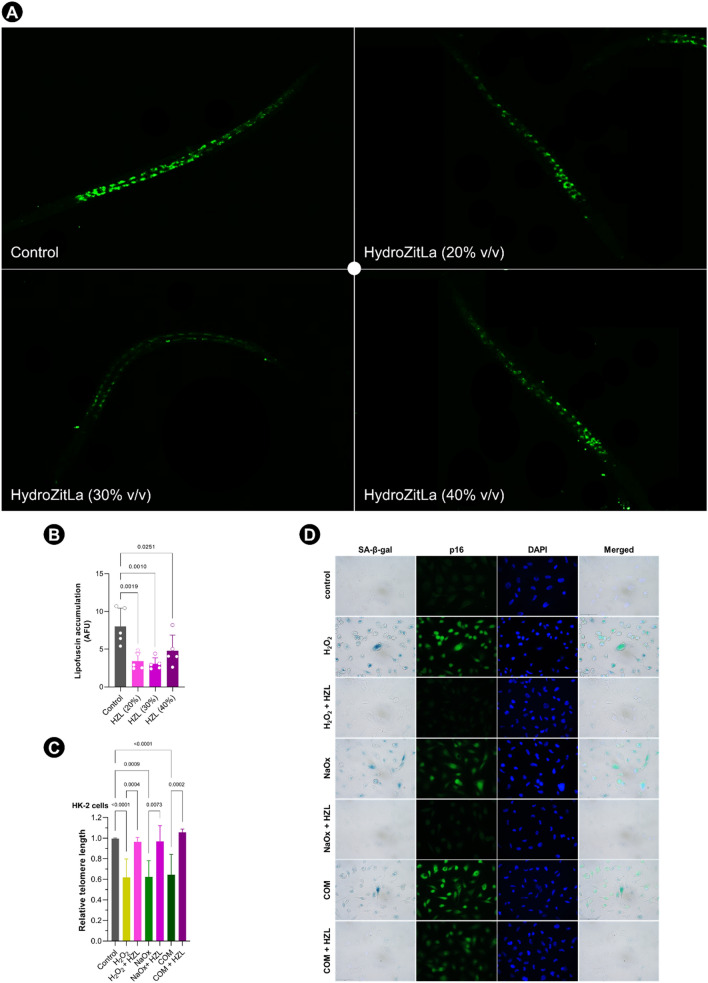
Low fluid intake, low urinary citrate excretion, and high oxidative stress are main causative factors of calcium oxalate (CaOx) nephrolithiasis. HydroZitLa contains citrate and natural antioxidants and is developed to correct these three factors simultaneously. Antioxidants theoretically can prolong the lifespan of organisms. In this study, we preclinically investigated the antilithogenic, lifespan-extending and anti-aging effects of HydroZitLa in HK-2 cells, male Wistar rats, and Caenorhabditis elegans. HydroZitLa significantly inhibited CaOx crystal aggregation in vitro and reduced oxidative stress in HK-2 cells challenged with lithogenic factors. For experimental nephrolithiasis, rats were divided into four groups: ethylene glycol (EG), EG + HydroZitLa, EG + Uralyt-U, and untreated control. CaOx deposits in kidneys of EG + HydroZitLa and EG + Uralyt-U rats were significantly lower than those of EG rats. Intrarenal expression of 4-hydroxynonenal in EG + HydroZitLa rats was significantly lower than that of EG rats. The urinary oxalate levels of EG + HydroZitLa and EG + Uralyt-U rats were significantly lower than those of EG rats. The urinary citrate levels of EG + HydroZitLa and EG + Uralyt-U rats were restored to the level in normal control rats. In C. elegans, HydroZitLa supplementation significantly extended the median lifespan of nematodes up to 34% without altering feeding ability. Lipofuscin accumulation in HydroZitLa-supplemented nematodes was significantly lower than that of non-supplemented control. Additionally, HydroZitLa inhibited telomere shortening, p16 upregulation, and premature senescence in HK-2 cells exposed to lithogenic stressors. Conclusions, HydroZitLa inhibited oxidative stress and CaOx formation both in vitro and in vivo. HydroZitLa extended the lifespan and delayed the onset of aging in C. elegans and human kidney cells. This preclinical evidence suggests that HydroZitLa is beneficial for inhibiting CaOx stone formation, promoting longevity, and slowing down aging.


Conflict of interest statement

C.B., N.C. and N.L. are inventors of HydroZitLa. Chulalongkorn University and the inventors own the intellectual property for HydroZitLa. None of the other authors have any conflict of interest to declare in relation to the present work.

Figures

Figure 1
Figure 1
COM aggregation inhibition and oxidative stress mitigation by HydroZitLa. (A) HydroZitLa (HZL) significantly decreased aggregation of calcium oxalate monohydrate (COM) crystals compared with distilled water (DW) control. (B) H2O2 (10 µM) significantly increased ROS generation in HK-2 cells compared with control. Co-treatments with tocopheryl acetate (TA, 300 µM) and HydroZitLa (10% v/v) significantly decreased ROS generation in HK-2 cells treated with H2O2. (C) Protein carbonyl contents in HK-2 cells treated with H2O2 (1,000 µM) and COM (150 µg/cm2) were significantly higher than that in control. HydroZitLa co-treatment significantly decreased levels of ROS generation and protein carbonyl content in HK-2 cells treated with H2O2 and COM. (D) Representative micrographs showing reduction of cell survival and induction of morphological change in HK-2 cells treated with H2O2 (1000 µM) and COM (black precipitates, 150 µg/cm2). HydroZitLa co-treatment rescued cells from apoptosis as well as restored cell morphology and proliferation. *P < 0.05 vesrus Control, #P < 0.05 versus H2O2, $P < 0.05 versus COM. Micrograph magnification: × 100.
Figure 2
Figure 2
Kidney appearance and H&E staining of rat renal sections. (A) Kidney of EG rats were pale, enlarged, and swelled (upper panel). Renal tissues of EG rats had a robust sign of inflammation, but renal tissues of EG + HydroZitLa and EG + Uralyt-U rats appeared normal, similar to that in normal control rats (lower panel). (B) The average kidney weights of EG rats were significantly greater than that of EG + HydroZitLa and EG + Uralyt-U rats. Micrograph magnification: × 100.
Figure 3
Figure 3
CaOx crystal deposits in rat renal sections detected by polarized light microscopy and Yasue staining. (A) The birefringent CaOx crystals were obviously accumulated in the renal sections of EG rats (upper panel). By contrast, the birefringent CaOx deposits were apparently vanished in the kidneys of EG + HydroZitLa and EG + Uralyt-U rats. Yasue staining confirmed that CaOx deposits were substantially deposited in EG rats’ kidneys, but they almost disappeared in kidneys of EG + HydroZitLa and EG + Uralyt-U rats (lower panel). These CaOx deposits were not observed in normal control rats. (B) CaOx deposits were counted in Yasue-stained sections. Numbers of intrarenal CaOx deposits in EG + HydroZitLa and EG + Uralyt-U rats were significantly lower than in EG rats. Micrograph magnification: × 100.
Figure 4
Figure 4
Expression of 4-HNE in rat renal sections and levels of oxalate, citrate, uric acid, and iCOCI in 24-h urine samples (day 35). (A) 4-HNE expression was obviously increased in EG rat renal tissues relative to normal control rat. Intrarenal expression of 4-HNE in EG + HydroZitLa rats was significantly lower than that in EG rats. (B) Urinary oxalate was elevated in EG rats relative to normal control rats. Levels of urinary oxalate in EG + HydroZitLa and EG + Uralyt-U rats were significantly lower than that in EG rats. (C) Urinary citrate was declined in EG rats relative to normal control rats. Levels of urinary citrate excretion in rats treated with HydroZitLa and Uralyt-U were higher than that in EG rats, and they could reach the normal levels found in normal control rats. (D) Urinary uric acid levels among EG, EG + HydroZitLa, and EG + Uralyt-U rats were comparable. (E) Similar to urinary oxalate, levels of urinary iCOCI of EG + HydroZitLa and EG + Uralyt-U rats were significantly lower than that of EG rats. Micrograph magnification: × 400.
Figure 5
Figure 5
Lifespan extension and food intake behavior of C. elegans supplemented with HydroZitLa (HZL). (A) Kaplan–Meier survival curves compared between C. elegans supplemented with HydroZitLa (10%–40% v/v) and non-supplemented control. Supplementation with 10%, 20%, 30%, and 40% v/v HydroZitLa significantly increased the median lifespan of nematodes compared with non-supplemented control. The highest lifespan extension was with 30% v/v HydroZitLa supplementation. Median survival time of non-supplemented control worms was 16 days. Supplementations with HydroZitLa at 10%, 20%, 30%, and 40% significantly increased the median survivals to 18.5 (15.6% increase), 19.5 (21.9% increase), 21.5 (34.4% increase), and 21.0 (31.3% increase) days, respectively. (B) Pharyngeal pumping rates measured at days 0, 5, 10, and 15 were not significantly different between supplemented and non-supplemented worms. NS: not significant.
Figure 6
Figure 6
Anti-aging effect of HydroZitLa (HZL) tested in C. elegans and HK-2 cells. (A) HydroZitLa supplementations at 20%, 30%, and 40% v/v clearly decreased lipofuscin accumulation in C. elegans. (B) Autofluorescent intensities of lipofuscin in HydroZitLa-supplemented nematodes (for all tested concentrations) were significantly lower than that in non-supplemented control. (C) Relative telomere lengths in HK-2 cells treated with H2O2, sodium oxalate (NaOx), and calcium oxalate monohydrate (COM) were significantly lower than untreated control. HydroZitLa co-treatment significantly inhibited shortening of telomere in HK-2 cells treated with H2O2, NaOx, and COM. (D) Double staining of SA-β-gal and p16 demonstrated that H2O2, NaOx, and COM induced premature senescence (as indicated by increased proportion of SA-β-gal positive cells) and upregulation of p16 expression. p16 was strongly upregulated in those SA-β-gal positive cells. HydroZitLa co-treatment effectively inhibited upregulation of p16 and onset of premature senescence in HK-2 cells exposed to lithogenic stressors. Magnifications: × 100 (A), × 400 (D).

Similar articles

Cited by

References

    1. Michell AR. Urolithiasis–historical, comparative and pathophysiological aspects: A review. J. R. Soc. Med. 1989;82:669–672. - PMC - PubMed
    1. Shah J, Whitfield HN. Urolithiasis through the ages. BJU Int. 2002;89:801–810. - PubMed
    1. Boonla C, Hunapathed C, Bovornpadungkitti S, Poonpirome K, Tungsanga K, Sampatanukul P, Tosukhowong P. Messenger RNA expression of monocyte chemoattractant protein-1 and interleukin-6 in stone-containing kidneys. BJU Int. 2008;101:1170–1177. - PubMed
    1. Boonla C, Krieglstein K, Bovornpadungkitti S, Strutz F, Spittau B, Predanon C, Tosukhowong P. Fibrosis and evidence for epithelial-mesenchymal transition in the kidneys of patients with staghorn calculi. BJU Int. 2011;108:1336–1345. - PubMed
    1. Chuang TF, Hung HC, Li SF, Lee MW, Pai JY, Hung CT. Risk of chronic kidney disease in patients with kidney stones-a nationwide cohort study. BMC Nephrol. 2020;21:292. - PMC - PubMed

Publication types

Supplementary concepts