. 2022 Mar 24;5(1):260.
doi: 10.1038/s42003-022-03221-2.

Multiscale reconstruction of various vessels in the intact murine liver lobe

Affiliations

Multiscale reconstruction of various vessels in the intact murine liver lobe

Qi Zhang et al. Commun Biol. .

Abstract

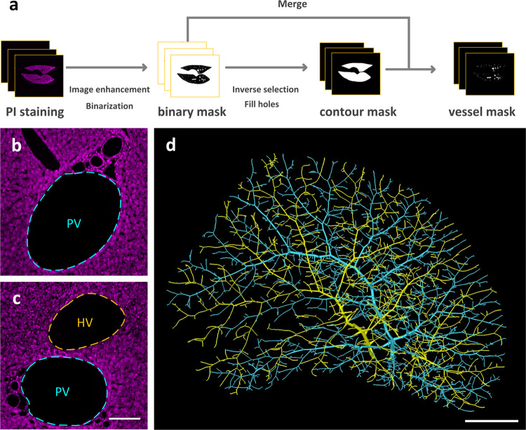
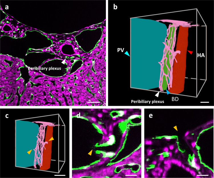
The liver contains a variety of vessels and participates in miscellaneous physiological functions. While past studies generally focused on certain hepatic vessels, we simultaneously obtained all the vessels and cytoarchitectural information of the intact mouse liver lobe at single-cell resolution. Here, taking structural discrepancies of various vessels into account, we reconstruct and visualize the portal vein, hepatic vein, hepatic artery, intrahepatic bile duct, intrahepatic lymph of an intact liver lobe and peribiliary plexus in its selected local areas, providing a technology roadmap for studying the fine hepatic vascular structures and their spatial relationship, which will help research into liver diseases and evaluation of medical efficacies in the future.


Conflict of interest statement

The authors declare no competing interests.

Figures

Fig. 1
Fig. 1. The data acquisition pipeline and acquired results.
a The procedure of acquiring dual-wavelength image dataset by high-definition fluorescent micro-optical sectioning tomography (HD-fMOST). b The slice image from the vascular channel of the intact mouse liver dataset. c The slice image from the cytoarchitecture channel of the intact mouse liver dataset. d The zoom-in of the merged channel images indicated in the white boxes in (b) and (c). e, f The zoom-in of the area indicated by the white boxes in (d). The yellow arrows indicate sinusoids. g The zoom-in of the merged channel images indicated in the white boxes in (b). The cyan arrow indicates portal vein. The red arrow indicates hepatic artery. h The zoom-in of the merged channel images indicated with a white box in (g). i Portal triads in vessel channel image. j Portal triads in merged dual-channel image. The blue arrow indicates bile ducts. The projection thickness of (b, c, g) is 100 µm, and the thickness of (df, hj) is 1 µm. Scale bars, b, c 1 mm; d, gj 50 μm; e, f 20 μm.
Fig. 2
Fig. 2. Reconstruction of the portal vein and hepatic vein.
a The reconstruction pipeline. b, c The cytoarchitecture channel images of the local areas in the liver. The portal vein and hepatic vein are respectively marked with cyan and yellow dashed lines. d 3D reconstruction of the portal vein and hepatic vein. The portal vein is marked with cyan. The hepatic vein is marked with yellow. PV portal vein, HV hepatic vein. Scale bar, b, c 50 μm; d 1 mm.
Fig. 3
Fig. 3. The reconstruction of the hepatic artery, bile duct, and lymphatic vessel.
a Portal triads in merged channel images. b Portal triads in vessel channel images. The red arrow indicates the hepatic artery. The yellow arrows indicate the lymphatic vessels. c Portal triads in cytoarchitecture channel images. The blue arrow indicates the bile ducts. d The combination of the reconstructed blood vessels, bile ducts, and lymphatic vessels in the liver. Portal vein is marked with cyan. Hepatic vein is marked with yellow. Bile ducts is marked with purple. Hepatic artery is marked with red. Intrahepatic lymphatic vessel is marked with orange. eg Structure of hepatic artery, intrahepatic bile ducts, and intrahepatic lymphatic vessel are shown separately. HA hepatic artery, BD bile duct. Scale bar, ac 20 µm, dg 1 mm.
Fig. 4
Fig. 4. Quantitative analysis of hepatic sinusoids.
a A typical hepatic sinusoidal structure, marked by yellow dash-line. b The reconstruction of hepatic sinusoidal in the local area. c 3D visualization of sinusoid network, the diameter is encoded by different colors. Scale bar, 50 μm (a), 20 μm (b, c).Color bar, unit: μm (c).
Fig. 5
Fig. 5. The reconstruction of the peribiliary plexus.
a The vessel distribution of the portal triads. Bile ducts and peribiliary are pointed with the white arrow. b Reconstruction of vessels in a selected local block. Portal vein, hepatic artery, bile ducts, and peribiliary plexus are respectively marked in blue, red, green, and pink. The cyan arrow indicates portal vein. The red arrow indicates hepatic artery. The white arrow indicates peribiliary plexus. The blue arrow indicates bile ducts. c The intersection of the peribiliary vascular plexus with the hepatic artery and portal vein is marked using yellow arrows. d The merged channel image at the intersection of hepatic artery and peribiliary vascular plexus indicated with the upper right arrow in (c). The intersection is marked with the yellow arrows. e The local area of merged channel images at the intersection between portal vein and peribiliary vascular plexus indicated with the lower left arrow in (c). The intersection is marked with yellow arrow. PV portal vein, HA hepatic vein, BD bile duct. Scale bar, 25 μm (a, b), 50 μm (c), 10 μm (d, e).

References

    1. Kiernan F. The anatomy and physiology of the liver. Philos. Trans. R. Soc. 1833;123:711–770. doi: 10.1098/rstl.1833.0031. - DOI
    1. Teutsch HF, Schuerfeld D, Groezinger E. Three-dimensional reconstruction of parenchymal units in the liver of the rat. Hepatology. 1999;29:494–505. doi: 10.1002/hep.510290243. - DOI - PubMed
    1. Burt, A. D., Ferrell, L. D. & Hübscher, S. G. MacSween’s Pathology of the Liver (Elsevier, 2017).
    1. Michels NA. The hepatic, cystic, and retroduodenal arteries and their relations to the biliary ducts with samples of the entire celiacal blood supply. Ann. Surg. 1951;133:503–524. doi: 10.1097/00000658-195104000-00009. - DOI - PMC - PubMed
    1. Michels NA. Variational anatomy of the hepatic, cystic, and retroduodenal arteries; a statistical analysis of their origin, distribution, and relations to the biliary ducts in two hundred bodies. AMA Arch. Surg. 1953;66:20–34. doi: 10.1001/archsurg.1953.01260030031003. - DOI - PubMed

Publication types