Review
. 2022 May;9(15):e2103222.
doi: 10.1002/advs.202103222. Epub 2022 Mar 25.

Exosome Processing and Characterization Approaches for Research and Technology Development

Affiliations
Review

Exosome Processing and Characterization Approaches for Research and Technology Development

James J Lai et al. Adv Sci (Weinh). 2022 May.

Abstract

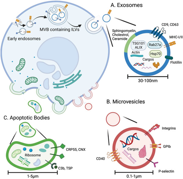
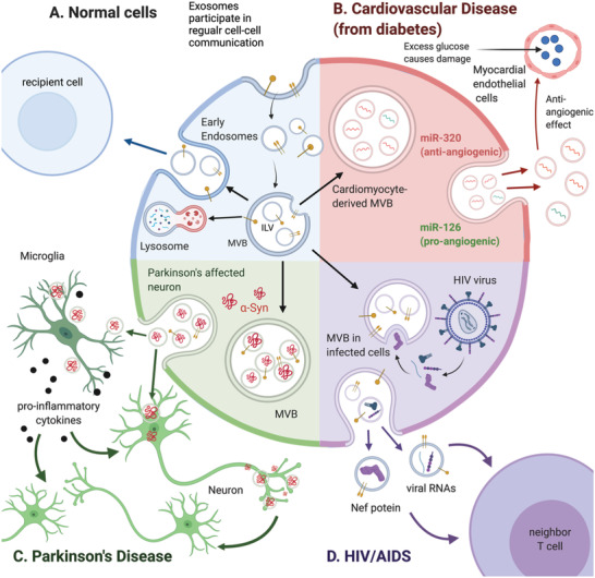
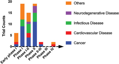
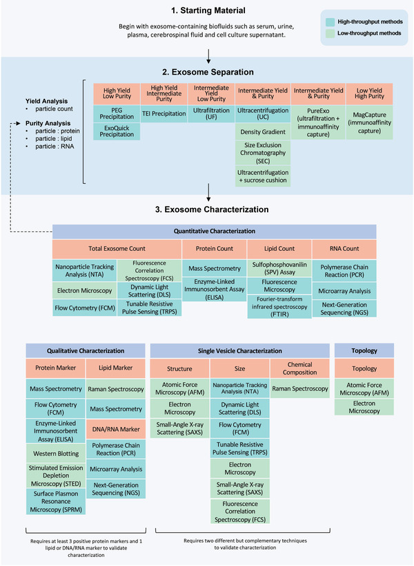
Exosomes are extracellular vesicles that share components of their parent cells and are attractive in biotechnology and biomedical research as potential disease biomarkers as well as therapeutic agents. Crucial to realizing this potential is the ability to manufacture high-quality exosomes; however, unlike biologics such as proteins, exosomes lack standardized Good Manufacturing Practices for their processing and characterization. Furthermore, there is a lack of well-characterized reference exosome materials to aid in selection of methods for exosome isolation, purification, and analysis. This review informs exosome research and technology development by comparing exosome processing and characterization methods and recommending exosome workflows. This review also provides a detailed introduction to exosomes, including their physical and chemical properties, roles in normal biological processes and in disease progression, and summarizes some of the on-going clinical trials.

Keywords: Good Manufacturing Practices; MISEV2018 guidelines; analytical characterizations; exosome clinical trials; exosomes; extracellular vesicles; isolation processes.


Conflict of interest statement

The authors declare no conflict of interest.

Figures

Scheme 1
Scheme 1
The biogenesis pathways and biochemical composition of A) exosomes, B) microvesicles, and C) apoptotic bodies. (A) Proteins, lipids, and genetic material are loaded into ILVs which are eventually released from the parent cell as exosomes. (B) Microvesicles are formed by directly budding off of the plasma membrane and contain proteins, lipids, and genetic materials. (C) Apoptotic bodies bud directly from the plasma membrane during apoptosis, and consequently, contain higher amounts of disintegrated organelle content. Created with BioRender.com.
Scheme 2
Scheme 2
Exosome biogenesis. A) Normal biogenesis pathway. B) Exosomes derived from cardiomyocytes may play a role in the development of cardiovascular disease from diabetes by upregulating antiangiogenic miR‐320 and downregulating proangiogenic miR‐126 in neighboring endothelial cells. C) In Parkinson's disease pathogenesis, exosomes from infected neurons and microglia act as carriers to transmit α‐synuclein (α‐syn) and proinflammatory cytokines. D) HIV‐infected cells secrete exosomes containing viral genomes, antigens, and other HIV‐associated proteins. The transfer of these viral products to other cells facilitates HIV infection. Created with BioRender.com.
Figure 1
Figure 1
Clinical trials involving exosomes, based on an analysis of clinical trials listed in clinicaltrial.gov (May 25th, 2021; search bar: “Other terms”; keyword: “Exosome”; exclusion: trials without FDA‐defined phases). More than 40% of the trials are cancer‐related (phase I–III); other trials (color coded) are investigating exosomes as diagnostic and therapeutic agents for mild cognitive impairment, Alzheimer's disease, heart failure, stroke, periodontitis, and other conditions.
Scheme 3
Scheme 3
Exosome processing and characterization flowchart. The exosome separation portion of the workflow includes common processing approaches with yields and purity levels. The characterization portion of the flowchart contains quantitative characterization, qualitative characterization, single vesicle characterization, and topology. The workflow starts with measuring and recording parameters of the starting materials such as cell count, fluid volume, and quantity of non‐EV molecules. Next, an appropriate processing method is selected based on the starting material and the target yield and purity. After exosome isolation, characterization begins with quantitation of the purity and yield of particles, proteins, lipids, and nucleic acids. If the sample does not meet the desired purity and yield, additional processing is required. Appropriate methods are then selected for bulk and single vesicle characterization to finish the characterization process.
Figure 2
Figure 2
Relative yield, purity, and throughput of different exosome isolation techniques. ‐ Qualitative data were obtained from comparison studies, A–E.[ 137 , 142 , 148 , 164 , 172 ] Bubble size indicates the throughput of each method relative to ultracentrifugation, the gold‐standard for exosome isolation. Precipitation methods analyzed include polyethylene glycol (PEG), Exoquick‐TC (System Biosciences), and total exosome isolation (TEI, Invitrogen).

References

    1. a) Cheng H. L., Fu C. Y., Kuo W. C., Chen Y. W., Chen Y. S., Lee Y. M., Li K. H., Chen C. C., Ma H. P., Huang P. C., Wang Y. L., Lee G. B., Lab Chip 2018, 18, 2917; - PubMed
    2. b) Kura B., Kalocayova B., Devaux Y., Bartekova M., Int. J. Mol. Sci. 2020, 21, 700; - PMC - PubMed
    3. c) Osada‐Oka M., Shiota M., Izumi Y., Nishiyama M., Tanaka M., Yamaguchi T., Sakurai E., Miura K., Iwao H., Hypertens. Res. 2017, 40, 353. - PubMed
    1. Bellin G., Gardin C., Ferroni L., Chachques J. C., Rogante M., Mitrecic D., Ferrari R., Zavan B., Cells 2019, 8, 166. - PMC - PubMed
    1. Chen C. C., Liu L., Ma F., Wong C. W., Guo X. E., Chacko J. V., Farhoodi H. P., Zhang S. X., Zimak J., Ségaliny A., Riazifar M., Pham V., Digman M. A., Pone E. J., Zhao W., Cell Mol. Bioeng. 2016, 9, 509. - PMC - PubMed
    1. Rastogi S., Sharma V., Bharti P. S., Rani K., Modi G. P., Nikolajeff F., Kumar S., Int. J. Mol. Sci. 2021, 22, 440. - PMC - PubMed
    1. a) Welch J. L., Stapleton J. T., Okeoma C. M., J. Gen. Virol. 2019, 100, 350; - PMC - PubMed
    2. b) Sims B., Farrow A. L., Williams S. D., Bansal A., Krendelchtchikov A., Matthews Q. L., Arch. Virol. 2018, 163, 1683; - PMC - PubMed
    3. c) Ouattara L. A., Anderson S. M., Doncel G. F., Andrologia 2018, 50, e13220; - PMC - PubMed
    4. d) Cheruiyot C., Pataki Z., Ramratnam B., Li M., Proteomics: Clin. Appl. 2018, 12, 1700142; - PubMed
    5. e) Pulliam L., Sun B., Mustapic M., Chawla S., Kapogiannis D., J. Neurovirol. 2019, 25, 702. - PMC - PubMed

Publication types

LinkOut - more resources