. 2022 May;33(9-10):499-517.
doi: 10.1089/hum.2021.245. Epub 2022 Mar 22.

Efficacy and Safety of a Krabbe Disease Gene Therapy

Affiliations

Efficacy and Safety of a Krabbe Disease Gene Therapy

Juliette Hordeaux et al. Hum Gene Ther. 2022 May.

Abstract

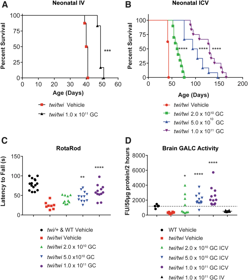
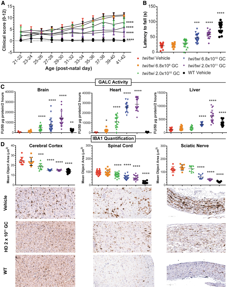
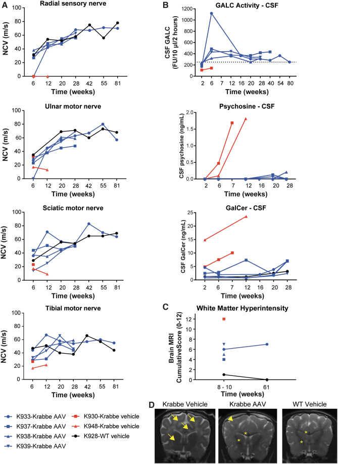
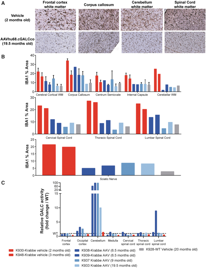
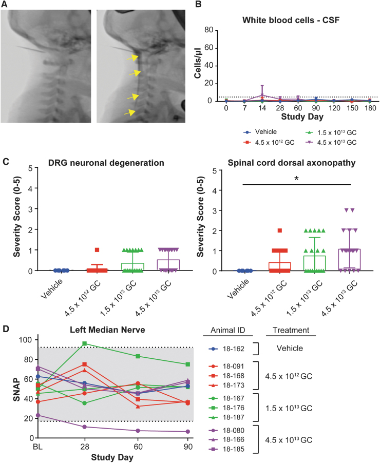
Krabbe disease is a lysosomal storage disease caused by mutations in the gene that encodes galactosylceramidase, in which galactosylsphingosine (psychosine) accumulation drives demyelination in the central and peripheral nervous systems, ultimately progressing to death in early childhood. Gene therapy, alone or in combination with transplant, has been developed for almost two decades in mouse models, with increasing therapeutic benefit paralleling the improvement of next-generation adeno-associated virus (AAV) vectors. This effort has recently shown remarkable efficacy in the canine model of the disease by two different groups that used either systemic or cerebrospinal fluid (CSF) administration of AAVrh10 or AAV9. Building on our experience developing CSF-delivered, AAV-based drug products for a variety of neurodegenerative disorders, we conducted efficacy, pharmacology, and safety studies of AAVhu68 delivered to the CSF in two relevant natural Krabbe animal models, and in nonhuman primates. In newborn Twitcher mice, the highest dose (1 × 10¹¹ genome copies [GC]) of AAVhu68.hGALC injected into the lateral ventricle led to a median survival of 130 days compared to 40.5 days in vehicle-treated mice. When this dose was administered intravenously, the median survival was 49 days. A single intracisterna magna injection of AAVhu68.cGALC at 3 × 10¹³ GC into presymptomatic Krabbe dogs increased survival for up to 85 weeks compared to 12 weeks in controls. It prevented psychosine accumulation in the CSF, preserved peripheral nerve myelination, ambulation, and decreased brain neuroinflammation and demyelination, although some regions remained abnormal. In a Good Laboratory Practice-compliant toxicology study, we administered the clinical candidate into the cisterna magna of 18 juvenile rhesus macaques at 3 doses that displayed efficacy in mice. We observed no dose-limiting toxicity and sporadic minimal degeneration of dorsal root ganglia (DRG) neurons. Our studies demonstrate the efficacy, scalability, and safety of a single cisterna magna AAVhu68 administration to treat Krabbe disease. ClinicalTrials.Gov ID: NCT04771416.

Keywords: AAV; Krabbe; Twitcher mouse; cisterna magna; demyelination; galactosylceramidase (GALC); galactosylceramide; lysosomal storage disease; psychosine.


Conflict of interest statement

J.M.W. is a paid advisor to and holds equity in iECURE, Scout Bio, Passage Bio, and the Center for Breakthrough Medicines (CBM). He also holds equity in the G2 Bio-associated asset companies. He has sponsored research agreements with Amicus Therapeutics, Biogen, CBM, Elaaj Bio, FA212, G2 Bio, G2 Bio-associated asset companies, iECURE, Janssen, Passage Bio, and Scout Bio, which are licensees of Penn technology. J.M.W. and J.H. are inventors on patents that have been licensed to various biopharmaceutical companies and for which they may receive payments.

A.M.B. is a beneficiary of a licensing agreement with Axovant Gene Therapies (royalties); has received income from Neurogene (consulting and honorarium); and is an inventor on a patent application (Gray SJ, Lykken E, Vite CH, Bradbury AM. Optimized GALC Genes and Expression Cassettes and Their Use. PCT/US2019/067727). All other authors have no competing financial interests exist.

Figures

Figure 1.
Figure 1.
Proof-of-concept efficacy in neonatal Twitcher mice. (A) Survival curve of Twitcher mice injected intravenously on PND0 with 50 μL of PBS (vehicle, n = 8; median survival 40.5 days) or with 1 × 1011 GC of AAVhu68.CB7.hGALCco.rBG diluted in 50 μL PBS (n = 6; median survival 49 days). ***p < 0.001 Log-rank (Mantel-Cox) test. (B) Survival curve of Twitcher mice injected in the lateral cerebral ventricle (ICV) on PND0 with 2 μL of PBS (vehicle, n = 8; median survival 43 days), with 2 × 1010 GC of AAVhu68.CB7.hGALCco.rBG in 2 μL PBS (low dose, n = 10; median survival 62 days), with 5 × 1010 GC of AAVhu68.CB7.hGALCco.rBG in 2 μL PBS (mid dose, n = 12; median survival 99 days), or with 1 × 1011 GC of AAVhu68.CB7.hGALCco.rBG in 2 × 2 μL PBS (bilateral ICV, high dose, n = 12; median survival 130 days). ****p < 0.0001 Log-rank (Mantel-Cox) test. (C) Neuromotor function assessed by the accelerated rotarod test on PND35 in mice treated via neonatal ICV administration [same animals as in (B)]. **p < 0.01, ***p < 0.001, ****p < 0.0001, Kruskal-Wallis test followed by post hoc Dunn's multiple comparison test, alpha = 0.05, comparison to vehicle Twitcher mice. (D) GALC enzyme activity in brain lysate from tissue obtained at humane endpoint in animals administered IV or ICV [same animals as in (A, B)]. *p < 0.05, ****p < 0.0001, Kruskal-Wallis test followed by post hoc Dunn's multiple comparison test, alpha = 0.05, comparison to vehicle Twitcher mice. GALC, galactosylceramidase; GC, genome copies; ICV, intracerebroventricular; IV, intravenous; PBS, phosphate-buffered saline; PND, postnatal day.
Figure 2.
Figure 2.
MED study in juvenile postdisease onset Twitcher mice. (A) Compound clinical severity score in Twitcher mice treated on PND12–14 ICV with either 4 μL of artificial CSF (vehicle, n = 17), or 4 μL of AAVhu68.CB7.hGALCco.rBG at the following doses: 6.8 × 109 GC (n = 16), 2 × 1010 GC (n = 17), 6.8 × 1010 (n = 17), or 2 × 1011 GC (n = 16). WT littermates were treated ICV with 4 μL of artificial CSF (n = 17). The operator was blinded to the mice genotype and treatment. ****p < 0.0001 linear mixed-effect modeling comparing the clinical score change over time compared to the vehicle Twitcher group, alpha = 0.05. (B) Neuromotor function assessed by the accelerated rotarod test on PND35 in same mice as in (A) **p < 0.01, ***p < 0.001, ****p < 0.0001, Kruskal-Wallis test followed by post hoc Dunn's multiple comparison test, alpha = 0.05, comparison to vehicle Twitcher mice. (C) GALC enzyme activity in brain (sagittal half), heart (sagittal half), and liver (half of the left lobe) lysate from tissue obtained either at scheduled necropsy (PND40, half of the animals) or humane endpoint (half of the animals) in the same animals as in (A) *p < 0.05, ****p < 0.0001, Kruskal-Wallis test followed by post hoc Dunn's multiple comparison test, alpha = 0.05, comparison to vehicle Twitcher mice. (D) Neuroinflammation quantification in the cerebral cortex, spinal cord, and sciatic nerve measured by mean object area of IBA1-positive cells stained by immunohistochemistry in tissues collected at the fixed necropsy time PND40. ***p < 0.001, ****p < 0.0001, Kruskal-Wallis test followed by post hoc Dunn's multiple comparison test, alpha = 0.05, comparison to vehicle-treated Twitcher mice. Representative images of IBA1 IHC from the vehicle groups (Twitcher and WT mice) and the HD group (2 × 1011 GC, ICV PND12–14). CSF, cerebrospinal fluid; MED, minimum effective dose; WT, wild type.
Figure 3.
Figure 3.
ICM efficacy study in Krabbe dogs, nerve-conduction studies, CSF biomarkers, and MRI. (A) Nerve-conduction velocities in the radial (sensory), sciatic (motor), ulnar (motor), and tibial (motor) nerves in 2- to 3-week-old Krabbe dogs treated ICM with either (1) 1 mL of artificial CSF (vehicle, n = 2); or (2) 3 × 1013 GC of AAVhu68.CB7.cGAMCco.rBG in 1 mL (n = 4). A WT littermate that received 1 mL of artificial CSF ICM was used as a control. Two treated dogs were sacrificed at the scheduled timepoint of 6 months postinjection for tissue collection. Two treated dogs and the vehicle-treated Krabbe dogs were followed until humane endpoint. (B) Quantification of CSF psychosine and GalCer levels and CSF GALC enzyme activity. The dotted line represent the average GALC activity value from the serial CSF timepoints of the WT control (K928). (C) Brain MRI WM intensity semiquantitative scoring. (D) Brain MRI examples from one Krabbe vehicle dog (8 weeks), one Krabbe dog treated with AAV (10 weeks), and one WT control littermate (10 weeks). A dark hypointense WM represents normal myelination (stars); a grey isointense signal represents mild demyelination (arrowhead); a white hyperintense signal represents marked demyelination (arrows). AAV, adeno-associated virus; GalCer, galactosylceramide; ICM, intracisterna magna; MRI, magnetic resonance imaging; WM, white matter.
Figure 4.
Figure 4.
ICM efficacy study in Krabbe dogs, neuroinflammation and GALC levels. (A) Representative pictures of IBA1 staining by IHC illustrating the macrophage/microglial neuroinflammation in Krabbe dogs treated with vehicle control or with AAV ICM. (B) Quantification of the mean area of the IBA1-positive signal in different neuroanatomical regions of the brain, spinal cord, and sciatic nerve of Krabbe dogs treated with AAV or vehicle. Each bar represents an animal. Error bars when present (cerebral cortical WM, corpus callosum, centrum semiovale, and internal capsule), represent the standard deviation from quantification of multiple slides when the neuroanatomical region was present on more than one brain section. (C) GALC activity in tissue lysate (50 μg protein per reaction, 2 h incubation) relative to the WT control (dotted line). Each bar represents one animal. WM, white matter.
Figure 5.
Figure 5.
GLP toxicology study in rhesus macaques, safety. (A) Fluoroscopic images of the neck region, before and after contrast ICM administration, in juvenile rhesus macaques (animal 18–167, mid-dose group). Contrast injection was used to confirm needle placement before AAV administration. Arrows show the diffusion of the contrast in the cisterna magna and spinal canal. (B) WBC counts in the CSF of juvenile rhesus macaques treated ICM with artificial CSF (vehicle, n = 2) or AAVhu68.CB7.hGALCco.rBG at the dose of 4.5 × 1012 GC (low dose, n = 6), 1.5 × 1013 GC (mid dose, n = 6), or 4.5 × 1013 (high dose, n = 6). Half of the animals were euthanized for tissue collection 3 months postdosing; the other half were euthanized 6 months postdosing. Dotted line represents the upper limit of normal (C) Histopathological analysis, DRG neuronal degeneration, and spinal cord dorsal column axonopathy severity grades (0 = normal, 1 = minimal, 2 = mild, 3 = moderate, 4 = marked, and 5 = severe). Three and six months cohorts are included, each data point represents 1 segment per animal (cervical, thoracic, and lumbar). *p < 0.05 Mann-Whitney rank test, alpha = 0.05. (D) Median nerve SNAP amplitudes at BL (before dosing), 28-, 60-, and 90-day postdosing in the 3 months cohorts. BL, baseline; DRG, dorsal root ganglia; GLP, Good Laboratory Practice; SNAP, sensory nerve action potential; WBC, white blood cell.

References

    1. Wenger DA, Rafi MA, Luzi P, et al. . Krabbe disease: genetic aspects and progress toward therapy. Mol Genet Metab 2000;70:1–9. - PubMed
    1. Svennerholm L, Vanier MT, Mansson JE. Krabbe disease: a galactosylsphingosine (psychosine) lipidosis. J Lipid Res 1980;21:53–64. - PubMed
    1. Escolar ML, Kiely BT, Shawgo E, et al. . Psychosine, a marker of Krabbe phenotype and treatment effect. Mol Genet Metab 2017;121:271–278. - PMC - PubMed
    1. Voccoli V, Tonazzini I, Signore G, et al. . Role of extracellular calcium and mitochondrial oxygen species in psychosine-induced oligodendrocyte cell death. Cell Death Dis 2014;5:e1529. - PMC - PubMed
    1. Potter GB, Santos M, Davisson MT, et al. . Missense mutation in mouse GALC mimics human gene defect and offers new insights into Krabbe disease. Hum Mol Genet 2013;22:3397–3414. - PMC - PubMed

Publication types

Associated data