. 2022 May 5;221(1):iyac048.
doi: 10.1093/genetics/iyac048.

The Mnn10/Anp1-dependent N-linked outer chain glycan is dispensable for Candida albicans cell wall integrity

Affiliations

The Mnn10/Anp1-dependent N-linked outer chain glycan is dispensable for Candida albicans cell wall integrity

Neta Dean et al. Genetics. .

Abstract

Candida albicans cell wall glycoproteins, and in particular their mannose-rich glycans, are important for maintaining cellular integrity as well as host recognition, adhesion, and immunomodulation. The asparagine (N)-linked mannose outer chain of these glycoproteins is produced by Golgi mannosyltransferases (MTases). The outer chain is composed of a linear backbone of ∼50 α1,6-linked mannoses, which acts as a scaffold for addition of ∼150 or more mannoses in other linkages. Here, we describe the characterization of C. albicans OCH1, MNN9, VAN1, ANP1, MNN10, and MNN11, which encode the conserved Golgi MTases that sequentially catalyze the α1,6 mannose outer chain backbone. Candida albicans och1Δ/Δ, mnn9Δ/Δ, and van1Δ/Δ mutants block the earliest steps of backbone synthesis and like their Saccharomyces cerevisiae counterparts, have severe cell wall and growth phenotypes. Unexpectedly, and in stark contrast to S. cerevisiae, loss of Anp1, Mnn10, or Mnn11, which together synthesize most of the backbone, have no obvious deleterious phenotypes. These mutants were unaffected in cell morphology, growth, drug sensitivities, hyphal formation, and macrophage recognition. Analyses of secreted glycosylation reporters demonstrated that anp1Δ/Δ, mnn10Δ/Δ, and mnn11Δ/Δ strains accumulate glycoproteins with severely truncated N-glycan chains. This hypo-mannosylation did not elicit increased chitin deposition in the cell wall, which in other yeast and fungi is a key compensatory response to cell wall integrity breaches. Thus, C. albicans has evolved an alternate mechanism to adapt to cell wall weakness when N-linked mannan levels are reduced.

Keywords: ANP1; Candida albicans; MNN10; MNN11; MNN9; N-linked glycosylation; OCH1; VAN1; Golgi; cell wall; mannosyltransferase.


Figures

Fig. 1.
Fig. 1.
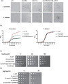
Phenotypic analysis of C. albicans and S. cerevisiae single and double mnn10 and mnn11 deletion mutants. a) Light microscopy (40×) of wild-type, mnn10Δ/Δ, mnn11Δ/Δ, mnn10Δ/Δ/mnn11Δ/Δ, and och1Δ/Δstrains. Saccharomyces cerevisiae strains are in the upper panel and corresponding C. albicans Δ/Δ mutants are in lower panel. b) Wild-type or mnn10Δ/Δ/mnn11Δ/Δ strains were diluted to 0.20 OD600 units/ml in liquid YPAD. Growth at 30°C was monitored over time (up till 27 h) by taking aliquots and measuring the absorbance at OD600. Triplicate measurements were collected for each time point. Means and standard deviations were plotted using GraphPad Prism. c) Hygromycin sensitivity of wild-type or C. albicans mnn10Δ/Δ mnn11Δ/Δ strains. Yeast were grown to early logarithmic stage, adjusted to 1 OD600 units/ml and 10-fold serially diluted. Three microliters of each dilution (104, 103, 102, 101, and 10°) were plated on YPAD containing hygromycin B at the indicated concentrations. Note that concentrations of hygromycin B above 100 µg/ml inhibit the growth of the wild-type parental C. albicans strain. d) Complementation of the hygromycin B-sensitive phenotype of S. cerevisiae mnn10Δ and mnn11Δ mutants by CaMNN10 and CaMNN11. Saccharomyces cerevisiae WT (SEY6210), mnn10Δ, and mnn11Δ harboring a vector, or plasmid-borne CaMNN10 or CaMNN11 were serially diluted as described in c), and plated on YPAD plus or minus 50 µg/ml hygromycin B. Plates were incubated at 30°C for 3 days.
Fig. 2.
Fig. 2.
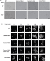
MNN10 and MNN11 are not required for hyphal formation Hyphal formation was examined in wild-type parental (BWP17) or isogenic mnn10Δ/Δ, mnn11Δ/Δ, mnn10Δ/Δ mnn11Δ/Δ, or och1Δ/Δ strains. Cultures were grown to early logarithmic stage (0.5 OD600 units/ml) and induced to form hyphae by the addition of 20% calf serum and a temperature shift from 30°C to 37°C. Aliquots were removed at the indicated times and viewed by light microscopy (a) or incubated for 1 min with calcofluor white and viewed by fluorescence microscopy (×40 magnification) (b).
Fig. 3.
Fig. 3.
Mnn10 is required for macrophage uptake of S. cerevisiae but not C. albicans. Phagocytosis of yeast by macrophage was quantitated by competition assays as described in Materials and Methods. RFP or GFP-expressing yeast strains of differing genotypes were mixed in equal numbers and added to J774 macrophages at an MOI of 5. After 90 min, calcofluor white was added, and red and green yeast were scored by fluorescence as being inside (calcofluor white negative) or outside (calcofluor white positive) of macrophages. Top and bottom panels show competition assays between C. albicans or between S. cerevisiae strains, respectively.
Fig. 4.
Fig. 4.
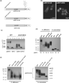
MNN10 and MNN11 are required for N-glycosylation. a) Schematic diagram of N-linked glycosylation RFP reporter. ssRFP-N-HA encodes yEmRFP tagged with an N-terminal signal sequence, a C-terminal HA tag and a single N/D/T N-glycan attachment recognition motif. ssRFP-X-HA is identical except it lacks the N/D/T recognition motif and therefore is not an acceptor for the N-linked glycan. RFP-X-HA lacks both the N/D/T and N-terminal signal sequences and therefore cannot enter the secretory pathway. b) Localization of ssRFP-N-HA in C. albicans. Yeast cells expressing ssRFP-N-HA, or ssRFP-HDEL that carries a C-terminal HDEL ER retention sequence instead of the N/D/T-HA, were grown to mid-log phase and viewed by fluorescence microscopy (using 100× magnification). Arrow denotes perinuclear ER staining. c) Western blot analysis of total (T) and secreted (s) ssRFP-N-HA or RFP-X-HA protein isolated from C. albicans wild-type or mnn10Δ/Δ. Cells were grown to mid-log phase and proteins were extracted by precipitation of proteins in an aliquot of the culture (T) or from culture supernatants (S), as described in Materials and Methods. Equal cell equivalents of protein extracts were analyzed by 10% SDS-PAGE and detected by chemiluminescence with anti-HA antibody. d) Western blot analysis of ss-RFP-N-HA and ss-RFP-X-HA in wild-type and och1 mutants of C. albicans and S. cerevisiae. C. albicans or S. cerevisiae wild-type and och1 mutants expressing secreted RFP (ssRFP) with (N) or without (X) a N-glycosylation site are indicated below each lane. Secreted proteins in culture supernatants were acetone precipitated as described in Materials and Methods. Protein extracts (2.25 µg/lane for C. albicans samples and 0.45 µg/lane for S. cerevisiae samples) were separated by 10% SDS-PAGE and detected by western blotting with anti-HA. e) N-glycosylation defects in C. albicans and corresponding S. cerevisiae α1,6 MTase mutant strains. Secreted ssRFP-N-HA was isolated from the culture supernatants of S. cerevisiae or C. albicans wild-type, mnn10Δ/Δ, mnn11Δ/Δ, mnn10Δ/Δ mnn11Δ/Δ, and och1Δ/Δ strains by acetone precipitation as described in Materials and Methods. Protein extracts (2.25 µg/lane for C. albicans samples and 0.45 µg/lane for S. cerevisiae samples) were separated by 10% SDS-PAGE and detected by western blotting with anti-HA antibody as in (d).
Fig. 5.
Fig. 5.
Colony morphology and glycosylation phenotypes of C. albicans Golgi α1,6 MTase mutants. a) Wild-type or homozygous C. albicans mutants defective in each of the Golgi α1,6 MTase genes (mnn11Δ/Δ, mnn10Δ/Δ, anp1Δ/Δ, van1Δ/Δ, mnn9Δ/Δ, and och1Δ/Δ) were spotted on YPAD solid media. Individual colonies were incubated for 3 days at 30°C before photography. b) Western blot analyses of secreted proteins from C. albicans (left panel) and S. cerevisiae (right panel) MTase mutants expressing ss-RFP-N-HA. Proteins from culture supernatants were acetone precipitated as described in Materials and Methods. Protein extracts (2.25 µg/lane for C. albicans samples and 0.45 µg/lane for S. cerevisiae samples) were separated by 10% SDS-PAGE and detected by western blotting with anti-HA antibody. Note that gels with C. albicans samples were run longer than S. cerevisiae.
Fig. 6.
Fig. 6.
Analysis of cell wall chitin C. albicans and S. cerevisiae α1,6 MTase mutants. a) Candida albicans strains were stained with calcofluor white and imaged by fluorescence microscopy as described in Materials and Methods; ×40 images were taken with the same exposure settings. b) Saccharomyces cerevisiae strains were stained with calcofluor white and imaged as in (a). c) Calcofluor white fluorescence intensity of cells [as described in (a) and (b)] was measured by microscopy, as described in Materials and Methods. d) Cell wall chitin was measured by the Morgan–Elson assay (Leloir and Cardini 1953). After purifying cell wall, absolute levels of glucosamine were measured spectrophotometrically by absorbance at 520 nm (see Materials and methods).
Fig. 7.
Fig. 7.
Schematic diagram of N-linked backbone extension in Golgi of S. cerevisiae and C. albicans. After core synthesis and transfer to proteins in ER, the N-linked glycan is extended by MTases in the Golgi. In S. cerevisiae, loss of α1,6 MTases Och1, Mnn, Van1, Mnn10, or Anp1 leads to increases in cell wall chitin. In C. albicans, loss of Och1, Mnn9, or Van 1 also stimulates cell wall chitin deposition but backbone truncations beyond Van1 do not trigger the cell wall integrity pathway.

Similar articles

Cited by

References

    1. Ballou CE. Isolation, characterization and properties of Saccharomyces cerevisiae mnn mutants with nonconditional protein glycosylation defects. Methods Enzymol. 1990;185:440–470. - PubMed
    1. Bates S, Hall RA, Cheetham J, Netea MG, MacCallum DM, Brown AJP, Odds FC, Gow NAR.. Role of the Candida albicans MNN1 gene family in cell wall structure and virulence. BMC Res Notes. 2013;6(294):294. - PMC - PubMed
    1. Bates S, Hughes HB, Munro CA, Thomas WPH, MacCallum DM, Bertram G, Atrih A, Ferguson MAJ, Brown AJP, Odds FC, et al.Outer chain N-glycans are required for cell wall integrity and virulence of Candida albicans. J Biol Chem. 2006;281(1):90–98. - PubMed
    1. Bulik DA, Olczak M, Lucero HA, Osmond BC, Robbins PW, Specht CA.. Chitin synthesis in Saccharomyces cerevisiae in response to supplementation of growth medium with glucosamine and cell wall stress. Eukaryot Cell. 2003;2(5):886–900. - PMC - PubMed
    1. Chapman RE, Munro S.. The functioning of the yeast Golgi apparatus requires an ER protein encoded by ANP1, a member of a new family of genes affecting the secretory pathway. Eur J Mol Biol. 1994;13:4896–4907. - PMC - PubMed

MeSH terms