. 2022 May 24;145(4):1285-1298.
doi: 10.1093/brain/awab417.

Topographic divergence of atypical cortical asymmetry and atrophy patterns in temporal lobe epilepsy

Bo-Yong Park  1   2   3 Sara Larivière  1 Raul Rodríguez-Cruces  1 Jessica Royer  1 Shahin Tavakol  1 Yezhou Wang  1 Lorenzo Caciagli  4   5   6 Maria Eugenia Caligiuri  7 Antonio Gambardella  7   8 Luis Concha  9 Simon S Keller  10   11 Fernando Cendes  12 Marina K M Alvim  12 Clarissa Yasuda  12 Leonardo Bonilha  13 Ezequiel Gleichgerrcht  14 Niels K Focke  15 Barbara A K Kreilkamp  15 Martin Domin  16 Felix von Podewils  17 Soenke Langner  18 Christian Rummel  19 Michael Rebsamen  19 Roland Wiest  19 Pascal Martin  20 Raviteja Kotikalapudi  20   21 Benjamin Bender  21 Terence J O'Brien  22   23 Meng Law  22 Benjamin Sinclair  22   23 Lucy Vivash  22   23 Patrick Kwan  22   23 Patricia M Desmond  24 Charles B Malpas  23 Elaine Lui  24 Saud Alhusaini  25   26 Colin P Doherty  27   28 Gianpiero L Cavalleri  25   28 Norman Delanty  25   28 Reetta Kälviäinen  29   30 Graeme D Jackson  31 Magdalena Kowalczyk  31 Mario Mascalchi  32 Mira Semmelroch  31 Rhys H Thomas  33 Hamid Soltanian-Zadeh  34   35 Esmaeil Davoodi-Bojd  36 Junsong Zhang  37 Matteo Lenge  38   39 Renzo Guerrini  38 Emanuele Bartolini  40 Khalid Hamandi  41   42 Sonya Foley  41 Bernd Weber  43 Chantal Depondt  44 Julie Absil  45 Sarah J A Carr  46 Eugenio Abela  46 Mark P Richardson  46 Orrin Devinsky  47 Mariasavina Severino  48   49 Pasquale Striano  48   49 Costanza Parodi  48   49 Domenico Tortora  48   49 Sean N Hatton  50 Sjoerd B Vos  4   5   51 John S Duncan  4   5 Marian Galovic  4   5   52 Christopher D Whelan  53 Núria Bargalló  54   55 Jose Pariente  54 Estefania Conde-Blanco  56 Anna Elisabetta Vaudano  57   58 Manuela Tondelli  57   58 Stefano Meletti  57   58 Xiang-Zhen Kong  59   60 Clyde Francks  59   61 Simon E Fisher  59   61 Benoit Caldairou  62 Mina Ryten  63   64   65 Angelo Labate  66 Sanjay M Sisodiya  4   5 Paul M Thompson  67 Carrie R McDonald  68 Andrea Bernasconi  62 Neda Bernasconi  62 Boris C Bernhardt  1
Affiliations

Topographic divergence of atypical cortical asymmetry and atrophy patterns in temporal lobe epilepsy

Bo-Yong Park et al. Brain. .

Abstract

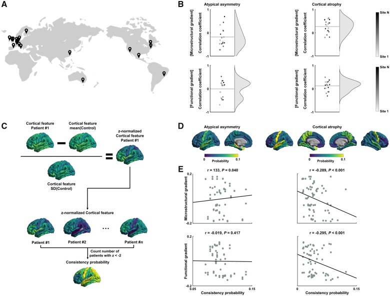
Temporal lobe epilepsy, a common drug-resistant epilepsy in adults, is primarily a limbic network disorder associated with predominant unilateral hippocampal pathology. Structural MRI has provided an in vivo window into whole-brain grey matter structural alterations in temporal lobe epilepsy relative to controls, by either mapping (i) atypical inter-hemispheric asymmetry; or (ii) regional atrophy. However, similarities and differences of both atypical asymmetry and regional atrophy measures have not been systematically investigated. Here, we addressed this gap using the multisite ENIGMA-Epilepsy dataset comprising MRI brain morphological measures in 732 temporal lobe epilepsy patients and 1418 healthy controls. We compared spatial distributions of grey matter asymmetry and atrophy in temporal lobe epilepsy, contextualized their topographies relative to spatial gradients in cortical microstructure and functional connectivity calculated using 207 healthy controls obtained from Human Connectome Project and an independent dataset containing 23 temporal lobe epilepsy patients and 53 healthy controls and examined clinical associations using machine learning. We identified a marked divergence in the spatial distribution of atypical inter-hemispheric asymmetry and regional atrophy mapping. The former revealed a temporo-limbic disease signature while the latter showed diffuse and bilateral patterns. Our findings were robust across individual sites and patients. Cortical atrophy was significantly correlated with disease duration and age at seizure onset, while degrees of asymmetry did not show a significant relationship to these clinical variables. Our findings highlight that the mapping of atypical inter-hemispheric asymmetry and regional atrophy tap into two complementary aspects of temporal lobe epilepsy-related pathology, with the former revealing primary substrates in ipsilateral limbic circuits and the latter capturing bilateral disease effects. These findings refine our notion of the neuropathology of temporal lobe epilepsy and may inform future discovery and validation of complementary MRI biomarkers in temporal lobe epilepsy.

Keywords: asymmetry; cortical thickness; gradients; multi-site; temporal lobe epilepsy.


Figures

Figure 1
Figure 1
Topography of atypical cortical asymmetry and atrophy patterns in TLE. (A) Atypical inter-hemispheric asymmetry of cortical thickness and regional cortical atrophy between individuals with TLE relative to controls, calculated using ENIGMA-Epilepsy dataset. Blue regions indicate significant ipsilateral versus contralateral cortical thickness asymmetry/atrophy in TLE relative to controls. Patient hemispheres are sorted into ipsilateral/contralateral to the seizure focus. (B) Effects (i.e. asymmetry index and cortical thickness) are stratified according to seven intrinsic functional communities and major lobes. (C) Associations between epilepsy-related findings and microstructural/functional gradients calculated using HCP dataset. Cortex-wide microstructural profile similarity matrix and scree plot describing connectome variance after identification of principal eigenvectors are shown. The first principal eigenvector (microstructural gradient) is shown on the cortical surface. Spatial correlations between the principal microstructural gradient and TLE-related effects (i.e. atypical cortical asymmetry and atrophy) are reported with scatter plots. (D) Identical analysis to C but based on functional gradients. Cing = cingulate; DAN = dorsal attention network; DMN = default mode network; FPN = frontoparietal control network; Front = frontal; Ins = insular; LBN = limbic network; Occ = occipital; Par = parietal; SMN = somatomotor network; Temp = temporal; VAN = ventral attention network; VN = visual network.
Figure 2
Figure 2
Consistency of atypical cortical asymmetry and atrophy. (A) World map of data acquisition sites. (B) Spatial correlations between topographic gradients and atypical cortical asymmetry/atrophy patterns of all sites. (C) Schema describing the computation of patient-wise consistency probability. The number of patients with large deviations of cortical features (i.e. atypical inter-hemispheric asymmetry or regional cortical atrophy) was counted. (D) Consistency probability of atypical cortical asymmetry and atrophy. (E) Spatial correlations between consistency probability and topographic gradients.
Figure 3
Figure 3
Associations between cortical features and clinical variables. (A) Probability of regions being selected across 5-fold nested cross-validation and 100 repetitions for predicting duration of epilepsy using atypical asymmetry index (left) and regional atrophy (right). Correlations between actual and predicted values of epilepsy duration are reported in the scatter plots. Black lines indicate the mean correlation and grey lines represent the 95% CI for 100 iterations with different training/test datasets. (B) Linear correlations between gradients and selected probability. (C) Spatial correlations between duration of epilepsy and atypical asymmetry index (left), as well as cortical atrophy (right) in highly probable (selected probability > 0.5) regions. (DF) Identical analysis to A–C, but with respect to age at seizure onset. MAE = mean absolute error.

References

    1. Falconer MA, Serafetinides EA, Corsellis JAN.. Etiology and pathogenesis of temporal lobe epilepsy. Arch Neurol. 1964;10(3):233–248. - PubMed
    1. Margerison JH, Corsellis JAN.. Epilepsy and the temporal lobes. Brain. 1966;89(3):499–530. - PubMed
    1. Blanc F, Martinian L, Liagkouras I, Catarino C, Sisodiya SM, Thom M.. Investigation of widespread neocortical pathology associated with hippocampal sclerosis in epilepsy: A postmortem study. Epilepsia. 2011;52(1):10–21. - PubMed
    1. Blümcke I, Thom M, Aronica E, et al. . International consensus classification of hippocampal sclerosis in temporal lobe epilepsy: A Task Force report from the ILAE Commission on Diagnostic Methods. Epilepsia. 2013;54(7):1315–1329. - PubMed
    1. Thom M. Review: Hippocampal sclerosis in epilepsy: A neuropathology review. Neuropathol Appl Neurobiol. 2014;40(5):520–543. - PMC - PubMed

Publication types

LinkOut - more resources