

Ureaplasma-Driven Neonatal Neuroinflammation: Novel Insights from an Ovine Model
- PMID: 35334011
- PMCID: PMC9957905
- DOI: 10.1007/s10571-022-01213-8
Ureaplasma-Driven Neonatal Neuroinflammation: Novel Insights from an Ovine Model
Abstract
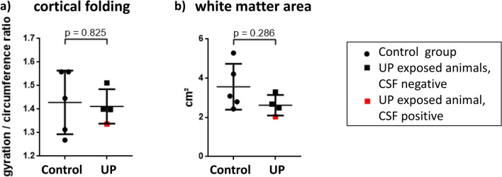
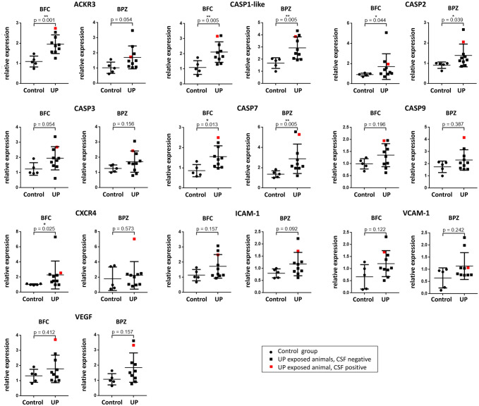
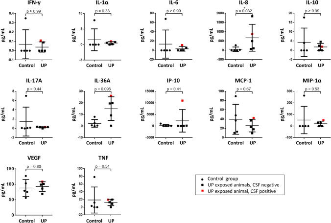
Ureaplasma species (spp.) are considered commensals of the adult genitourinary tract, but have been associated with chorioamnionitis, preterm birth, and invasive infections in neonates, including meningitis. Data on mechanisms involved in Ureaplasma-driven neuroinflammation are scarce. The present study addressed brain inflammatory responses in preterm lambs exposed to Ureaplasma parvum (UP) in utero. 7 days after intra-amniotic injection of UP (n = 10) or saline (n = 11), lambs were surgically delivered at gestational day 128-129. Expression of inflammatory markers was assessed in different brain regions using qRT-PCR and in cerebrospinal fluid (CSF) by multiplex immunoassay. CSF was analyzed for UP presence using ureB-based real-time PCR, and MRI scans documented cerebral white matter area and cortical folding. Cerebral tissue levels of atypical chemokine receptor (ACKR) 3, caspases 1-like, 2, 7, and C-X-C chemokine receptor (CXCR) 4 mRNA, as well as CSF interleukin-8 protein concentrations were significantly increased in UP-exposed lambs. UP presence in CSF was confirmed in one animal. Cortical folding and white matter area did not differ among groups. The present study confirms a role of caspases and the transmembrane receptors ACKR3 and CXCR4 in Ureaplasma-driven neuroinflammation. Enhanced caspase 1-like, 2, and 7 expression may reflect cell death. Increased ACKR3 and CXCR4 expression has been associated with inflammatory central nervous system (CNS) diseases and impaired blood-brain barrier function. According to these data and previous in vitro findings from our group, we speculate that Ureaplasma-induced caspase and receptor responses affect CNS barrier properties and thus facilitate neuroinflammation.
Keywords: Animal model; CNS Integrity; Immaturity; Neonatal meningitis; Preterm birth; Ureaplasma parvum.
© 2022. The Author(s).
Conflict of interest statement
All authors declare that they have no conflict of interest.
Figures
References
MeSH terms
Substances
LinkOut - more resources
Full Text Sources
Miscellaneous
