Comparative Study
. 2022 Sep;16(3):763-772.
doi: 10.1007/s12105-022-01440-x. Epub 2022 Mar 25.

Comparison of Interleukin-1 Ligand Expression by Human Papilloma Virus Status in HNSCCs

Affiliations
Comparative Study

Comparison of Interleukin-1 Ligand Expression by Human Papilloma Virus Status in HNSCCs

Ishrat Nourin Khan et al. Head Neck Pathol. 2022 Sep.

Abstract

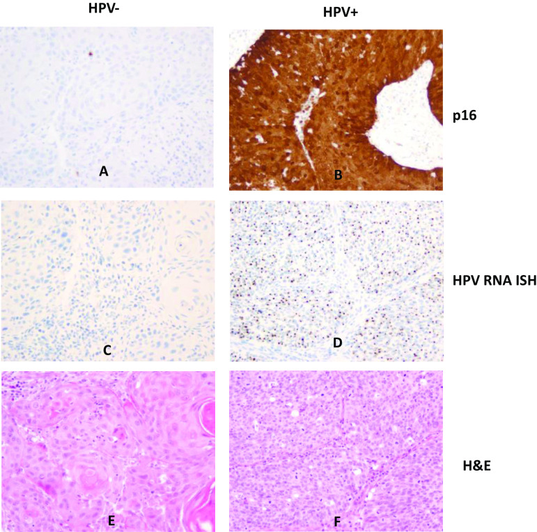
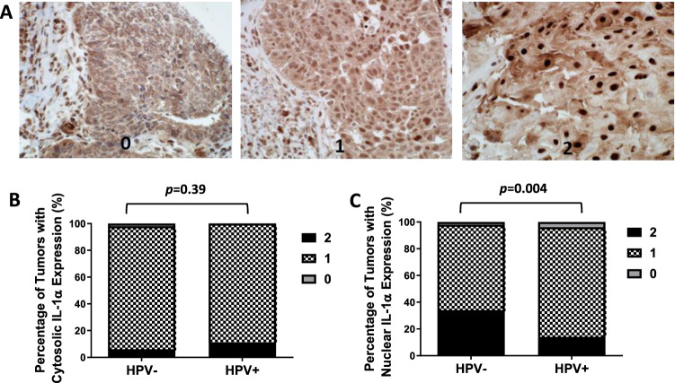
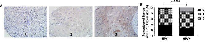
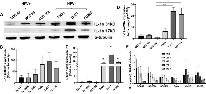
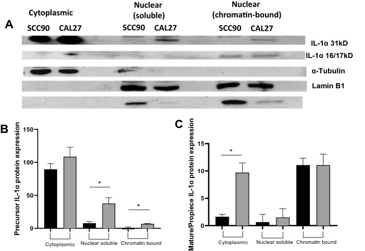
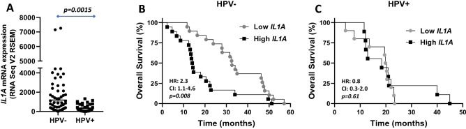
Interleukin-1 alpha (IL-1α) is a cytokine involved in the acute phase immune response and its expression is upregulated in a variety of solid tumors including head and neck squamous cell carcinomas (HNSCCs). Tumor expression of IL-1α is associated with increased tumor aggressiveness in HNSCCs, but this has yet to be studied in the context of human papilloma virus (HPV) status. This study is aimed at determining differences in tumor expression and subcellular localization of IL-1α in HPV-positive (HPV+) and HPV-negative (HPV-) HNSCC tumors. Tissue microarrays (TMAs) containing HPV+ (n = 31) and HPV- (n = 47) primary and metastatic HNSCCs were analyzed for IL-1α expression using immunohistochemical (IHC) staining. HPV status was confirmed using p16 IHC staining and RNA in situ hybridization (RNA ISH). Differences in IL-1α protein expression and secretion in HPV+ and HPV- HNSCC cell lines were determined by western blot and ELISA respectively. Associations between tumor IL1A expression and survival outcomes were assessed in HPV+ and HPV- HNSCC patients from publicly available gene expression datasets. Tumor expression of IL-1α was significantly increased in HPV- tumors and cell lines (as detected by IHC and western blot respectively) compared to HPV+ tumors and cell lines. There was no difference in IL-1α release between HPV+ and HPV- cell lines. IL-1α was expressed in both nuclear and cytoplasmic compartments, with predominant expression in the nucleus. Gene expression of IL1A was significantly increased in HPV-tumors/cell lines compared to HPV+ tumors/cell lines. Lastly, increased IL1A gene expression was significantly associated with worse survival in HPV- tumors but not in HPV+ tumors. Overall IL-1α expression particularly in the nucleus may possess more prognostic significance in HPV- tumors rather than HPV+ tumors. This work warrants further investigation into the role of intracellular IL-1α ligand expression in HNSCCs and may have important implications in IL-1 pathway blockade as therapeutic strategy.

Keywords: HPV; Head and neck squamous cell carcinoma; IL-1α; IL-1β; Tissue microarray.


Conflict of interest statement

No conflicts of interest to disclose.

Figures

Fig. 1
Fig. 1
Representative of positive and negative p16 immunostaining and high-risk HPV RNA ISH in HNSCCs. Shown are images (×200) representing p16 IHC (A, B), high-risk HPV RNA ISH (C, D) and H&E (E, F) in HPV− (A, C, E) and HPV+ (B, D, F) HNSCCs
Fig. 2
Fig. 2
IL-1α immunostaining and expression scores in HNSCCs. A Shown are images (×20) of negative [0], moderate [1], and strong [2] nuclear IL-1α expression. A, C Percentage of tumors with strong [2], moderate [1] or no [0] cytoplasmic (B) and nuclear (C) IL-1α expression based on HPV status
Fig. 3
Fig. 3
Representative examples of IL-1β immunostaining and expression scores in HNSCCs. A Shown are images (×20) of negative [0], moderate [1], and strong [2] IL-1β expression. B Shown are the percentage of tumors with strong [2], moderate [1] or no [0] IL-1β expression based on HPV status
Fig. 4
Fig. 4
IL-1α protein expression in HPV+ and HPV− HNSCC cell lines. A Lysates isolated from three HPV+ HNSCC cell lines (SCC47, SCC90, SCC152) and three HPV− HNSCC cell lines (FaDu, Cal-27, SQ20B) were analyzed for IL-1α by Western blot. Alpha-tubulin was used as a loading control. B, C Volumetric analysis was conducted on 31 kDa (B) and 16/17 kDa (C) IL-1α bands and normalized to α-tubulin. D RNA isolated from HNSCC cell lines were analyzed for IL1A expression by RT-PCR. E Secreted IL-1α from HNSCC-derived cell culture media at the indicated time points was measured by ELISA. n = 3; errors bars = SEM. *p < 0.05, **p < 0.01, ***p < 0.001 vs SCC152
Fig. 5
Fig. 5
IL-1α cytoplasmic and nuclear protein expression in SCC90 and CAL27 HNSCC cell lines. A Cytoplasmic and nuclear (soluble and chromatin-bound) fractions from lysates isolated from SCC90 (HPV+) and Cal-27 (HPV−) cell lines were analyzed for IL-1α by Western blot. Alpha-tubulin, lamin B1 and HP-1 was used as loading controls for the cytoplasmic, nuclear (soluble) and nuclear (chromatin-bound) fractions respectively. B, C Volumetric analysis was conducted on 31 kDa (B) and 17 kDa (C) IL-1α bands and normalized to the respective loading controls. n = 3; errors bars = SEM. *:p < 0.05
Fig. 6
Fig. 6
Association of IL1A gene expression in HPV+ and HPV− tumors with clinical outcomes. A Gene expression of IL1A (A) from HPV− (n = 84) and HPV+ HNSCC s (n = 40) was plotted for HPV-tested HNSCC patients from the TCGA database. Error bars represent standard error of the mean. BC Shown are Kaplan-Meier estimates of the overall survival of HPV− (B) and HPV+ (C) HNSCC patients according to Low (lower quartile) and High (upper quartile) IL1A tumor gene expression. HR hazard ratio, CI 95% confidence interval

References

    1. Dinarello CA. Overview of the interleukin-1 family of ligands and receptors. Semin Immunol. 2013;25(6):389–393. doi: 10.1016/j.smim.2013.10.001. - DOI - PubMed
    1. Dinarello CA. Interleukin-1 in the pathogenesis and treatment of inflammatory diseases. Blood. 2011;117(14):3720–3732. doi: 10.1182/blood-2010-07-273417. - DOI - PMC - PubMed
    1. Baker KJ, Houston A, Brint E. IL-1 family members in cancer; two sides to every story. Front Immunol. 2019;10:1197. doi: 10.3389/fimmu.2019.01197. - DOI - PMC - PubMed
    1. Eisenthal A, Rosenberg SA. The effect of various cytokines on the in vitro induction of antibody-dependent cellular cytotoxicity in murine cells. Enhancement of IL-2-induced antibody-dependent cellular cytotoxicity activity by IL-1 and tumor necrosis factor-alpha. J Immunol. 1989;142(7):2307–2313. - PubMed
    1. Pullyblank AM, Guillou PJ, Monson JR. Interleukin 1 and tumour necrosis factor alpha may be responsible for the lytic mechanism during anti-tumour antibody-dependent cell-mediated cytotoxicity. Br J Cancer. 1995;72(3):601–606. doi: 10.1038/bjc.1995.380. - DOI - PMC - PubMed

Publication types