. 2022 May 9;32(9):1924-1936.e6.
doi: 10.1016/j.cub.2022.03.005. Epub 2022 Mar 24.

Isolation, profiling, and tracking of extracellular vesicle cargo in Caenorhabditis elegans

Affiliations

Isolation, profiling, and tracking of extracellular vesicle cargo in Caenorhabditis elegans

Inna A Nikonorova et al. Curr Biol. .

Abstract

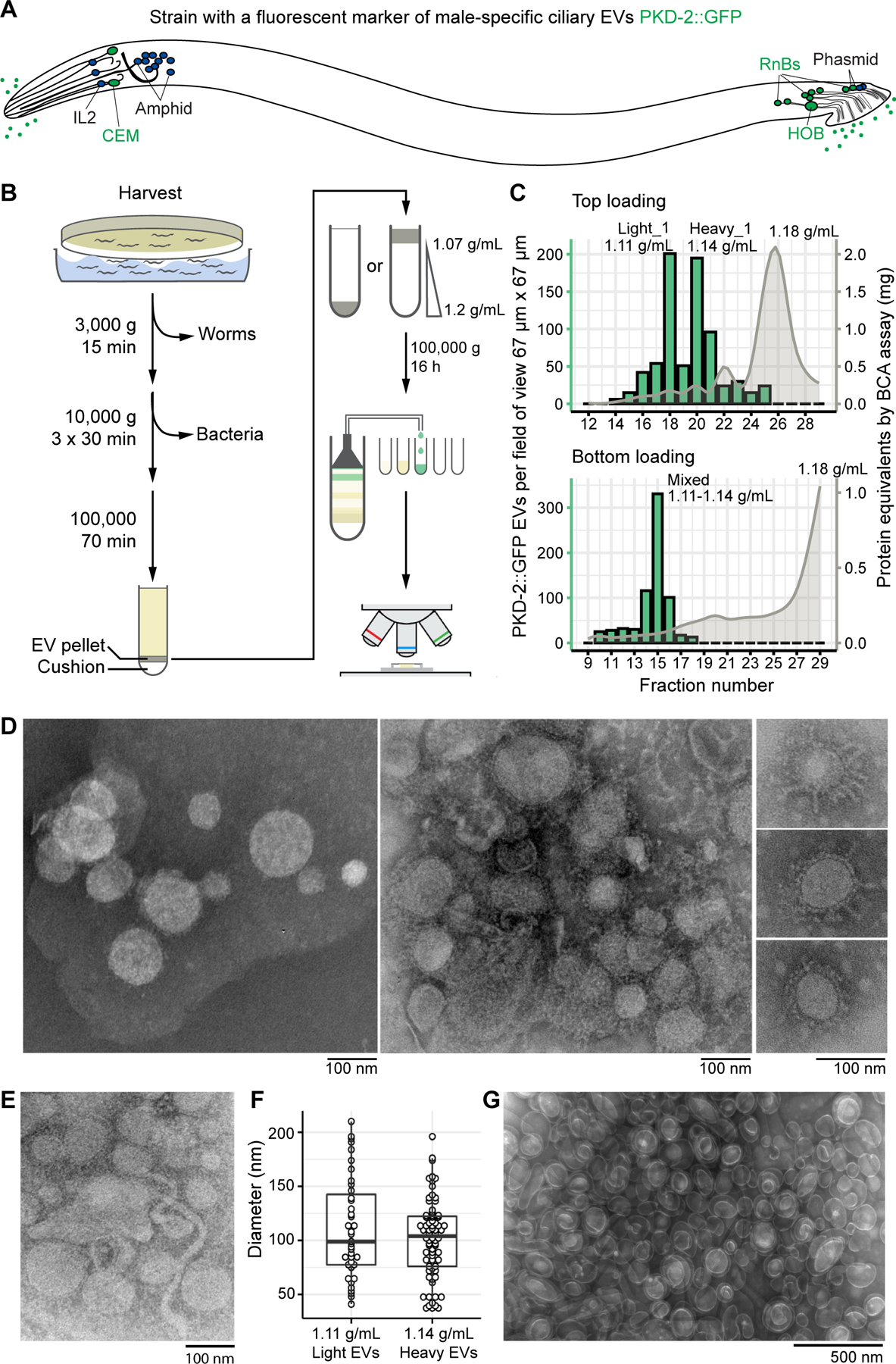
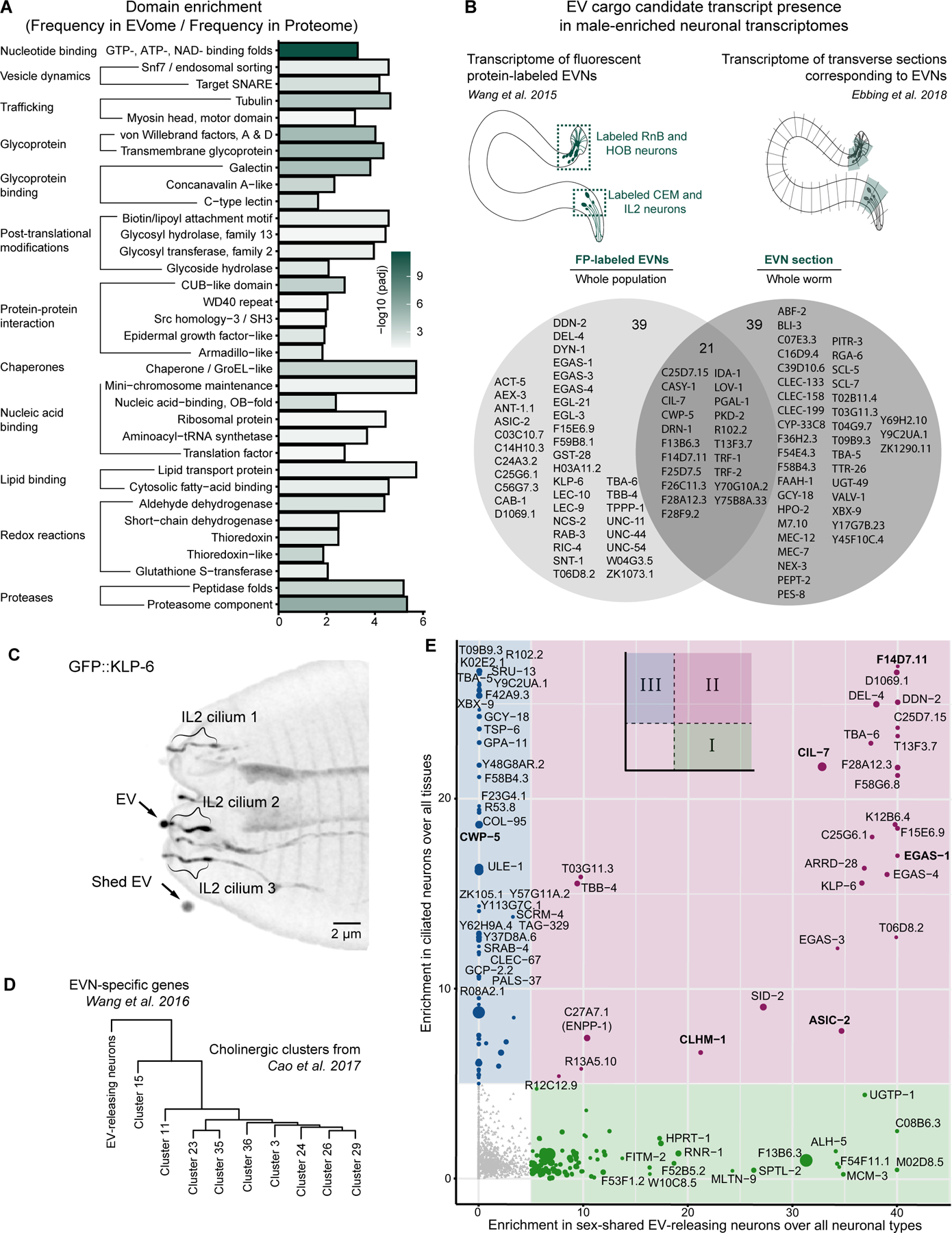
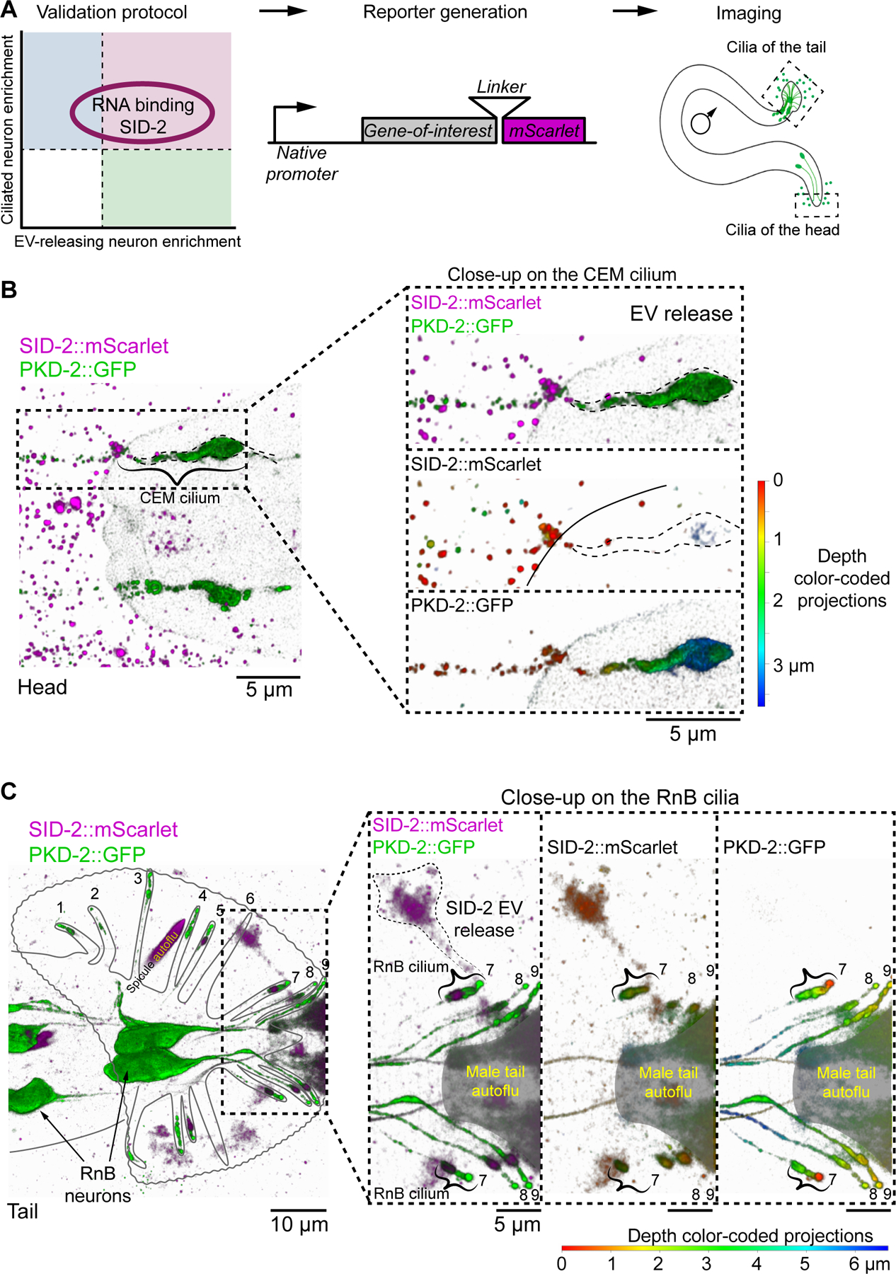
Extracellular vesicles (EVs) may mediate intercellular communication by carrying protein and RNA cargo. The composition, biology, and roles of EVs in physiology and pathology have been primarily studied in the context of biofluids and in cultured mammalian cells. The experimental tractability of C. elegans makes for a powerful in vivo animal system to identify and study EV cargo from its cellular source. We developed an innovative method to label, track, and profile EVs using genetically encoded, fluorescent-tagged EV cargo and conducted a large-scale isolation and proteomic profiling. Nucleic acid binding proteins (∼200) are overrepresented in our dataset. By integrating our EV proteomic dataset with single-cell transcriptomic data, we identified and validated ciliary EV cargo: CD9-like tetraspanin (TSP-6), ectonucleotide pyrophosphatase/phosphodiesterase (ENPP-1), minichromosome maintenance protein (MCM-3), and double-stranded RNA transporter SID-2. C. elegans EVs also harbor RNA, suggesting that EVs may play a role in extracellular RNA-based communication.

Keywords: ENPP1; MCM3; PKD2, polycystin; SID-2; cilia; ectosome; exosome; extracellular vesicle; seminal fluid.


Conflict of interest statement

Declaration of interests The authors declare no competing interests.

Figures

Figure 1.
Figure 1.. Buoyant density centrifugation enriches for ciliary PKD-2::GFP EVs more than 100-fold and resolves them into two populations.
(A) Location of environmentally exposed cilia of sensory neurons. Male-specific cilia are labeled in green. Neurons: CEM – cephalic male, IL2 – inner labial type 2, HOB – hook type B, RnBs – ray neurons type B. (B) Schematic of the EV enrichment workflow. (C) Combination charts showing number of the PKD-2::GFP EVs and the amount of protein in the collected fractions. Centrifugation of the top-loaded sample resolved PKD-2 EVs in two types with densities 1.11 g/ml (light) and 1.14 g/mL (heavy), respectively. Bottom-loaded samples failed to resolve the PKD-2 EV subtypes but reached a similar level of enrichment (more than 100-fold). (D) Transmission electron microscopy (TEM) of the negatively stained PKD-2 EV enriched fractions revealed presence of vesicles with coatings. (E) Tubular structures of 17–20 nm in diameter reminiscent of the stem of a budding vesicle. (F) Diameters of EVs recovered from the light and heavy fractions fall in the range of 50 – 200 nm as measured by TEM. (G) EVs from the fraction with most protein, presumably bacterial OMVs. Refer to Figure S1 for images of PKD-2::GFP EVs as seen with the Airyscan detection system, protein and EV profiling of fractions for every replicate in the study, and other information on the samples.
Figure 2.
Figure 2.. Identified EV proteome is enriched in nucleic acid binding proteins and ciliary EV cargo.
(A) Consolidated groups of InterPro domains enriched in the identified EV proteome (for the full list see Data S2). (B) Identified EVome contained proteins encoded by previously identified transcripts specific to EV-releasing neurons (EVNs). The Venn diagram shows EV cargo candidates that overlap with two transcriptomic datasets [8,26]. (C) GFP::KLP-6 EV shedding from the IL2 cilia of a wild-type hermaphrodite, orthogonal projection. GFP::KLP-6 was observed in EVs released outside the animal. (D) The transcriptome of EV-releasing neurons is most similar to the Cholinergic cluster 15 from [14] likely representing the IL2 neurons. (E) EV cargo candidates were categorized into three groups based on their relative enrichment in the IL2 sex-shared EV-releasing neurons (x-axis) and in the ciliated neurons (y-axis). Three categories are indicated by colors. Previously validated ciliary EV cargoes are bolded. Refer to Figure S2 for comparative analysis between biological and technical replicates and protein size distribution histograms; and to Data S1-S3 for full list of identified proteins, enrichment analyses, and comparisons to other transcriptomic studies.
Figure 3.
Figure 3.. Nucleic acid binding transporter SID-2 is specifically enriched in the male-specific ciliated EV-releasing neurons (EVNs) and is shed in the form of EVs to the environment.
(A) Validation protocol for putative ciliary EV cargo. (B-C) EV release of SID-2::mScarlet and PKD-2::GFP from cilia of CEM neuron of the head (B) and RnB neuron of the tail (C). Panel C shows ray #7 in the process of shedding SID-2::mScarlet as evidenced by continuous fluorescence signal spanning the ciliary base, ciliary shaft and the outside cloud of EVs. Other clouds on panel C are not labeled as EV release events, because it was not clear from which cilium they originated. Resolution of the imaging technique was 140 nm in lateral direction and 400 nm in axial direction, thus smaller EVs appear as clouds rather than individual EVs. Areas of intense inherent autofluorescence of the male tail are shaded with gray and labeled as autoflu. Images show 3D renderings: on merged images green and magenta colors represent different reporters, on images showing individual channels colors code for Z-depth - 3.5 μm for (B) and 6.5 μm for (C). Ci- cilia, CEM – cephalic male neuron, HOB – hook neuron type B, RnB – Ray neuron type B. Black and white maximum intensity projections of (B-C) and other data related to SID-2::mScarlet expression are included in Figure S3.
Figure 4.
Figure 4.. Phosphodiesterase ENPP-1 (C27A7.1) is a ciliary EV cargo.
(A) Release of ENPP-1::mScarlet EVs from the IL2 (inner labial 2) cilia labeled as #1, 2 and 3. The IL2 cilia #1 and #2 are in a state of pre-EV release. The cilium #3 is captured in a post-EV release state with the released ENPP-1::mScarlet EVs forming a “stream” of smaller EVs (small arrowheads) that is preceded by 2 larger EVs located at the front end of the “EV stream” (large arrowheads). See Video S1 for time-lapse imaging of ENPP-1::mScarlet EV protruding from the IL2 cilia. (B) Simultaneous release of PKD-2::GFP EVs (green dashed circles) and ENPP-1::mScarlet EVs (magenta dashed circles) from the CEM (cephalic male) cilia. PKD-2::GFP serves as a marker of the CEM cilia and EVs; Ci – cilia. Images show 3D renderings: on merged images green and magenta colors represent different reporters, on images showing individual channels colors code for Z-depth - 2.5 μm for (A) and 4.0 μm for (B). Black and white maximum intensity projections for (A-B) and other data related to ENPP-1:mScarlet expression are included in Figure S4. Refer to Video S1 for live imaging of ENPP-1::mScarlet EV release from the IL2 cilia.
Figure 5.
Figure 5.. Different neuronal types carry distinct EV cargo; a single neuronal cilium may shed multiple cargoes.
(A) MCM-3::mScarlet is present in cilia of PKD-2-expressing and IL2 neurons. Circles indicate MCM-3::Scarlet (magenta) and PKD-2::GFP EVs (B-C) TSP-6::mScarlet is present in amphid channel and AWA neurons of the head (C) and is released in ciliary EVs into the environment (D). Amphid channel is outline with white dashed line. Images are shown as projected 3D objects with the use of actual pixel values in a transparent volume. Images show 3D renderings: on merged images green and magenta colors represent different reporters, on images showing individual channels colors code for Z-depth - 5.0 μm for (A) and 6.0 μm for (C). Black and white maximum intensity projections for (A-C) and other data related to MCM-3::mScarlet and TSP-6::mScarlet expression patterns are included in Figure S5.
Figure 6.
Figure 6.. ENPP-1::mScarlet is transferred from the male reproductive tract to the hermaphrodite uterus during copulation.
(A) ENPP-1::mScarlet is present in the somatic part of the male reproductive tract in the vacuoles of cuboidal cells of the vas deferens. Zoomed inset shows a single plane through the giant vacuoles. Also see Video S2 for full Z-stack through the giant vacuoles showing appearance of speckled material with ENPP-1::mScarlet. (B) Male-to-hermaphrodite transfer experiment where ENPP-1::mScarlet males were mated to non-fluorescent hermaphrodites. (C) Imaging of the non-fluorescent hermaphrodite after copulation revealed presence of malederived ENPP-1::mScarlet in the uterus. Yellow asterisks indicate intestinal auto fluorescent droplets. See Video S3 for real-time imaging of male transferring ENPP-1::mScarlet into the hermaphrodite uterus. (D) Venn diagram showing overlap of the identified EV proteome with products of the male-enriched transcripts [47]. (E) Scatter plot showing EV cargo predicted to be of either neuronal or glial origin. See Figure S6 for enrichment of identified EV cargo candidates in other tissues (hypodermis and germline) and for RNA profiling of PKD-2::GFP EV fractions.

References

    1. Maas SLN, Breakefield XO, and Weaver AM (2017). Extracellular Vesicles: Unique Intercellular Delivery Vehicles. Trends Cell Biol 27, 172–188. - PMC - PubMed
    1. Théry C, Witwer KW, Aikawa E, Alcaraz MJ, Anderson JD, Andriantsitohaina R, Antoniou A, Arab T, Archer F, Atkin-Smith GK, et al. (2018). Minimal information for studies of extracellular vesicles 2018 (MISEV2018): a position statement of the International Society for Extracellular Vesicles and update of the MISEV2014 guidelines. J. Extracell. Vesicles 7, 1535750. - PMC - PubMed
    1. Sung BH, Parent CA, and Weaver AM (2021). Extracellular vesicles: Critical players during cell migration. Dev. Cell 56, 1861–1874. - PMC - PubMed
    1. Foot NJ, and Kumar S (2021). The Role of Extracellular Vesicles in Sperm Function and Male Fertility. In Subcellular Biochemistry, pp. 483–500. - PubMed
    1. Lizarraga-Valderrama LR, and Sheridan GK (2021). Extracellular vesicles and intercellular communication in the central nervous system. FEBS Lett 595, 1391–1410. - PubMed

Publication types

LinkOut - more resources