. 2022 May 5;82(9):1643-1659.e10.
doi: 10.1016/j.molcel.2022.03.007. Epub 2022 Mar 24.

Structural basis of SARM1 activation, substrate recognition, and inhibition by small molecules

Affiliations

Structural basis of SARM1 activation, substrate recognition, and inhibition by small molecules

Yun Shi et al. Mol Cell. .

Abstract

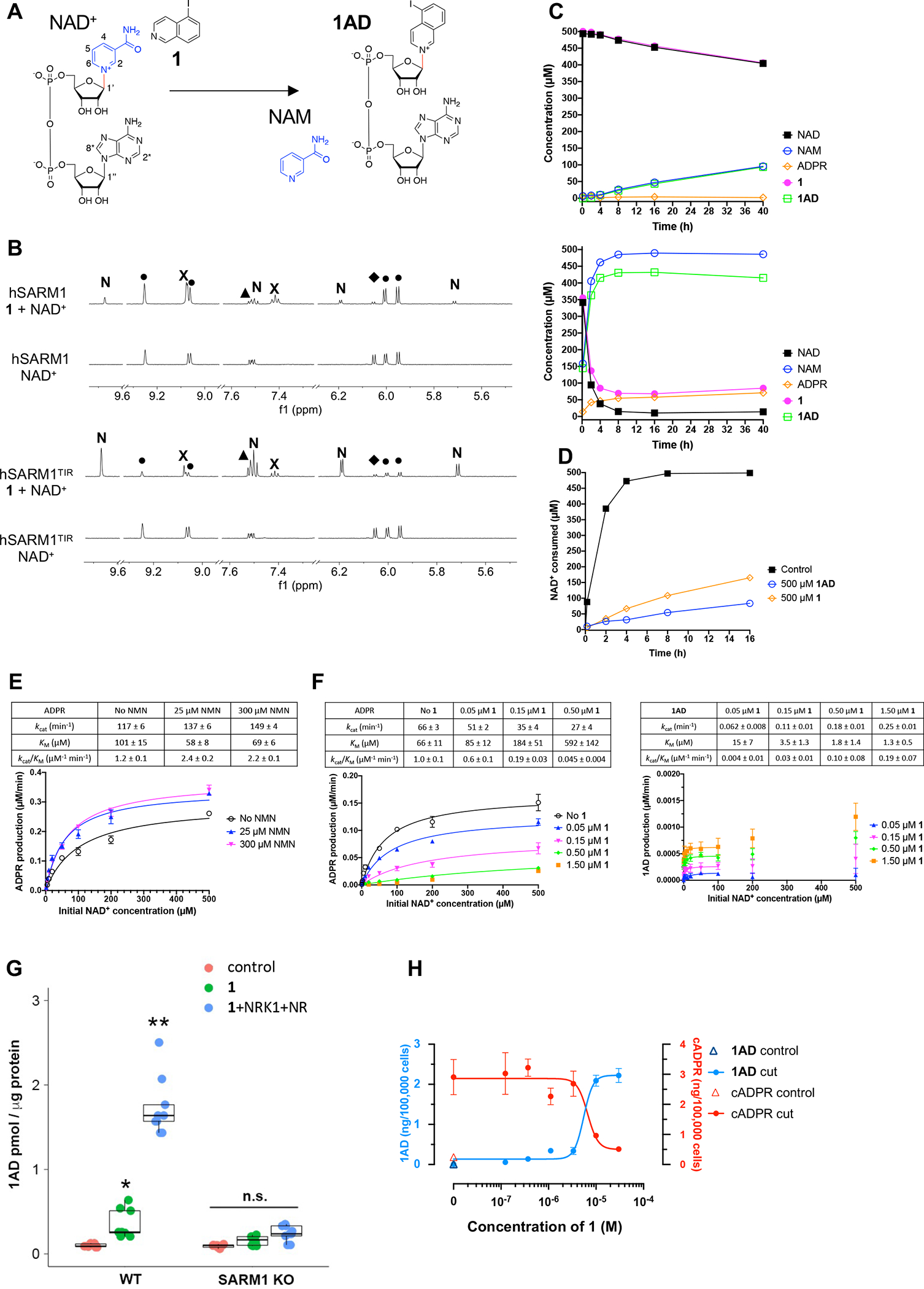
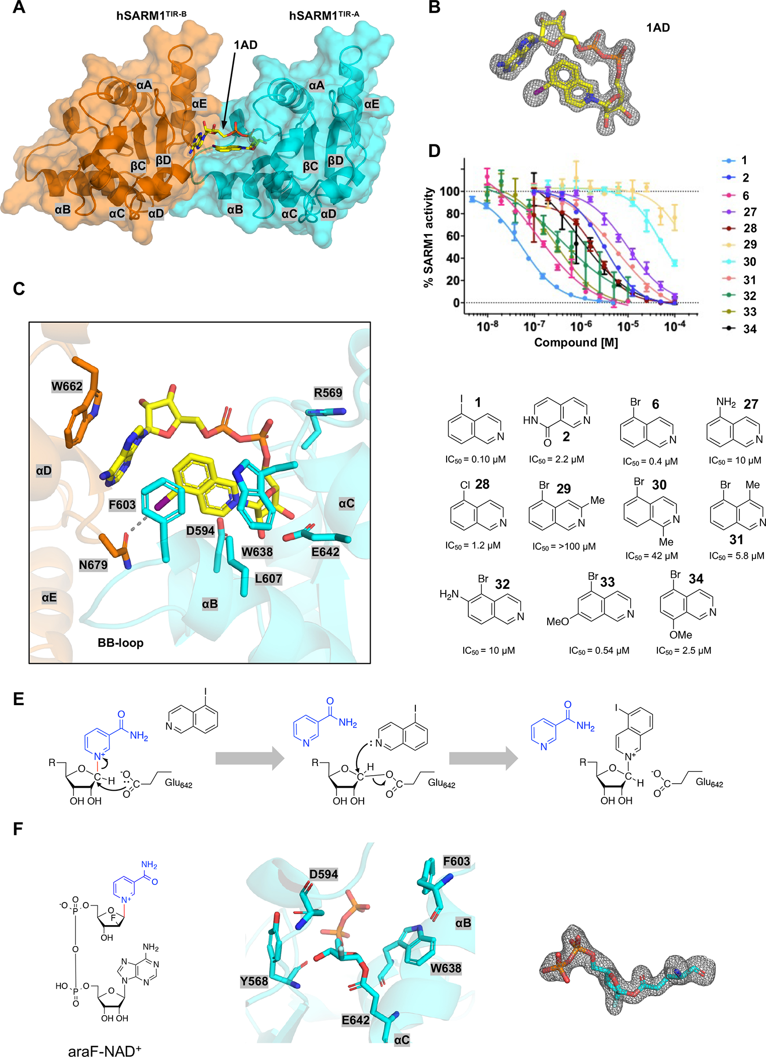
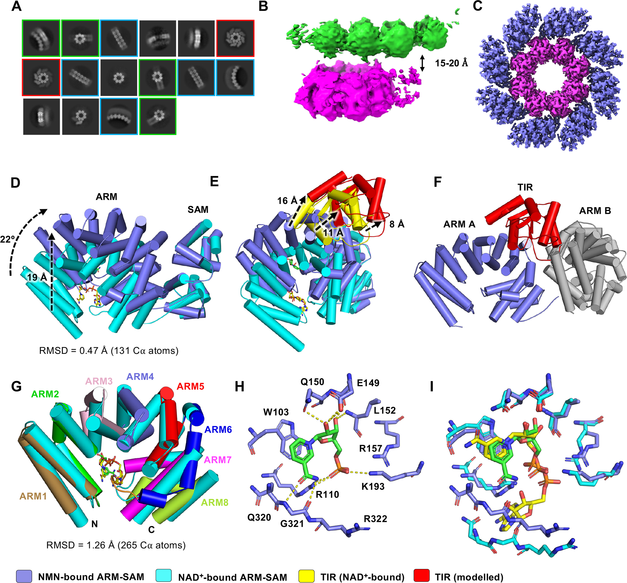
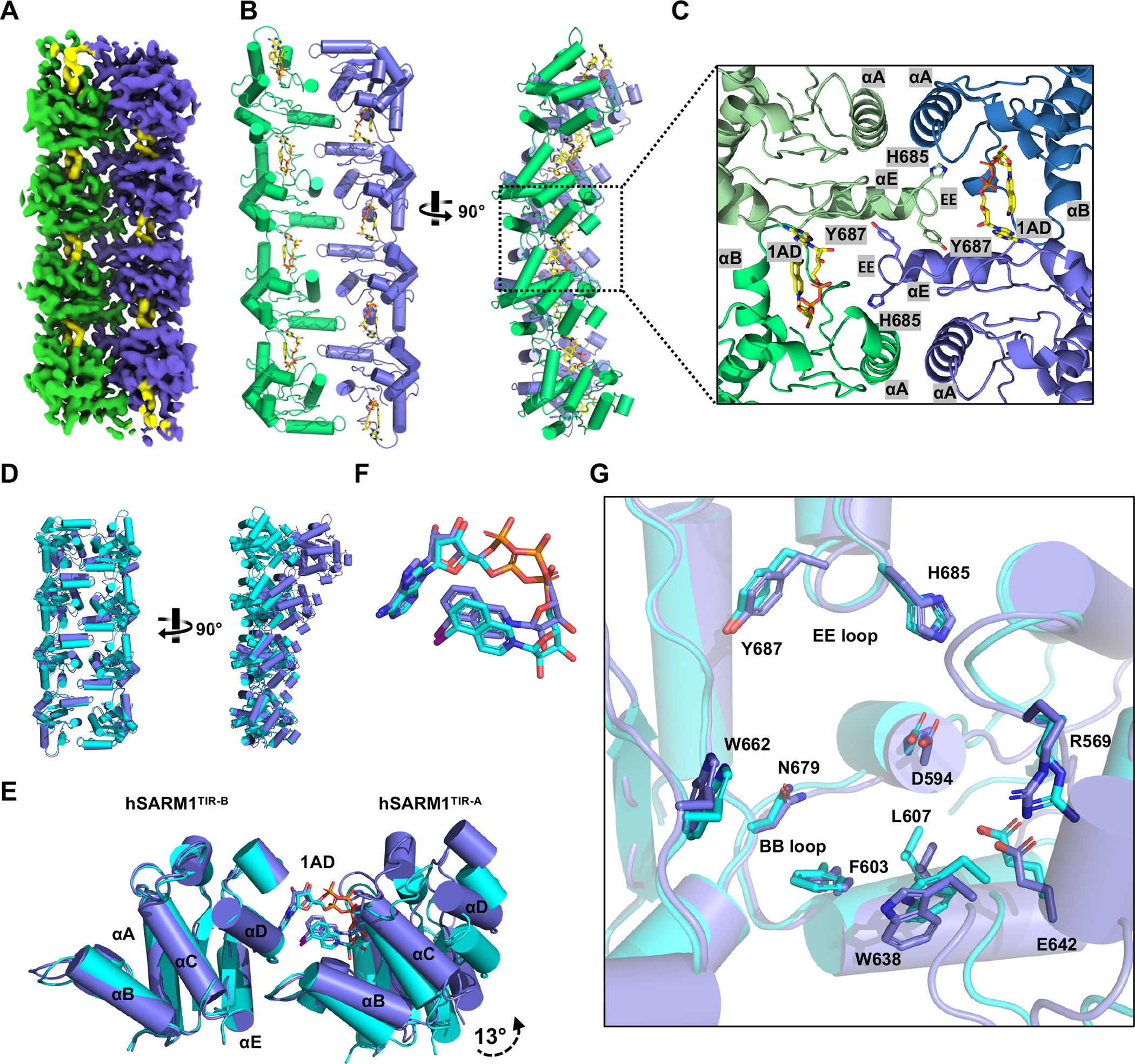
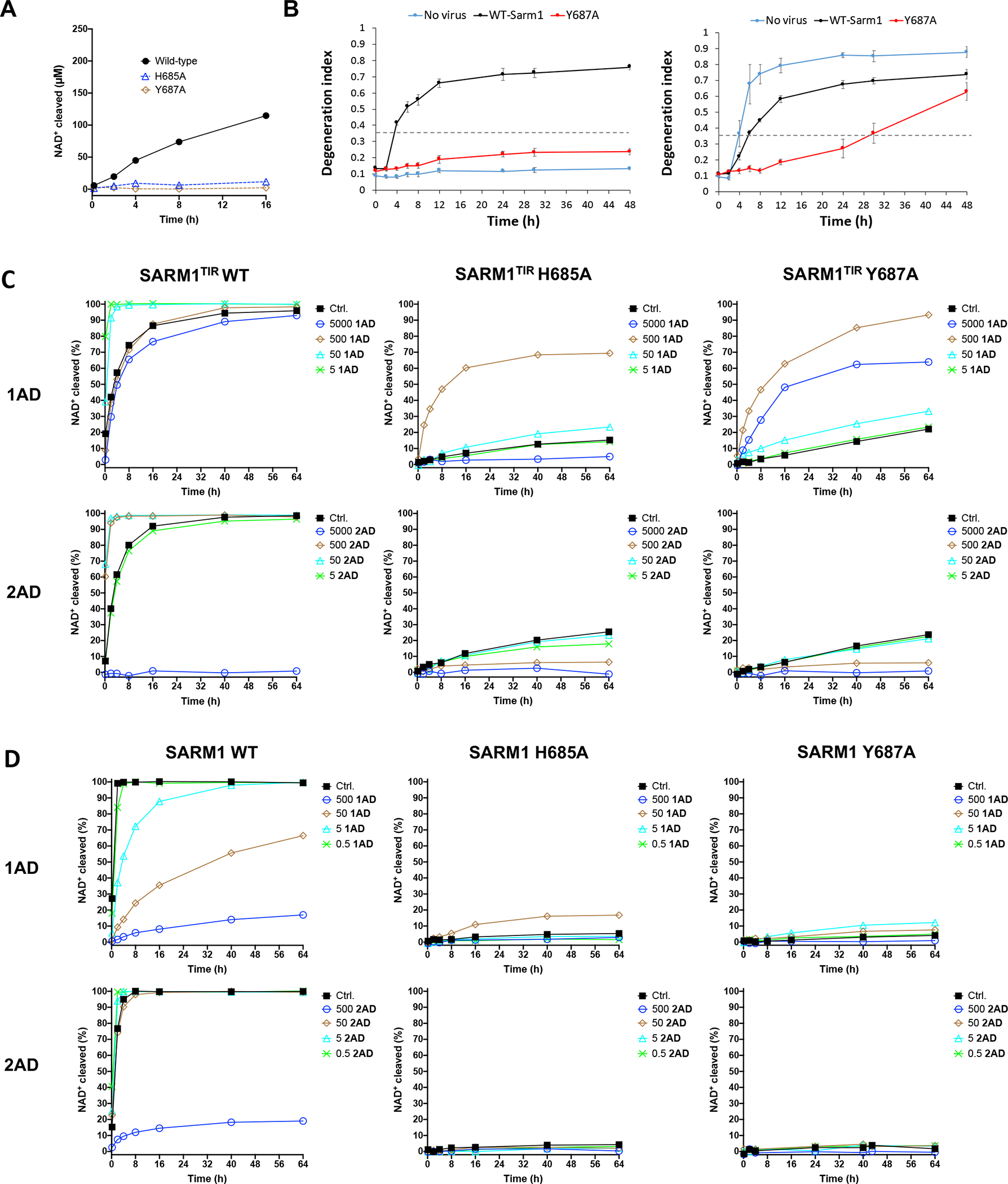
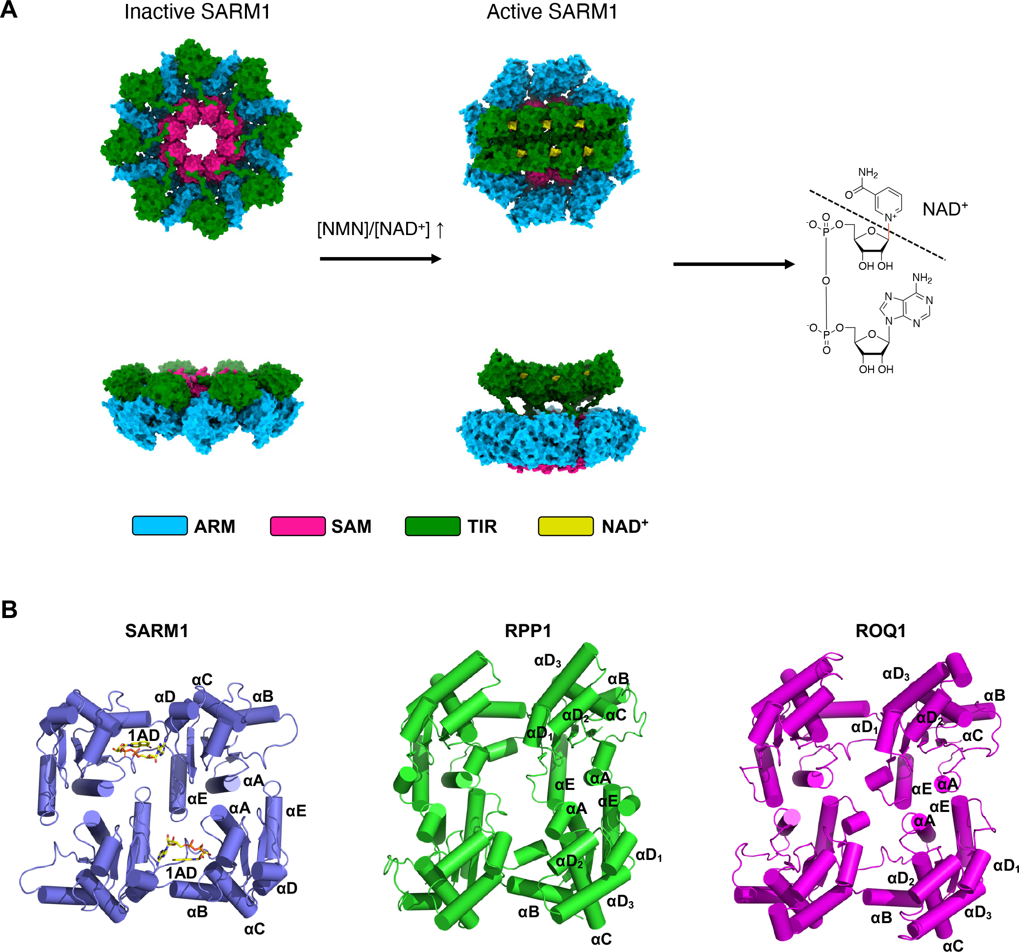
The NADase SARM1 (sterile alpha and TIR motif containing 1) is a key executioner of axon degeneration and a therapeutic target for several neurodegenerative conditions. We show that a potent SARM1 inhibitor undergoes base exchange with the nicotinamide moiety of nicotinamide adenine dinucleotide (NAD+) to produce the bona fide inhibitor 1AD. We report structures of SARM1 in complex with 1AD, NAD+ mimetics and the allosteric activator nicotinamide mononucleotide (NMN). NMN binding triggers reorientation of the armadillo repeat (ARM) domains, which disrupts ARM:TIR interactions and leads to formation of a two-stranded TIR domain assembly. The active site spans two molecules in these assemblies, explaining the requirement of TIR domain self-association for NADase activity and axon degeneration. Our results reveal the mechanisms of SARM1 activation and substrate binding, providing rational avenues for the design of new therapeutics targeting SARM1.

Keywords: ARM domain; NADase; TIR domain; X-ray crystallography; allosteric activator; base exchange; cryo-EM; orthosteric inhibitor.


Conflict of interest statement

Declaration of interests A.D. and J.M. are co-founders, scientific advisory board members, and shareholders of Disarm Therapeutics. B.K. is a shareholder of Disarm Therapeutics. Y. Sasaki and B.K. are consultants to Disarm Therapeutics. B.K. and T.V. receive research funding from Disarm Therapeutics. R.O.H., R.K., and T.B. are employees and shareholders in Disarm Therapeutics. S.E.A., M.F., K.C., A.B., and P.S.K. are employees of Evotec SE. R.O.H., T.B., and A.B. are inventors on a patent related to isoquinoline inhibitors of SARM1 (WO 2019/236879 Al). The authors have no additional competing financial interests.

Figures

Figure 1.
Figure 1.. Mechanism of orthosteric inhibition.
A Proposed reaction replacing NAM with 1. B Expansions of NMR spectra showing the appearance of new resonances. Top: 16 h spectra of 0.5 μM hSARM1 + NAD+ and 0.5 μM hSARM1 + 1 + NAD+. Bottom: 16 h spectra of 10 μM hSARM1TIR + NAD+ and 10 μM hSARM1TIR 1 + NAD+. Resonance peaks from NAD+, ADPR, NAM, 1, and the proposed new product 1AD are labelled as “”, “◆”, “”, “X”, and “N”, respectively. The initial concentration of compounds (1 and NAD+) was 500 μM. C Reaction progression curves of 0.5 μM hSARM1 + 500 μM 1 + 500 μM NAD + (top) and 10 μM hSARM1TIR + 500 μM 1 + 500 μM NAD +. D Inhibition of hSARM1 NAD+ consumption by 500 μM 1AD and 500 μM 1. 0.5 μM hSARM1 was activated by 100 min NMN (500 μM) pre-incubation. Initial NAD + concentration was 500 μM. E Kinetic analyses based on ADPR production. 2.5 nM hSARM1 was activated by 120 min preincubation with varying concentrations of NMN. Reported values are mean ± SEM, n = 3. F Kinetic analyses of hSARM1, showing ADPR and 1AD production with varying initial concentrations of 1. 2.5 nM of SARM1 was pre-incubated with 25 μM NMN for 120 min. Reported kinetic property values are mean ± SEM, n = 3. No stable fit can be obtained for ADPR production in the presence of 1.5 μM 1. When fitting for 1AD production, data with more than 100 μM NAD+ were not used due to an atypical biphasic like behavior that may originate from non-uniform formation of multiple active sites within the SARM1 octamer and their different binding to NAD+/1AD. G SARM1-dependent production of 1AD in DRG neurons. Statistical analysis was performed by two-way ANOVA with Tukey post-hoc test. F(2, 48)=111.5, p<2×10⁻¹⁶ among treatments and genotypes (n=12). *p<0.05 and ** p<2×10⁻¹⁶ denotes statistical significances and n.s. denotes no statistical significance compared with the wild-type protein without 1 (control). H Production of 1AD in axotomized DRG neurons. DRG neurons were treated with 1 at the concentrations indicated. Axonal 1AD and cADPR were measured 4h after axotomy. Data represent means ± SEM, n=3. See also Figure S1.
Figure 2.
Figure 2.. Structural basis of orthosteric inhibition and catalysis.
A Structure of the BB-loop mediated hSARM1TIR asymmetric dimer (cartoon and transparent surface; hSARM1TIR-A, orange; hSARM1TIR-B, cyan) in complex with 1AD (yellow stick). B Standard omit mFo-DFc map of 1AD, contoured at 3.0 σ. C Enlarged cutaway of the 1AD binding pocket in the hSARM1TIR structure. D Inhibition activities of selected isoquinoline-derived compounds. Values represent mean ± SEM; n = 2 (2, 29, 30, 31), 4 (6, 28, 32, 33, 34), 5 (1), and 6 (27). E A simplified model showing a putative catalytic mechanism for hSARM1 base-exchange reactions. F Left, chemical structure of araF-NAD+; middle, enlarged cutaway view of the partially-resolved catalytic intermediate with araF-ADPR covalently linked to E642 of hSARM1TIR; right, standard omit mFo-DFc map of araF-ADPR, contoured at 3.0 σ. Electron density for the leaving NAM group and the adenosine moiety was not observed. See also Figure S1
Figure 3.
Figure 3.. Structural basis of substrate recognition.
A Chemical structures of 2, 2AD, 3, and 3AD. B Reaction progression of 0.5 μM hSARM1 + 500 μM 2 + 500 μM NAD + (left) and 0.5 μM hSARM1 + 500 μM 3 + 500 μM NAD + (right). C Enlarged cutaway view of the 2AD binding pocket in the hSARM1TIR structure. D Enlarged cutaway view of the 3AD binding pocket in the hSARM1TIR structure. E Binding modes of 1AD vs 2AD (top), 1AD vs 3AD (middle), and 2AD vs 3AD (bottom). F A summary table of NADase and base-exchange activities of hSARM1 and hSARM1TIR mutants from Figure S2. The activities are shown in percentage relative to the wild-type proteins for 40 h (hSARM1) and 4 h (hSARM1TIR) time points. See also Figure S1 and S2.
Figure 4.
Figure 4.. Cryo-EM structures of hSARM1 SAM-TIR:1AD and ARM-SAM:NMN.
A Representative 2D class averages from hSARM1 incubated with NMN and 1AD. 2D classes consisting of octameric rings and double-stranded TIR domain assemblies with and without octameric rings are highlighted by red, green and blue boxes, respectively. B Electrostatic potential density map of SAM-TIR:1AD. Map areas corresponding to the TIR and SAM domains are shown in green and magenta, respectively. C Electrostatic potential density map of the ARM-SAM:NMN. Map areas corresponding to the ARM and SAM domains are displayed in slate and pink, respectively. D-F Structural superposition of ARM-SAM:NMN (slate) and hSARM1:NAD+ (ARM-SAM, cyan; TIR, yellow; PDB: 7ANW). ARM-SAM subunits were aligned by the SAM domain. A hypothetical TIR domain (red) was modelled in ARM-SAM:NMN based on the ARM:TIR interaction observed in hSARM:NAD+ (PDB: 7ANW). Movement of the ARM and modelled TIR (red) domains induced by NMN binding is indicated by black dashed arrows. NMN and NAD+ are shown in green and yellow stick representation, respectively. In (D), the superposition shows that the ARM-SAM linker region acts as a hinge to facilitate the translation and rotation of the ARM domain upon NMN binding. In (E), the effect of NMN-induced ARM domain reorientation on the TIR domain (red) is highlighted, while in (F), the resulting clash between the TIR domain and the next ARM domain (grey) in the octameric ring is shown. G Structural superposition of the ARM domains in ARM-SAM:NMN (coloured by ARM motif; 310–325 region highlighted in orange) and hSARM1:NAD+ (cyan; PDB: 7ANW). H Interactions between ARM domain and NMN (stick representation) in ARM-SAM:NMN. Polar contacts are shown as yellow dashed lines. I Comparison of the allosteric sites in ARM-SAM:NMN (slate) and hSARM1:NAD+ (cyan; PDB: 7ANW). The superposition shows that several of the NMN and NAD+ interacting residues adopt similar conformations. Notable exceptions are residues 320–322, which move approximately 4 Å, to accommodate NMN; these conformations appear incompatible with NAD+ binding. See also Figure S3, S4, S5 and S7.
Figure 5.
Figure 5.. Cryo-EM structure of hSARM1 TIR:1AD.
A Electrostatic potential density map of the two-stranded TIR domain assembly. The two antiparallel strands are shown in green and slate, respectively. 1AD is shown in yellow. B Cartoon diagram of the two-stranded TIR domain assembly (helices are depicted as cylinders). C Enlarged cutaway of the TIR domain interfaces in the two-stranded assembly. 1AD and EE loop residues H685 and Y687 are highlighted in stick representation. D-G Comparison of the TIR-domain assemblies in the hSARM1TIR:1AD crystal (cyan) and hSARM1 TIR:1AD cryo-EM (slate) structures. (D) Top- (left) and side-view (right) of octameric assembly. (E) Orthosteric site. (F) Zoomed-in view of isolated 1AD from the orthosteric site. (G) Orthosteric site with residues involved in 1AD binding highlighted in stick representation. The hSARM1TIR-B chain was used for the alignments in (E-G). See also Figure S3, S4, and S6.
Figure 6.
Figure 6.. Characterisation of EE loop mutants.
A NADase activities of 0.5 μM wild-type, H685A, and Y687A hSARM1. The initial NAD + concentration was 500 μM. B DRG cultures from SARM1 knock out mice (left) or wild-type mice (right) were infected at two days in culture with lentivirus expressing either wild-type SARM1 or the SARM1 Y687A mutant. Axotomy was performed in mature DRG neurons. Axon degeneration was monitored by the axon degeneration index (Gerdts et al., 2013) over time. Results are mean ± SD, n=3. Axons with index values above 0.35 (dashed line) are considered degenerated. C-D Effects of 1AD and 2AD on enzymatic activity. (C) 10 μM wild-type, H685A, and Y87A hSARM1TIR with varying concentrations of 1AD and 2AD. (D) 0.5 μM wild-type, H685A, and Y87A hSARM1 activated by 100 min pre-incubation of 500 μM NMN with varying concentrations of 1AD and 2AD. The initial concentration of NAD+ was 500 μM. Concentrations of 1AD and 2AD are labelled (μM).
Figure 7.
Figure 7.. SARM1 activation mechanism.
A Upon NMN binding, conformational changes in the ARM domain result in their rotation relative to the octameric SAM domain ring, which in turn causes a clash with the TIR domains. As the TIR domains are released from the ring, they self-associate into a two-stranded antiparallel assembly to yield six catalytically-competent active sites per SARM1 octamer. Inactive structure corresponds to PDB ID 7ANW. The flexible region between the SAM and TIR domain (residues 546–560) in the activated structure was modelled using Coot. B Comparison of the TIR domain tetramers observed in the RPP1 (PDB: 7JLX) and ROQ1 (PDB: 7DFV) cryo-EM structures with the TIR-domain assembly observed in SARM1.

Similar articles

Cited by

References

    1. Afonine PV, et al. (2012). Towards automated crystallographic structure refinement with phenix.refine. Acta Crystallogr D 68, 352–367. - PMC - PubMed
    1. Afonine PV, Klaholz BP, Moriarty NW, Poon BK, Sobolev OV, Terwilliger TC, Adams PD, and Urzhumtsev A (2018). New tools for the analysis and validation of cryo-EM maps and atomic models. Acta Crystallogr D 74, 814–840. - PMC - PubMed
    1. Angeletti C, Amici A, Gilley J, Loreto A, Trapanotto AG, Antoniou C, Merlini E, Coleman MP, and Orsomando G (2022). SARM1 is a multi-functional NAD(P)ase with prominent base exchange activity, all regulated by multiple physiologically relevant NAD metabolites. iScience 25, 103812. - PMC - PubMed
    1. Bepler T, Morin A, Rapp M, Brasch J, Shapiro L, Noble AJ, and Berger B (2019). Positive-unlabeled convolutional neural networks for particle picking in cryo-electron micrographs. Nat Methods 16, 1153–1160. - PMC - PubMed
    1. Bratkowski M, et al. (2020). Structural and mechanistic regulation of the pro-degenerative NAD hydrolase SARM1. Cell Rep 32, 107999. - PubMed

Publication types

MeSH terms

LinkOut - more resources