Review
. 2022 May;21(5):465-479.
doi: 10.1016/S1474-4422(21)00414-2. Epub 2022 Mar 22.

Emerging insights into the complex genetics and pathophysiology of amyotrophic lateral sclerosis

Affiliations
Review

Emerging insights into the complex genetics and pathophysiology of amyotrophic lateral sclerosis

Stephen A Goutman et al. Lancet Neurol. 2022 May.

Abstract

Amyotrophic lateral sclerosis is a fatal neurodegenerative disease. The discovery of genes associated with amyotrophic lateral sclerosis, commencing with SOD1 in 1993, started fairly gradually. Recent advances in genetic technology have led to the rapid identification of multiple new genes associated with the disease, and to a new understanding of oligogenic and polygenic disease risk. The overlap of genes associated with amyotrophic lateral sclerosis with those of other neurodegenerative diseases is shedding light on the phenotypic spectrum of neurodegeneration, leading to a better understanding of genotype-phenotype correlations. A deepening knowledge of the genetic architecture is allowing the characterisation of the molecular steps caused by various mutations that converge on recurrent dysregulated pathways. Of crucial relevance, mutations associated with amyotrophic lateral sclerosis are amenable to novel gene-based therapeutic options, an approach in use for other neurological illnesses. Lastly, the exposome-the summation of lifetime environmental exposures-has emerged as an influential component for amyotrophic lateral sclerosis through the gene-time-environment hypothesis. Our improved understanding of all these aspects will lead to long-awaited therapies and the identification of modifiable risks factors.


Conflict of interest statement

Declaration of interests SAG declares consulting fees from Biogen and ITF Pharma, a patent “Methods for treating amyotrophic lateral sclerosis”, and participation on a Data Safety Monitoring Board for Watermark. OH declares consulting fees from Novartis, Cytokinetics, Denali Pharma, Stitching Foundation, and La Caixa; payment or honoraria from Biogen; participation on a Data Safety Monitoring Board for Accelsiors and steering committee for Cytokinetics; and is Editor-in-Chief for the journal Amyotrophic Lateral Sclerosis and Frontotemporal Dementia. AA-C declares consulting fees from Mitsubishi Tanabe Pharma, Biogen Idec, Cytokinetics, Wave Pharmaceuticals, Apellis, Amylyx, Novartis, and Eli Lilly. AC declares grants from Biogen to his institution, payments or honoraria from Biogen and Amylyx, and participation on a Data Safety Monitoring Board for Ely Lilly and ABScience and advisory board for Mitsubishi Tanabe, Roche, Denali Pharma, Cytokinetics, Biogen, and Amylyx. MCK has an honorary role as President of the Brain Foundation and as Editor-in-Chief of the Journal of Neurology, Neurosurgery and Psychiatry. ELF declares a patent “Methods for treating amyotrophic lateral sclerosis”. MGS declares no competing interests.

Figures

Figure 1.
Figure 1.. ALS genetic architecture.
ALS genetics is characterized by (A) monogenic, (B) oligogenic, and (C) polygenic risk. Only three representative chromosomes shown. (D) ALS genes are not fully penetrant and the pathogenicity of certain variants remains uncertain, complicating the full picture. Left: For a population of gene carriers, low penetrance variants lead to a low frequency of ALS onset (red figures). Right: For a population of gene carriers, high penetrance variants lead to a high frequency of ALS onset (red figures). (E) Overlaid over the genetic aspects are environmental factors, since heritability is incomplete. Thus, a multistep ALS model has emerged, which advocates that multiple “steps” are necessary for ALS onset. Left: Larger-effect mutations, e.g., mutant SOD1, require fewer steps for ALS onset (red figure). Right: Smaller-effect mutations, e.g., mutant TARDBP, require more steps for ALS onset (red figure). Future work is needed to precisely define a “step” and determine when one has occurred, e.g., genetic or environmental factors. (F) Several genetic therapies are in the clinical trial pipeline (umbrella trial, stratified by molecular profile) and tailored precision treatments are future goals; thus, molecular profiling of ALS patients could become standard clinical practice. SNP, single nucleotide variant. Created with BioRender.com.
Figure 2.
Figure 2.. ALS pathophysiology.
Shared ALS pathological pathways center on impaired RNA metabolism, altered proteostasis/ autophagy, cytoskeletal/ trafficking defects, mitochondrial dysfunction, and compromised DNA repair. Numbering from top left downwards: (1) Mutant RNA-binding proteins (RBPs), e.g., FUS, TDP-43, disrupt RNA transcription and splicing. C9orf72 repeat expansion RNAs aggregate into RNA foci, sequestering RBPs and impairing RNA metabolism. Additionally, haploinsufficiency from the single remaining normal C9orf72 allele leads to loss-of-function of native C9orf72 protein function, related to multiple aspects, trafficking, autophagy, DNA repair. (2) Mutant C9orf72, FUS, and TARDBP functionally impair nucleocytoplasmic transport (NCT) and induce nuclear envelope morphology defects and cytoplasmic inclusions of NCT components, e.g., nucleoporins, importins, and Ran (small GTPase Ras-related nuclear proteins). (3) Repeat-associated non-AUG translation of C9orf72 repeat expansions yields dipeptide repeats (DPRs), which are toxic through several pathways, including protein aggregates, chromatin alterations and DNA damage, impaired NCT and component sequestration. Additional cytoplasmic protein aggregation (e.g., TDP-43, SOD1) induces proteostasis and autophagy defects. Protein aggregates block the endoplasmic reticulum-associated protein degradation (ERAD) response and ubiquitin proteasome system (UPS), preventing aggregate clearance. Mutations to ubiquitination proteins (e.g., CCNF, UBQLN2) additionally dysregulate the UPS. Protein aggregates and RBPs also accumulate into stress granules, which become persistent in ALS. Mutations to vesicle-forming proteins (e.g., OPTN, VAPB, VCP) disrupt vesicular transport and distribution, leading to dysfunctional autophagy and proteostasis. (4) Mutations to the tubulin transport machinery (e.g., DCTN1, KIF5A, TUBA4A) and actin (e.g., PFN1) induce cytoskeletal/ trafficking defects, which impairs distribution of vital organelles throughout cells (e.g., mitochondria, cardo-laden vesicles). (5) Protein aggregates (e.g., TDP-43, SOD1) and mutations to mitochondrial protein components (e.g., CHCHD10) trigger mitochondrial and bioenergetics dysfunction and raise oxidative stress. (6) liquid-to-liquid phase separation of aggregation prone proteins (e.g., FUS, TDP-43) drives formation of stress granules. Created, in part, with BioRender.com.
Figure 3.
Figure 3.. ALS inflammatory pathways.
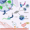
This pathophysiology in ALS is characterized by dysregulated peripheral immune cell counts, immune cell infiltration (trafficking) into the central nervous system, induction of an activated immune phenotype, and altered cytokine production. (A) Various peripheral immune cell populations in blood have differential levels in ALS, e.g., innate (neutrophils, natural killer (NK) cells) and adaptive (CD8 T cells). Circulating NK cells in ALS increase expression of surface markers of cytotoxic function (CD38, NKG2D, NKp30, NKp46) and trafficking (CD11a, CD11b, CXCR3, CX3CR1). Circulating monocytes and dendritic cells expressing mutant TARDBP and C9orf72 repeat expansions increase interferon gamma (IFNγ) production. (B) Peripheral immune cells traffic to the central nervous system (CNS) in ALS, e.g., neutrophils, NK cells. BM, basal membrane; CD11a, cluster of differentiation 11a; CD11b, cluster of differentiation 11b; CD38, cluster of differentiation 38; CXCR3, C-X-C motif chemokine receptor 3; CX3CR1, C-X3-C motif chemokine receptor 1; NKG2D, killer cell lectin like receptor K1 (KLRK1); NKp30, natural cytotoxicity triggering receptor 3 (NCR3); NKp46, Natural cytotoxicity triggering receptor 1 (NCR1); PVS, perivascular space. Created, in part, with BioRender.com.

Comment in

References

    1. Goutman SA. Diagnosis and Clinical Management of Amyotrophic Lateral Sclerosis and Other Motor Neuron Disorders. Continuum (Minneap Minn). 2017;23(5, Peripheral Nerve and Motor Neuron Disorders):1332–59. - PubMed
    1. Chia R, Chio A, Traynor BJ. Novel genes associated with amyotrophic lateral sclerosis: diagnostic and clinical implications. Lancet Neurol. 2018;17(1):94–102. - PMC - PubMed
    1. Ryan M, Heverin M, McLaughlin RL, Hardiman O. Lifetime Risk and Heritability of Amyotrophic Lateral Sclerosis. JAMA Neurol. 2019;76(11):1367–74. - PMC - PubMed
    1. Nicolas A, Kenna KP, Renton AE, Ticozzi N, Faghri F, Chia R, et al. Genome-wide Analyses Identify KIF5A as a Novel ALS Gene. Neuron. 2018;97(6):1268–83 e6. - PMC - PubMed
    1. Ryan M, Heverin M, Doherty MA, Davis N, Corr EM, Vajda A, et al. Determining the incidence of familiality in ALS: A study of temporal trends in Ireland from 1994 to 2016. Neurol Genet. 2018;4(3):e239. - PMC - PubMed

Publication types

MeSH terms