

Human vestibular schwannoma reduces density of auditory nerve fibers in the osseous spiral lamina
- PMID: 35334332
- PMCID: PMC11181009
- DOI: 10.1016/j.heares.2022.108458
Human vestibular schwannoma reduces density of auditory nerve fibers in the osseous spiral lamina
Abstract
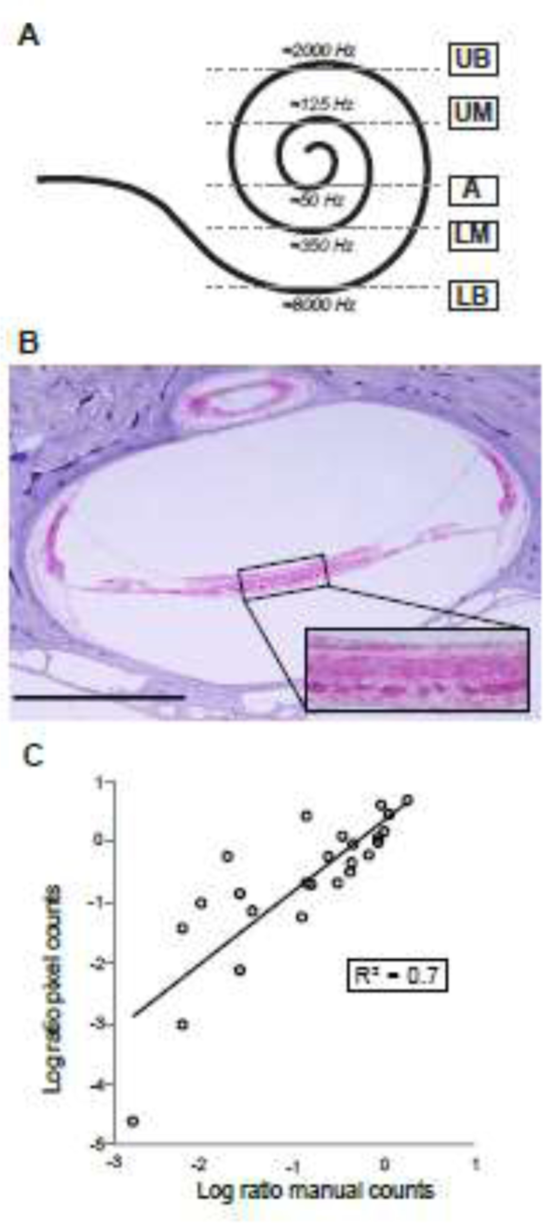
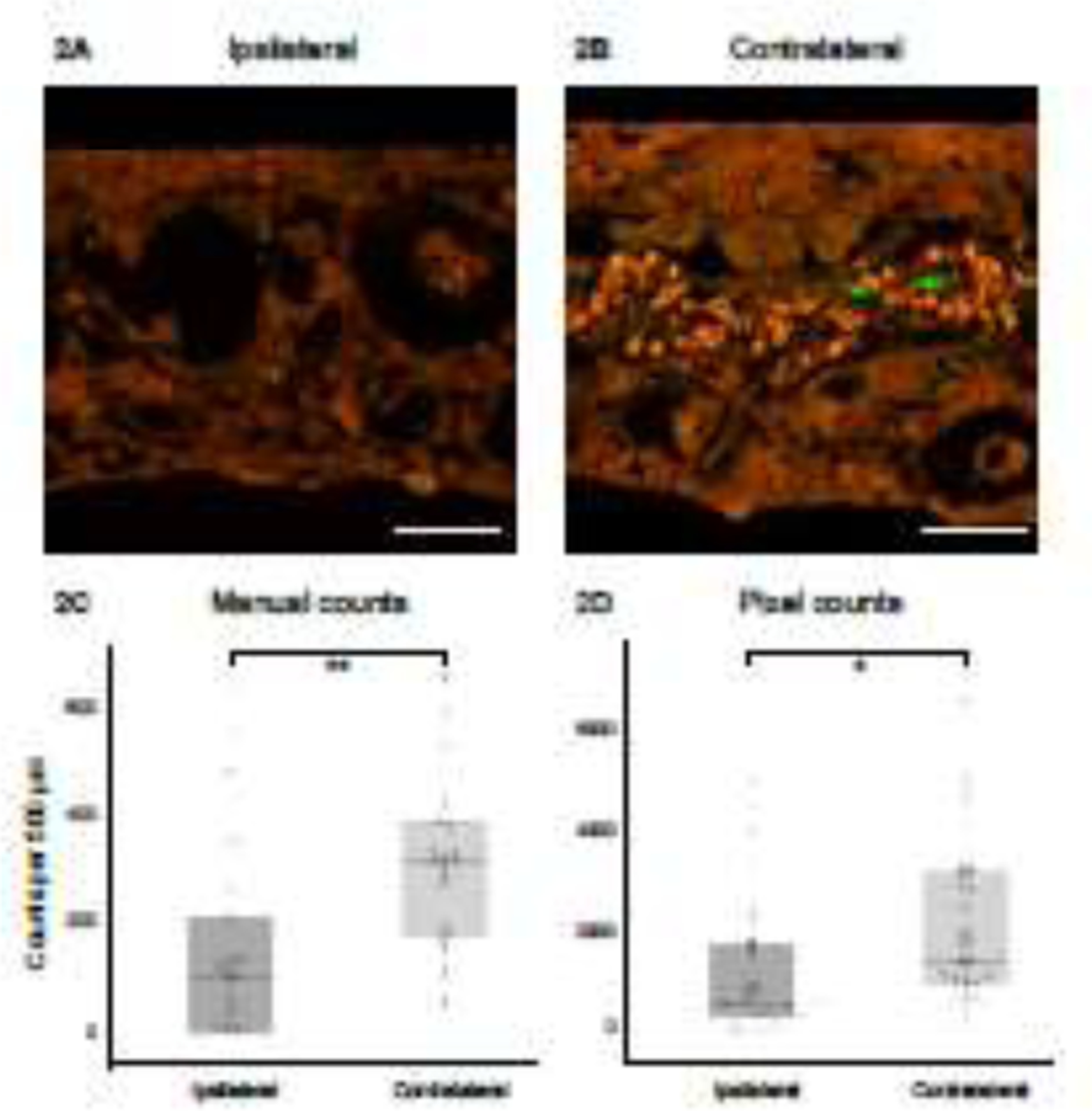
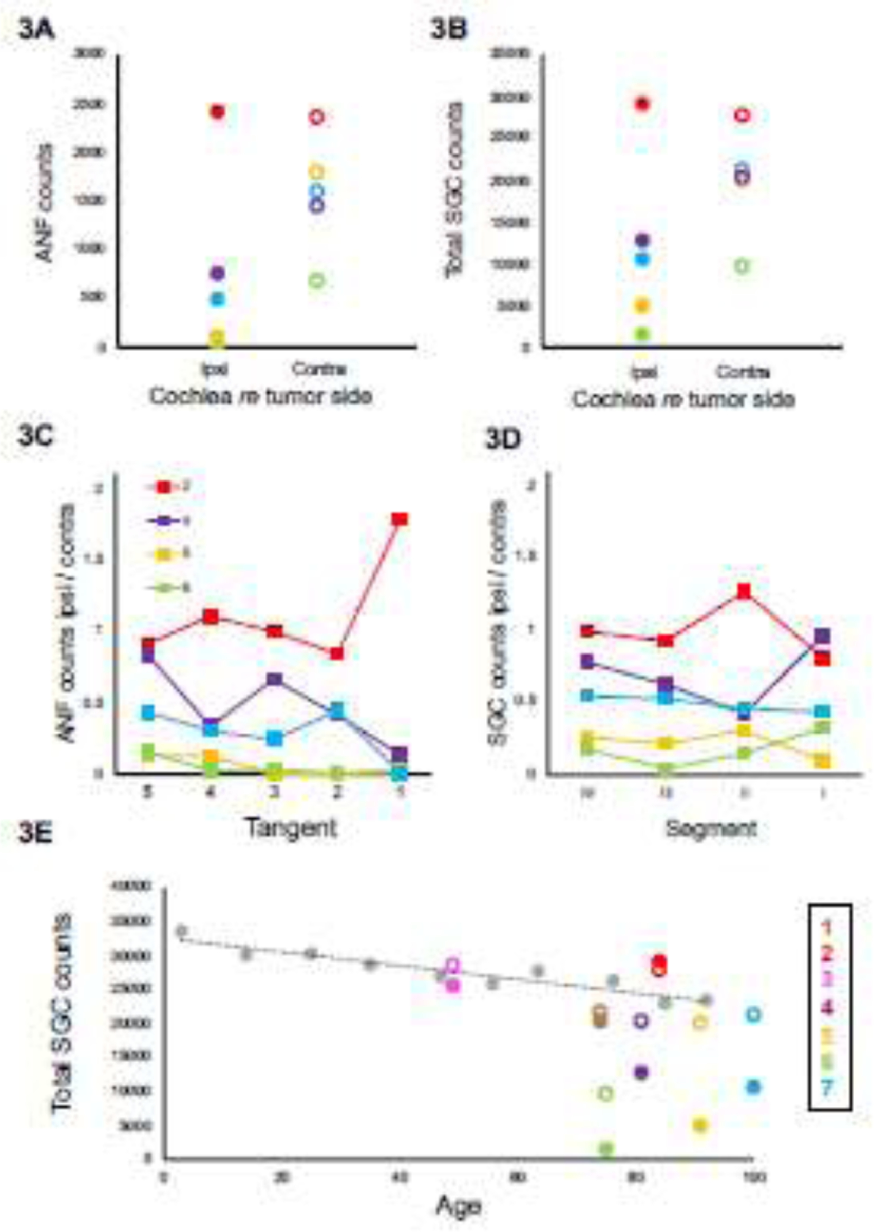
Hearing loss in patients with vestibular schwannoma (VS) is commonly attributed to mechanical compression of the auditory nerve, though recent studies suggest that this retrocochlear pathology may be augmented by cochlear damage. Although VS-associated loss of inner hair cells, outer hair cells, and spiral ganglion cells has been reported, it is unclear to what extent auditory-nerve peripheral axons are damaged in VS patients. Understanding the degree of damage VSs cause to auditory nerve fibers (ANFs) is important for accurately modeling clinical outcomes of cochlear implantation, which is a therapeutic option to rehabilitate hearing in VS-affected ears. A retrospective analysis of human temporal-bone histopathology was performed on archival specimens from the Massachusetts Eye and Ear collection. Seven patients met our inclusion criteria based on the presence of sporadic, unilateral, untreated VS. Tangential sections of five cochlear regions were stained with hematoxylin and eosin, and adjacent sections were stained to visualize myelinated ANFs and efferent fibers. Following confocal microscopy, peripheral axons of ANFs within the osseous spiral lamina were quantified manually, where feasible, and with a "pixel counting" method, applicable to all sections. ANF density was substantially reduced on the VS side compared to the unaffected contralateral side. In the upper basal turn, a significant difference between the VS side and unaffected contralateral side was found using both counting methods, corresponding to the region tuned to 2000 Hz. Even spiral ganglion cells (SGCs) contralateral to VS were affected by the tumor as the majority of contralateral SGC counts were below average for age. This observation provides histological insight into the clinical observation that unilateral vestibular schwannomas pose a long-term risk of progression of hearing loss in the contralateral ear as well. Our pixel counting method for ANF quantification in the osseous spiral lamina is applicable to other pathologies involving sensorineural hearing loss. Future research is needed to classify ANFs into morphological categories, accurately predict their electrical properties, and use this knowledge to inform optimal cochlear implant programming strategies.
Keywords: Auditory nerve fiber density; Cellmask®; Hearing loss; Human temporal bones; Osseous spiral lamina; Vestibular schwannoma.
Copyright © 2022. Published by Elsevier B.V.
Conflict of interest statement
Declaration of Competing Interest The authors disclose no conflict of interest.
Figures
Similar articles
-
Patterns of neural degeneration in the human cochlea and auditory nerve: implications for cochlear implantation.Otolaryngol Head Neck Surg. 1997 Sep;117(3 Pt 1):220-8. doi: 10.1016/s0194-5998(97)70178-5. Otolaryngol Head Neck Surg. 1997. PMID: 9334769
-
Analysis of the human auditory nerve.Hear Res. 1989 Dec;43(1):25-38. doi: 10.1016/0378-5955(89)90056-7. Hear Res. 1989. PMID: 2613564
-
Dysfunction of the cochlea contributing to hearing loss in acoustic neuromas: an underappreciated entity.Otol Neurotol. 2012 Apr;33(3):473-80. doi: 10.1097/MAO.0b013e318248ee02. Otol Neurotol. 2012. PMID: 22377650 Free PMC article.
-
Ipsilateral Cochlear Implantation in the Presence of Observed and Irradiated Vestibular Schwannomas.Ann Otol Rhinol Laryngol. 2020 Dec;129(12):1229-1238. doi: 10.1177/0003489420935482. Epub 2020 Jun 18. Ann Otol Rhinol Laryngol. 2020. PMID: 32551844 Review.
-
The Cochlear Spiral Ganglion Neurons: The Auditory Portion of the VIII Nerve.Anat Rec (Hoboken). 2019 Mar;302(3):463-471. doi: 10.1002/ar.23815. Epub 2018 May 4. Anat Rec (Hoboken). 2019. PMID: 29659185 Review.
Cited by
-
Neural Adaptation at Stimulus Onset and Speed of Neural Processing as Critical Contributors to Speech Comprehension Independent of Hearing Threshold or Age.J Clin Med. 2024 May 6;13(9):2725. doi: 10.3390/jcm13092725. J Clin Med. 2024. PMID: 38731254 Free PMC article.
-
Single-cell transcriptomic atlas reveals increased regeneration in diseased human inner ear balance organs.Nat Commun. 2024 Jun 6;15(1):4833. doi: 10.1038/s41467-024-48491-y. Nat Commun. 2024. PMID: 38844821 Free PMC article.
-
Evidence of cochlear neural degeneration in normal-hearing subjects with tinnitus.Sci Rep. 2023 Nov 30;13(1):19870. doi: 10.1038/s41598-023-46741-5. Sci Rep. 2023. PMID: 38036538 Free PMC article.
-
Predicting neural deficits in sensorineural hearing loss from word recognition scores.Sci Rep. 2022 Jun 23;12(1):8929. doi: 10.1038/s41598-022-13023-5. Sci Rep. 2022. PMID: 35739134 Free PMC article.
-
Long-Term Hearing Outcome For Vestibular Schwannomas After Microsurgery And Radiotherapy: A Systematic Review and Meta-Analysis.Otolaryngol Head Neck Surg. 2024 Dec;171(6):1670-1681. doi: 10.1002/ohn.910. Epub 2024 Jul 24. Otolaryngol Head Neck Surg. 2024. PMID: 39045727 Free PMC article.
References
-
- Chongsathidkiet P, Jackson C, Koyama S, Loebel F, Cui X, Farber SH, Woroniecka K, Elsamadicy AA, Dechant CA, Kemeny HR, Sanchez-Perez L, Cheema TA, Souders NC, Herndon JE, Coumans JV, Everitt JI, Nahed BV, Sampson JH, Gunn MD, Martuza RL, Dranoff G, Curry WT, Fecci PE, 2018. Sequestration of T cells in bone marrow in the setting of glioblastoma and other intracranial tumors. Nat. Med. 24. 10.1038/s41591-018-0135-2 - DOI - PMC - PubMed
-
- De Moura LF, 1967. Inner ear pathology in acoustic neurinoma. Arch. Otolaryngol. (Chicago, Ill. 1960) 85, 125–133. - PubMed
Publication types
MeSH terms
Grants and funding
LinkOut - more resources
Full Text Sources
Medical
Miscellaneous
