. 2022 Jun:231:111805.
doi: 10.1016/j.jinorgbio.2022.111805. Epub 2022 Mar 18.

In silico study of potential antiviral activity of copper(II) complexes with non-steroidal anti-inflammatory drugs on various SARS-CoV-2 target proteins

Affiliations

In silico study of potential antiviral activity of copper(II) complexes with non-steroidal anti-inflammatory drugs on various SARS-CoV-2 target proteins

Elena G Geromichalou et al. J Inorg Biochem. 2022 Jun.

Abstract

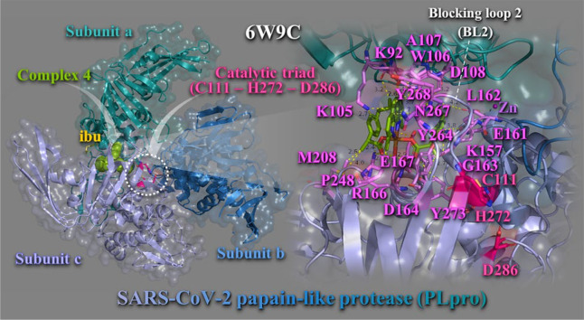
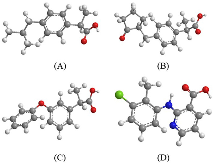
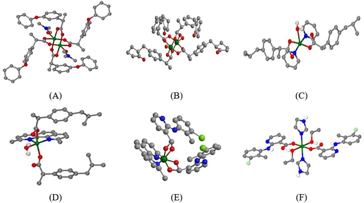
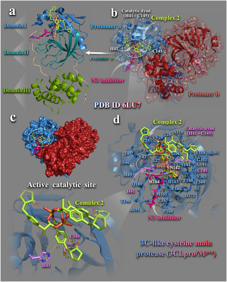
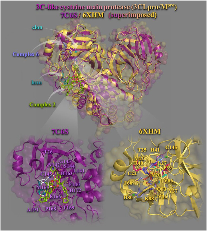
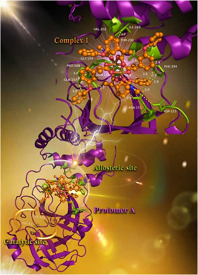
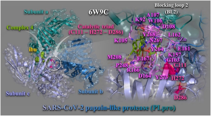
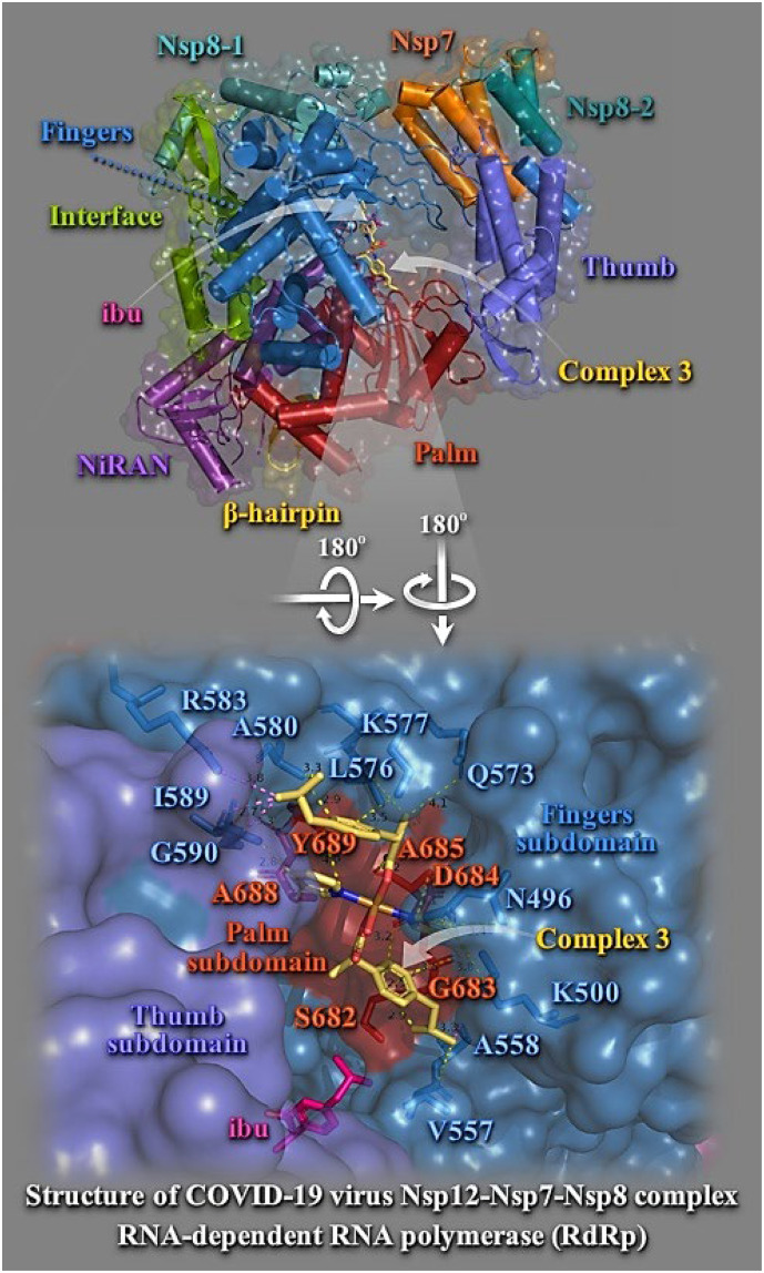
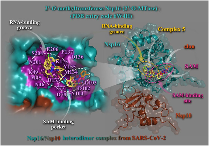
In silico molecular docking studies, in vitro toxicity and in silico predictions on the biological activity profile, pharmacokinetic properties, drug-likeness, ADMET (absorption, distribution, metabolism, excretion, and toxicity) physicochemical pharmacokinetic data, and target proteins and toxicity predictions were performed on six copper(II) complexes with the non-steroidal anti-inflammatory drugs ibuprofen, loxoprofen, fenoprofen and clonixin as ligands, in order to investigate the ability of these complexes to interact with the key therapeutic target proteins of SARS-CoV-2 (Severe Acute Respiratory Syndrome Coronavirus 2) 3C-like cysteine main protease (3CLpro/Mpro), viral papain-like protease (PLpro), RNA-dependent RNA polymerase (RdRp), and non-structural proteins (Nsps) Nsp16-Nsp10 2'-O-methyltransferase complex, and their capacity to act as antiviral agents, contributing thus to understanding the role they can play in the context of coronavirus 2019 (COVID-19) pandemic. Cytotoxic activity against five human cancer and normal cell lines were also evaluated.

Keywords: 3C–like cysteine main protease; Nsp16–Nsp10 2′–O–methyltransferase complex; Papain–like protease; RNA–dependent RNA polymerase; SARS–CoV–2 target proteins; in silico predictive tools.


Conflict of interest statement

None.

Figures

Unlabelled Image
Graphical abstract
Fig. 1
Fig. 1
(A) ibuprofen, (B) loxoprofen, (C) fenoprofen, (D) clonixin. (C: grey, O: red, H: white, N: blue, Cl: light green.)
Fig. 2
Fig. 2
(A) – (F) Structures of complexes 16, respectively (adapted from [1]). (C: grey, O: red, H: white, N: blue, Cu: dark green, Cl: light green.)
Fig. 3
Fig. 3
Docking pose orientation of best–bound complex 2 on the symmetric dimer unit of SARS-CoV-2 3CLpro/Mpro (PBD: 6LU7). Target enzyme illustrating its two subunits (protomers a and b) is depicted either in cartoon–colored by chain (protomers a and b in sky blue and firebrick red, respectively) with additional depiction of a semitransparent surface for the unit colored according to protomer colors (b), or in opaque surface of the homodimer of Mpro colored by chain according to b (c). Structural analysis, along with the binding pose of complex 2 superimposed with the co–crystallized protease inhibitor N3 in the protomer a of the enzyme structure is depicted in (a) illustrating the domains I (residues 8–101), II (residues 102–184), and III (residues 201–303) of the asymmetric unit in cartoon representation colored in sky blue, deep teal, and split pea green, respectively, while loop regions 1–7, 185–200, and 305–306 are shown in wheat color. A close–up view of the binding interaction architecture of 2 in the catalytic active site of the enzyme (protomer a in sky blue cartoon with additional depiction of selected contacting residues flanking the catalytic active cavity rendered in stick model and colored according to cartoon) is shown in (d). The catalytic dyad (H41–C145) is also indicated in dashed white ovals (b, d).Catalytic residues H41 and C145 are depicted in stick model and colored according to atom type in violet C atoms. Complex 2, its NSAID Hloxo, and N3 inhibitor are rendered in stick model and colored according to atom type in limon, salmon, and hot pink C atoms, respectively. Binding contacts of 2 and N3 are shown as dotted yellow and orange lines, respectively. Hydrogen atoms are omitted from all molecules for clarity. Heteroatom color–code: Cu: orange and O: red. The final structure was ray–traced with depth cue in the ray–tracing rendering of the cartoon and illustrated with the aid of PyMol Molecular Graphics Systems. (For interpretation of the references to color in this figure legend, the reader is referred to the web version of this article.)
Fig. 4
Fig. 4
Molecular docking of complexes 2 and 6, and their NSAIDs Hloxo and Hclon, as well as the protease inhibitors boceprevil (U5G) of 7C6S and V2M of 6XHM, on two SARS-COV-2 3CLpro/Mpro enzymes. The final alignment of structures derived by the superimposition of 7C6S and 6XHM proteases. The structures were aligned by PyMol Molecular Graphics System. The overlay of the two resolved structures of 3CLpro/Mpro 7C6S and 6XHM are illustrated in cartoon mode and colored in deep purple and yellow–orange, respectively, with additional depiction of a semitransparent surface colored according to cartoon representation. All compounds are rendered in stick model and colored according to atom type in split pea green (2), slate blue (6), deep teal (Hloxo), cyan (Hclon), white (boceprevil), and hot pink (V2M), C atoms. A close–up view of the binding interaction architecture of 2 and 6 in their binding pockets superimposed with Hloxo and Hclon, respectively, with additional depiction of selected contacting residues rendered in stick model and colored according to cartoon, are shown at the lower panel. Binding contacts are shown as dotted yellow and deep purple lines. Hydrogen atoms are omitted from all molecules for clarity. Heteroatom color–code: Cu: split pea green (in 2), brown (in 6), Cl: green, O: red, and N: blue. The final structure was ray–traced with depth cue in the ray–tracing rendering of the cartoon and illustrated with the aid of PyMol Molecular Graphics Systems. (For interpretation of the references to color in this figure legend, the reader is referred to the web version of this article.)
Fig. 5
Fig. 5
Docking pose orientation of best–bound complex 1 on protomer a of SARS-CoV-2 3CLpro/Mpro (PBD: 6LU7). Target enzyme is depicted in cartoon colored in deep purple. A close–up view of the binding interaction architecture of 1 in the allosteric binding site of the enzyme in a deep cleft between the catalytic and dimerization domain of the enzyme with additional depiction of selected contacting residues rendered in stick model and colored in split pea green is shown in the upper panel of the molecular structure. Binding residues of complex 1 in the allosteric binding pocket are Gln (Q107), Pro (P108), Gly (G109), Gln (Q110), Asn (N151), Asp (D153), Val (V202), Ile (I249), Phe (F294), and Thr (T292). The two critical residues of the catalytic site (H41–C145) are also indicated in yellow–orange sticks (lower panel). Complex 1 is rendered in ball–and–stick model and colored according to atom type in orange. Binding contacts are shown as dotted yellow lines. Hydrogen atoms are omitted from all molecules for clarity. Heteroatom color–code: Cu: brown, O: red, and N: blue. The final structure was ray–traced and illustrated with the aid of PyMol Molecular Graphics Systems. (For interpretation of the references to color in this figure legend, the reader is referred to the web version of this article.)
Fig. 6
Fig. 6
Docking pose orientation of best bound complex 4 and Hibu (rendered in sphere and stick model and colored by atom type in split pea green and yellow–orange C atoms, respectively) on the crystal structure of SARS–CoV–2 PLpro (PBD: 6W9C). Target protein is illustrated in cartoon colored by deep teal (subunit A), sky blue (subunit B), and light blue (subunit C), with additional depiction of a semitransparent surface colored according to cartoon representation and indication of the catalytic triad in dashed white oval. A close–up view of the binding interaction architecture of 4 in its binding pocket superimposed with Hibu and additional depiction of selected contacting residues rendered in stick model and colored according to atom type in violet C atoms, are shown at the right panel. Complex 4 and Hibu are found to be stabilized in close proximity to catalytic triad (C111, H272, and D286) colored by atom type in hot pink C atoms. Zn ion is shown as purple sphere. Binding contacts are shown as dotted yellow lines. Hydrogen atoms are omitted from all molecules for clarity. Heteroatom color–code: Cu: orange, O: red, and N: blue. The final structure was ray–traced with depth cue in the ray–tracing rendering of the cartoon and illustrated with the aid of PyMol Molecular Graphics Systems. (For interpretation of the references to color in this figure legend, the reader is referred to the web version of this article.)
Fig. 7
Fig. 7
(Upper panel) Docking pose orientation of best bound complex 3 and Hibu (rendered in stick model and colored by atom type in yellow–orange and hot pink C atoms, respectively) on the crystal structure of SARS–CoV–2 RdRp (PBD: 6M71). Target protein is illustrated in cartoon with α–helices shown as cylinders, β–strands as arrows, and loops as strands, colored according to structure characterization of subdomain components of the RdRp complex as follows: β–hairpin, yellow–orange; NiRAN, violet purple; Interface, split pea green; Fingers, sky blue; Palm, firebrick red; Thumb, slate blue; Nsp7, orange; Nsp8–1, teal; and Nsp8–2, deep teal, with additional depiction of a semitransparent surface colored according to cartoon representation. (Lower panel) A close–up view of the binding interaction architecture of 3 in its binding pocket superimposed with Hibu illustrated as semitransparent surface with additional depiction of selected contacting residues rendered in stick model and colored according to atom type in sky blue and firebrick red corresponding to Fingers and Palm, respectively. Binding contacts are shown as dotted yellow and violet lines. Hydrogen atoms are omitted from all molecules for clarity. Heteroatom color–code: Cu: orange, and O: red. The final structure was ray–traced and illustrated with the aid of PyMol Molecular Graphics Systems.
Fig. 8
Fig. 8
(Right panel) Docking pose orientation of best–bound complex 5 and Hclon (rendered in stick model and colored by atom type in yellow–orange and orange C atoms, respectively) on the crystal structure of SARS–CoV–2 2′OMTase (Nsp16–Nsp10 heterodimer complex illustrated as cartoon colored in deep teal and chocolate, respectively with additional depiction of semitransparent surface) superimposed with SAM (rendered in stick colored in hot pink C atoms) in the small unit crystal form (PDB ID: 6W4H). SAM–binding site and RNA–binding groove are also indicated in dashed yellow and hot pink ovals, respectively. (Left panel) A close–up view of the ligand binding architecture mapping of 5 in its binding pocket superimposed with Hclon and SAM, as well as the residues implicated in SAM and RNA binding pockets (highlighted in deep purple) onto the SARS–CoV–2 Nsp16 structure (opaque surface in deep teal) showing the relative positions of the SAM and RNA binding regions. Binding contacts are shown as dotted yellow lines. Hydrogen atoms are omitted from all molecules for clarity. Heteroatom color–code: Cu and Cl: green, N: blue, and O: red. The final structure was ray–traced and illustrated with the aid of PyMol Molecular Graphics Systems.

Similar articles

Cited by

References

    1. Malis G., Geromichalou E., Geromichalos G.D., Hatzidimitriou A.G., Psomas G. J. Inorg. Biochem. 2021;224 - PubMed
    1. Gorbalenya A.E., Baker S.C., Baric R.S., de Groot R.J., Drosten C., Gulyaeva A.A., Haagmans B.L., Lauber C., Leontovich A.M., Neuman B.W., Penzar D., Perlman S., Poon L.M., Samborskiy D.V., Sidorov I.A., Sola I., Ziebuhr J. Nat. Microbiol. 2020;5:536–544. - PMC - PubMed
    1. Lebedeva N.S., Gubarev Y.A., Mamardashvili G.M., Zaitceva S.V., Zdanovich S.A., Malyasova A.S., Romanenko J.V., Koifman M.O., Koifman O.I. Sci. Rep. 2021;11:19481. - PMC - PubMed
    1. Jin Z., Du X., Xu Y., Deng Y., Liu M., Zhao Y., Zhang B., Li X., Zhang L., Peng C., Duan Y., Yu J., Wang L., Yang K., Liu F., Jiang R., Yang X., You T., Liu X., Yang X., Bai F., Liu H., Liu X., Guddat L., Xu W., Xiao G., Qin C., Shi Z., Jiang H., Rao Z., Yang-Show H. Nature. 2020;582:289–293. - PubMed
    1. Osipiuk J., Azizi S., Dvorkin S., Endres M., Jedrzejczak R., Jones K.A., Kang S., Kathayat R., Kim Y., Lisnyak V., Maki S., Nicolaescu V., Taylor C., Tesar C., Zhang Y., Zhou Z., Randall G., Michalska K., Snyder S., Dickinson B., Joachimiak A. Nat. Commun. 2021;12:743. - PMC - PubMed