

Post-mortem histopathology of pituitary and adrenals of COVID-19 patients
- PMID: 35334433
- PMCID: PMC8873038
- DOI: 10.1016/j.legalmed.2022.102045
Post-mortem histopathology of pituitary and adrenals of COVID-19 patients
Abstract
Background: Knowledge of the exact organ manifestation is essential for a comprehensive understanding of COVID-19 infection. Here, the histopathological changes in the pituitary and adrenal glands were analyzed.
Methods: In this series, the formalin-fixed tissues of 63 pituitary glands and 50 adrenal glands were examined. We performed HE and PAS staining and examined COVID-19 nucleocapsid antibody immunohistochemically in the pituitary glands and adrenals.
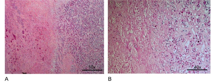
Results: Histologically, there was no evidence of COVID-19-specific changes in the pituitary and adrenal glands. Large pituitary necrosis may be interpreted as a shock reaction. Independent of infection, we found one T-cell lymphoma, two adenomas, and four Rathke-type cysts in the pituitary glands, and 70% of the adrenal glands showed decreased lipid content and an increase in compact cells as a stress response. In addition, a cortical adenoma in one adrenal gland and small cortical nodules in three adrenal glands were detected independently of COVID-19.
Conclusion: Pituitary and adrenal glands do not appear histologically predominant in the course of COVID-19.
Keywords: Adrenal; COVID-19 infection; Histopathology; Pituitary.
Copyright © 2022 Elsevier B.V. All rights reserved.
Conflict of interest statement
The authors declare that they have no known competing financial interests or personal relationships that could have appeared to influence the work reported in this paper.
Figures
References
-
- Braun F., Lütgehetmann M., Pfefferle S., Wong M.N., Carsten A., Lindenmeyer M.T., Nörz D., Heinrich F., Meißner K., Wichmann D., Kluge S., Gross O., Pueschel K., Schröder A.S., Edler C., Aepfelbacher M., Puelles V.G., Huber T.B. SARS-CoV-2 renal tropism associates with acute kidney injury. Lancet. 2020;396(10251):597–598. - PMC - PubMed
-
- H. Buurman, W. Saeger. Subclinical adenomas in postmortem pituitaries: classification and correlation to clinical data. Exp. Clin. Endocr. Metab. 114: S 29, abstract P04-044 (2006). - PubMed
-
- Casagrande M., Fitzek A., Spitzer M.S., Püschel K., Glatzel M., Krasemann S., Nörz D., Lütgehetmann M., Pfefferle S., Schultheiss M. Presence of SARS-CoV-2 RNA in the cornea of Viremic patients with COVID-19. JAMA Ophthalmol. 2021;139(4):383. doi: 10.1001/jamaophthalmol.2020.6339. - DOI - PMC - PubMed
-
- Ding Y., He L., Zhang Q., Huang Z., Che X., Hou J., Wang H., Shen H., Qiu L., Li Z., Geng J., Cai J., Han H., Li X., Kang W., Weng D., Liang P., Jiang S. Organ distribution of severe acute respiratory syndrome (SARS) associated coronavirus (SARS-CoV) in SARS patients: implications for pathogenesis and virus transmission pathways. J. Pathol. 2004;203(2):622–630. - PMC - PubMed
MeSH terms
LinkOut - more resources
Full Text Sources
Medical
