. 2022 Feb 23;58(3):335.
doi: 10.3390/medicina58030335.

The Anti-Endometriotic Effect of Cyperi Rhizoma Extract, Inhibiting Cell Adhesion and the Expression of Pain-Related Factors through Akt and NF-kB Pathways

Affiliations

The Anti-Endometriotic Effect of Cyperi Rhizoma Extract, Inhibiting Cell Adhesion and the Expression of Pain-Related Factors through Akt and NF-kB Pathways

Ji-Hye Ahn et al. Medicina (Kaunas). .

Abstract

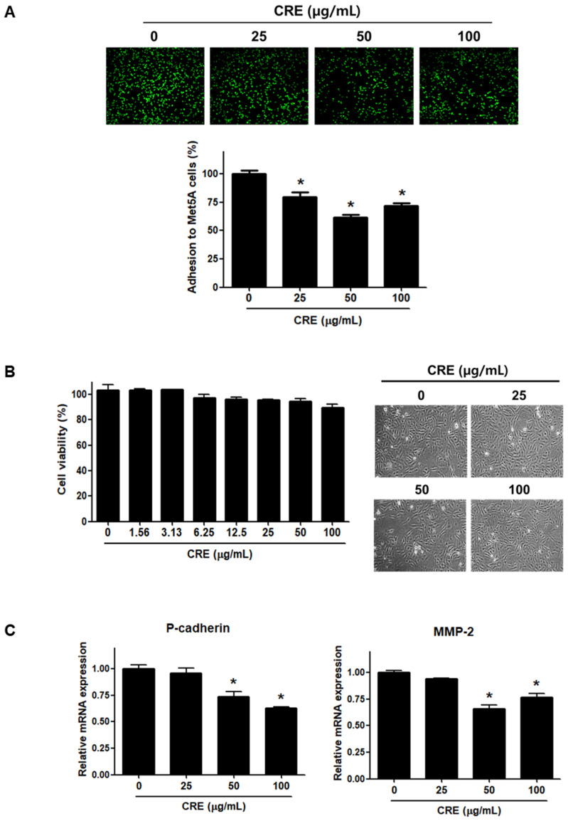
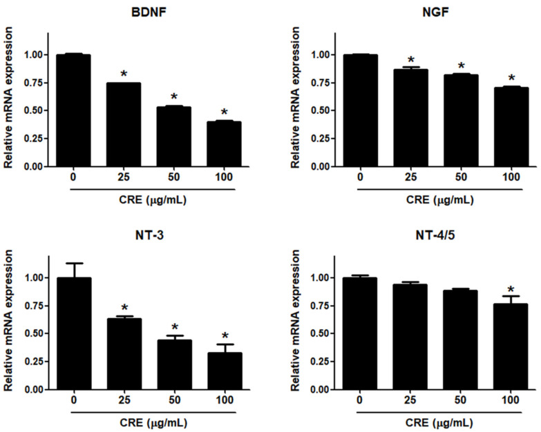
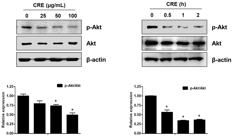
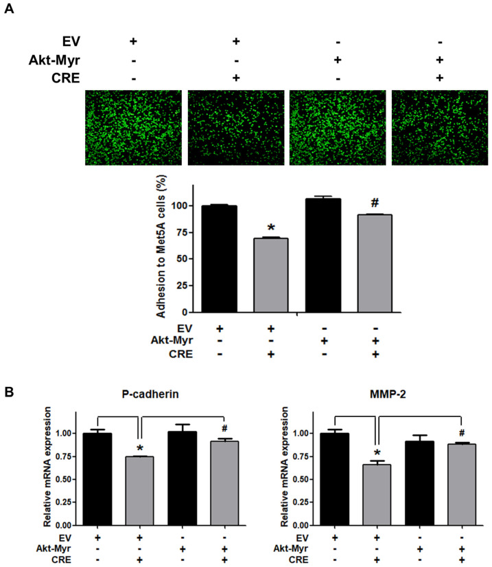
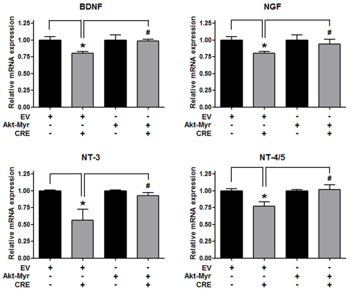
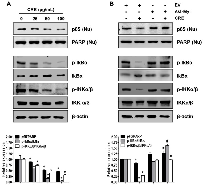
Rhizomes of Cyperus rotundus have been widely used as a traditional medicine in Asia for the treatment of gynecological diseases. However, there is no scientific evidence demonstrating the effect of C. rotundus rhizomes on endometriosis, which is characterized by the adhesion of endometrial tissues outside the uterus, resulting in chronic and severe pelvic pain. The aim of this study was to investigate the effects of Cyperi rhizoma extract (CRE) on cell adhesion and the expression of pain-related factors (neurotrophins) in endometriotic cells, and to elucidate the underlying molecular mechanisms. CRE inhibited the adhesion of human endometriotic 12Z cells to peritoneal mesothelial Met5A cells using by adhesion assays. The mRNA expression of adhesion molecules [P-cadherin and matrix metalloproteinase (MMP)-2] was downregulated by CRE treatment. In addition, CRE significantly inhibited the mRNA expression of neurotrophins (BDNF, NGF, NT-3 and NT-4/5) in 12Z cells. Moreover, Akt overexpression markedly neutralized the inhibition of cell adhesion by CRE and expression of neurotrophins in 12Z cells. Furthermore, it was found that CRE suppressed NF-kB activation through the Akt pathway. These data suggest that CRE exerts anti-endometriotic activities by the inhibition of cell adhesion and neurotrophin expression, through the negative regulation of the Akt and NF-kB pathways in endometriotic cells.

Keywords: Akt; Cyperi rhizome; NF-kB; adhesion; endometriosis; neurotrophins.


Conflict of interest statement

The authors have declared no conflict of interest.

Figures

Figure 1
Figure 1
Effect of CRE on endometriotic cell adhesion to mesothelial cells and expression of adhesion molecules in endometriotic cells. (A) Human endometriotic cells (12Z) were treated with CRE at indicated concentration (25, 50 and 100 µg/mL) for 24 h. The 12Z cells labeled with CellTrackerTM (10 µM) were cultured on Met5A cell layers in a 96-well plate for 1 h. The total fluorescence in each well was measured by fluorescence microphotography. (B) Cell viability was determined by MTT assay. Representative images show the cell morphology of 12Z cells treated with CRE at indicated concentration (25, 50 and 100 µg/mL) for 24 h. (C) The mRNA expression of P-cadherin and MMP-2 was measured by real-time RT-PCR. All expression levels were normalized to GAPDH. Results are the combined data (mean ± SD) from three independent experiments. * p < 0.05 compared with control group.
Figure 2
Figure 2
Effect of CRE on the expression of neurotrophins in endometriotic cells. The 12Z cells were treated with CRE (25, 50 and 100 µg/mL) for 24 h, and then the mRNA expression of neurotrophins (BDNF, NGF, NT-3 and NT-4/5) was measured by real-time RT-PCR. All expression levels were normalized to GAPDH. Results are the combined data (mean ± S.D.) from three independent experiments. * p < 0.05 compared with control group.
Figure 3
Figure 3
Effect of CRE on the phosphorylation of Akt in endometriotic cells. 12Z cells were treated with CRE (25, 50 and 100 µg/mL) for 2 h or with CRE (100 µg/mL) for 0.5, 1 and 2 h. Phosphorylated Akt and total Akt were measured by western blot analysis using specific antibodies. β-Actin was used as an internal control. Data are shown as mean band density. Data are presented as the means ± SD of three independent experiments; * p < 0.05 as compared with the control group.
Figure 4
Figure 4
Involvement of the Akt pathway in endometriotic cell adhesion to mesothelial cells and expression of adhesion molecules in endometriotic cells. 12Z cells were transfected with an empty vector (EV) or a constitutive active form (Akt-Myr), and then the cells were treated with CRE (100 µg/mL). After 24 h, (A) an adhesion assay and (B) a real-time RT-PCR were performed. The mRNA expression of P-cadherin and MMP-2 was normalized to GAPDH. * p < 0.05 compared with EV-transfected none treatment group and # p < 0.05 compared with EV-transfected CRE treatment group.
Figure 5
Figure 5
Involvement of the Akt pathway in the expression of neurotrophins in endometriotic cells. The 12Z cells were transfected with an empty vector (EV) or a constitutive active form (Akt-Myr), and then the cells were treated with CRE (100 µg/mL). After 24 h, real-time RT-PCR was performed to measure the mRNA expression of BDNF, NGF, NT-3 and NT-4/5. The expression levels were normalized to GAPDH. * p < 0.05 compared with EV-transfected none treatment group and # p < 0.05 compared with EV-transfected CRE treatment group.
Figure 6
Figure 6
Effect of CRE on the NF-kB activation in endometriotic cells. Nuclear (Nu) and whole cellular proteins were isolated from the cells (A) treated with CRE (25, 50 and 100 µg/mL) for 8 h and (B) transfected with EV or Akt-Myr. The levels of p65 (Nu), p-IkBα, IkBα, p-IKKα/β and IKKα/β were detected by western blot analysis using specific antibodies. PARP and β-actin were used as an internal control for nuclear fractions and whole cell lysates, respectively. Data are shown as mean band density. Data are presented as the means ± SD of three independent experiments; * p < 0.05 as compared with the control group, and # p < 0.05 as compared with EV-transfected CRE treatment group.

Similar articles

Cited by

References

    1. Burney R.O., Giudice L.C. Pathogenesis and pathophysiology of endometriosis. Fertil. Steril. 2012;98:511–519. doi: 10.1016/j.fertnstert.2012.06.029. - DOI - PMC - PubMed
    1. Giudice L.C., Kao L.C. Endometriosis. Lancet. 2004;364:1789–1799. doi: 10.1016/S0140-6736(04)17403-5. - DOI - PubMed
    1. Czyzyk A., Podfigurna A., Szeliga A., Meczekalski B. Update on endometriosis pathogenesis. Minerva Ginecol. 2017;69:447–461. doi: 10.23736/S0026-4784.17.04048-5. - DOI - PubMed
    1. Andres M.P., Arcoverde F.V.L., Souza C.C.C., Fernandes L.F.C., Abrao M.S., Kho R.M. Extrapelvic Endometriosis: A Systematic Review. J. Minim. Invasive Gynecol. 2020;27:373–389. doi: 10.1016/j.jmig.2019.10.004. - DOI - PubMed
    1. Young V.J., Brown J.K., Saunders P.T., Horne A.W. The role of the peritoneum in the pathogenesis of endometriosis. Hum. Reprod. Update. 2013;19:558–569. doi: 10.1093/humupd/dmt024. - DOI - PubMed