. 2022 Feb 27;58(3):355.
doi: 10.3390/medicina58030355.

NPS-1034 Induce Cell Death with Suppression of TNFR1/NF-κB Signaling in Testicular Cancer

Affiliations

NPS-1034 Induce Cell Death with Suppression of TNFR1/NF-κB Signaling in Testicular Cancer

Jian-Ting Chen et al. Medicina (Kaunas). .

Abstract

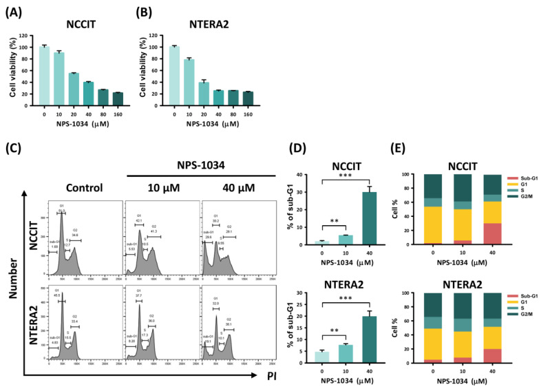
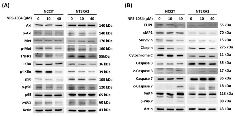
Background and objectives: NPS-1034 with a dual inhibitory effect on Met and Axl kinase receptors has exhibited therapeutic potential in previous models. However, no study on treating testicular cancer (TC) cell lines with NPS-1034 has been established. Materials and Methods: In this study, a series of in vitro examinations of the apoptotic effect induced by NPS-1034 in TC cell lines was conducted to clarify the molecular interactions involved. Results: A decrease in cell viability rate was observed following NPS-1034 treatment, as shown in the MTT assay. Induction of the apoptotic effect was observed in TC cells as the sub-G1 and Annexin-PI populations increased in a dose-dependent manner. The involvement of the tumor receptor necrosis factor receptor 1 (TNFR1) pathway was later determined by the proteome array and western blotting. A reduction in TNFR1 and NF-κB downstream protein expressions, an upregulation of cleaved caspase-3 and -7, and a downregulation of survivin and claspin all reassured the underlying mechanism of the TNFR1 involved in the apoptotic pathway induced by NPS-1034. Conclusions: Our findings provide evidence for a potential underlying TNFR1 pathway involved in NPS-1034 treatment. This study should offer new insights into targeted therapy for TC.

Keywords: NPS-1034; TNF receptor-1; apoptosis; p50; p65; testicular cancer.


Conflict of interest statement

The authors declare that they have no competing interest.

Figures

Figure 1
Figure 1
Analysis of TC cell survival with NPS-1034 treatment. The MTT assay was used to determine TC cell viability in (A) NCCIT and (B) NTERA2 cell lines treated with NPS-1034 (10, 20, 40, 80, and 160 µM) and DMSO (control) for 24 h. (C) Cell cycle analysis was performed to evaluate changes in each phase of NCCIT and NTERA2 cell lines treated with NPS-1034 at 10 and 40 µM for 48 h. (D) An increase in the sub-G1 phase can be observed in both cell lines after NPS-1034 treatment. (E) The proportion of the cell cycle was quantified in the segments of the bar chart. The data are presented as mean ± SD (** p < 0.01; *** p < 0.001).
Figure 2
Figure 2
NPS-1034 promotes apoptosis in TC cell lines. (A) Hoechst stain 33,342 was used to visualize the apoptotic cells of the NCCIT and NTERA2 cell lines treated with NPS-1034 (0, 10, and 40 µM) for 24 h. (B) The percentage of apoptotic cells is presented in the columns. (C) Flow cytometry was conducted to evaluate the cause of cell deaths with annexin v/PI dual stain. An examination was performed to determine the number of apoptotic NCCIT and NTERA2 cells treated with NPS-1034 for 48 h. (D) The proportion of cell distribution is shown in the column for NCCIT and NTERA2 cells. The data are presented as mean ± SD (*** p < 0.001).
Figure 3
Figure 3
An apoptosis array examined the expressions of proteins involved in cell death caused by NPS-1034. Both (A) NCCIT and (C) NTERA2 cell lines were tested with an apoptosis array. The expressions of proteins were quantified into bar charts, as shown in (B,D). The proteins were aligned based on the extent of changes in the protein expressions before and after NPS-1034 treatment.
Figure 4
Figure 4
NPS-1034 potentially triggered an apoptotic pathway in TC cell lines via the TNFR1 pathway. (A) NPS-1034 inhibits the p-Axl, p-Met and downstream pathway NF-κB of TNFR1, which might lead to certain gene expressions in both NCCIT and NTERA2 cell lines. (B) Proteins involved in the apoptotic pathways were determined following the treatment of NPS-1034 in TC cell lines.

Similar articles

Cited by

References

    1. Feldman D.R., Bosl G.J., Sheinfeld J., Motzer R.J. Medical treatment of advanced testicular cancer. JAMA. 2008;299:672–684. doi: 10.1001/jama.299.6.672. - DOI - PubMed
    1. Rajpert-De Meyts E. Developmental model for the pathogenesis of testicular carcinoma in situ: Genetic and environmental aspects. Hum. Reproduct. Update. 2006;12:303–323. doi: 10.1093/humupd/dmk006. - DOI - PubMed
    1. Le Cornet C., Lortet-Tieulent J., Forman D., Béranger R., Flechon A., Fervers B., Schüz J., Bray F. Testicular cancer incidence to rise by 25% by 2025 in Europe? Model-based predictions in 40 countries using population-based registry data. Eur. J. Cancer. 2014;50:831–839. doi: 10.1016/j.ejca.2013.11.035. - DOI - PubMed
    1. Park J.S., Kim J., Elghiaty A., Ham W.S. Recent global trends in testicular cancer incidence and mortality. Medicine. 2018;97:e12390. doi: 10.1097/MD.0000000000012390. - DOI - PMC - PubMed
    1. McGlynn K.A., Trabert B. Adolescent and adult risk factors for testicular cancer. Nat. Rev. Urol. 2012;9:339–349. doi: 10.1038/nrurol.2012.61. - DOI - PMC - PubMed