. 2022 Mar 5;58(3):389.
doi: 10.3390/medicina58030389.

Malunion of the Tibia: A Systematic Review

Affiliations

Malunion of the Tibia: A Systematic Review

Ishan Patel et al. Medicina (Kaunas). .

Abstract

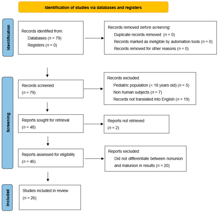
Background and Objectives: Tibial malunions are defined as tibial fractures that have healed in a clinically unacceptable position, resulting in deformity such as shortening, lengthening, abnormal rotation, or angulation. These deformities can have adverse effects on patients, such as pain and gait disturbance, as well as long term development of post-traumatic arthritis. This paper seeks to highlight some of the options for surgical management of malunions and detail the strategies and approaches used to manage these complicated cases. Materials and Methods: An exhaustive search was conducted on PubMed using the key search terms “Tibial” OR “Tibia” AND “Malunion” to be included in the title. Exclusions to the search included any article with patients aged < 18 years, any nonhuman subjects, and any article not published or translated into English. Results: A systematic review of the literature revealed 26 articles encompassing 242 patients who had undergone surgical correction for tibia malunion. A total of 19 patients suffered from complications. Methods of treatment included osteotomies, with plate and screws, external fixator, angled blade plate, intramedullary nails, Ilizarov fixator, Taylor Spatial Frame, Precise nail, and total knee arthroplasty. Restoring alignment and the articular surface led to overwhelmingly positive patient outcomes. Conclusions: Tibial malunions take many forms, and as such, there are many approaches to correcting deformities. The literature supports the following radiological parameters to diagnose tibial malunion: 5−10 degrees angulation, 1−2 cm shortening, 10−15 degrees internal rotation, and 10−20 degrees external rotation. Surgical plans should be customized to each individual patient, as there are many approaches to tibial malunion that have been shown to be successful in delivering excellent clinical outcomes.

Keywords: approach; malunion; outcomes; pilon; plateau; shaft; tibia; tibial.


Conflict of interest statement

The authors declare no conflict of interest.

Figures

Figure 1
Figure 1
Flowchart displaying methodology for literature review [3].
Figure 2
Figure 2
Part (A,D) show a proximal tibia extra-articular malunion in 6 degrees of varus and 24 degrees in procurvatum. Part (B,E) demonstrate patient after proximal tibial subtubercular osteotomy and dual plating, with the alignment restored. Part (C) depicts the contralateral limb.
Figure 3
Figure 3
Case of a 54-year-old female with intra-articular malunion of the proximal tibia with 5 mm of lateral depression and 13 degrees of varus deformity (first row of images). CT scan is shown in the second row of images. Patient was treated with a lateral parapatellar approach with a lateral tibial tubercle osteotomy and a trap door osteotomy. The depressed fragments were elevated followed by an opening-wedge tibial osteotomy. Medial and lateral plates were applied to hold the reduction (third row of images).
Figure 4
Figure 4
This is a case of a 53-year-old patient with post-traumatic tibial deformity complaining of knee and ankle pain. The deformity was assessed on long-leg standing alignment views, compared to the other tibia in AP and lateral views. CT scan revealed very little rotational deformity. An osteotomy was planned at the center of rotation and angulation (CORA), which was close to the same location on both AP and lateral. This is a single-plane deformity, which has been measured on both the AP and lateral images. The right two images show the patient post-transverse opening-wedge osteotomy fixed with an intramedullary nail and a plate. Healed in good alignment.
Figure 5
Figure 5
A case of a severe distal tibia deformity with obvious soft tissue damage on the lateral border of the deformity. Conventional X-rays were unable to fully assess or define the deformity that can be seen on a 3D reconstruction CT scan (B). Image (A) depicts a gross image. An osteotomy and fixation with a Tayler Spatial Frame (C) were used with a residual calculation to slowly correct the deformity. Slow correction would help protect the soft tissue envelope on the lateral border and stretch the medial soft tissues. Correction has resulted in a plantigrade foot with motion of the ankle and a healed tibia (D).

References

    1. Sarmiento A., Sobol P.A., Hoy A.L.S., Ross S.D., Racette W.L., Tarr R.R. Prefabricated functional braces for the treatment of fractures of the tibial diaphysis. J. Bone Jt. Surg Am. 1984;66:1328–1339. doi: 10.2106/00004623-198466090-00002. - DOI - PubMed
    1. Santoro D., Tantavisut S., Aloj D., Karam M.D. Diaphyseal osteotomy after post-traumatic malalignment. Curr. Rev. Musculoskelet. Med. 2014;7:312–322. doi: 10.1007/s12178-014-9244-9. - DOI - PMC - PubMed
    1. Page M.J., McKenzie J.E., Bossuyt P.M., Boutron I., Hoffmann T.C., Mulrow C.D., Shamseer L., Tetzlaff J.M., Akl E.A., Brennan S.E., et al. The PRISMA 2020 statement: An updated guideline for reporting systematic reviews. BMJ. 2021;372:n71. doi: 10.1136/bmj.n71. - DOI - PMC - PubMed
    1. Anglen J.O., Healy W.L. Tibial plateau fractures. Orthopedics. 1988;11:1527–1534. doi: 10.3928/0147-7447-19881101-05. - DOI - PubMed
    1. Krettek C., Hawi N., Jagodzinski M. Intracondylar segment osteotomy: Correction of intra-articular malalignment after fracture of the tibial plateau. Unfallchirurg. 2013;116:413–426. doi: 10.1007/s00113-013-2377-2. - DOI - PubMed

Publication types