. 2022 Mar 17;10(3):461.
doi: 10.3390/vaccines10030461.

Cytokine Adjuvants IL-7 and IL-15 Improve Humoral Responses of a SHIV LentiDNA Vaccine in Animal Models

Affiliations

Cytokine Adjuvants IL-7 and IL-15 Improve Humoral Responses of a SHIV LentiDNA Vaccine in Animal Models

Laury-Anne Leroy et al. Vaccines (Basel). .

Abstract

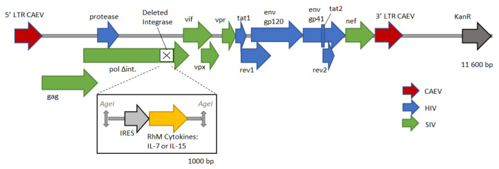
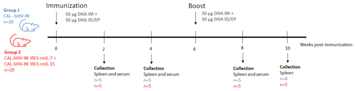
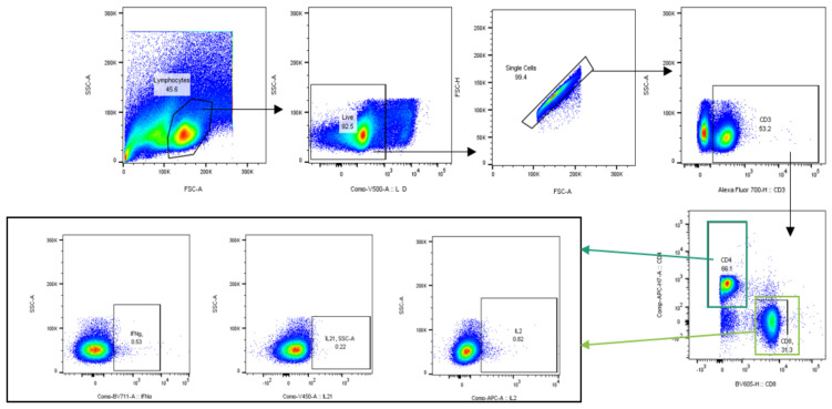
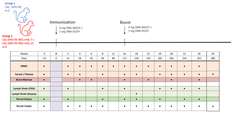
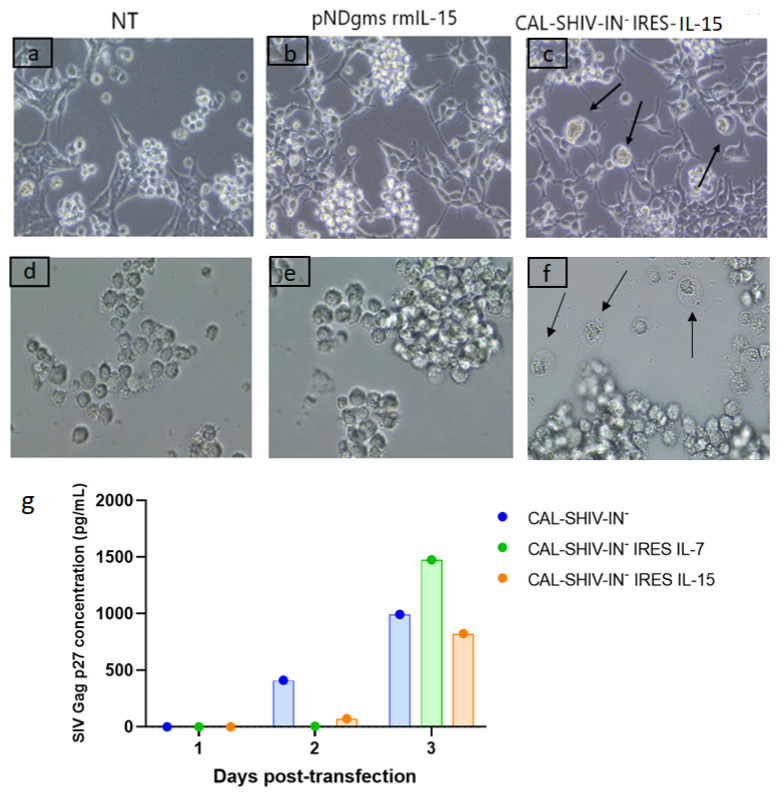
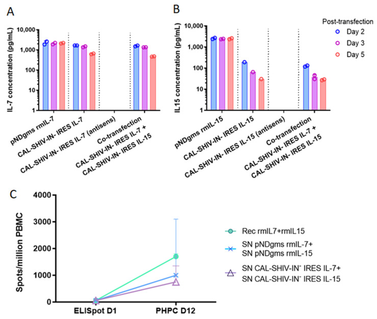
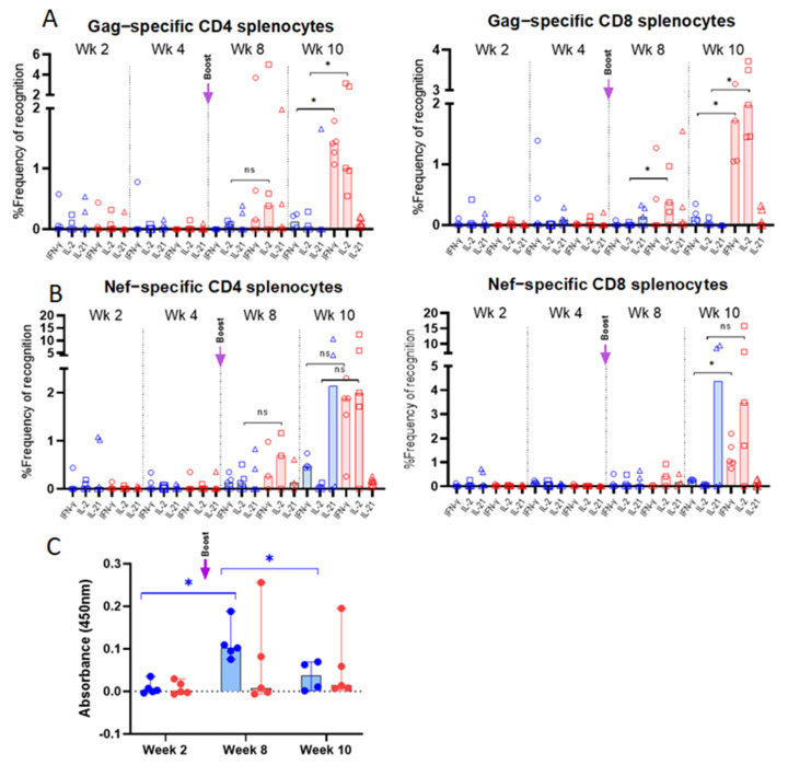
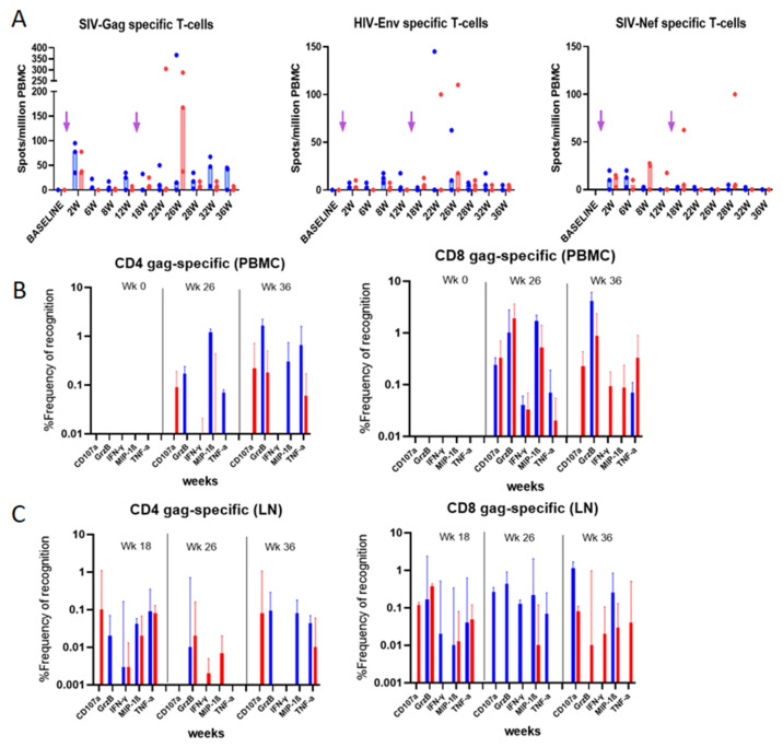
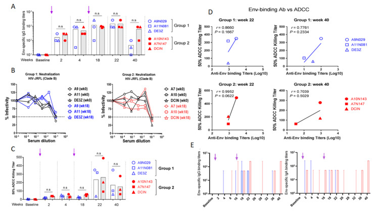
HIV-1 remains a major public health issue worldwide in spite of efficacious antiviral therapies, but with no cure or preventive vaccine. The latter has been very challenging, as virus infection is associated with numerous escape mechanisms from host specific immunity and the correlates of protection remain incompletely understood. We have developed an innovative vaccine strategy, inspired by the efficacy of live-attenuated virus, but with the safety of a DNA vaccine, to confer both cellular and humoral responses. The CAL-SHIV-IN- lentiDNA vaccine comprises the backbone of the pathogenic SHIVKU2 genome, able to mimic the early phase of viral infection, but with a deleted integrase gene to ensure safety precluding integration within the host genome. This vaccine prototype, constitutively expressing viral antigen under the CAEV LTR promoter, elicited a variety of vaccine-specific, persistent CD4 and CD8 T cells against SIV-Gag and Nef up to 80 weeks post-immunization in cynomolgus macaques. Furthermore, these specific responses led to antiviral control of the pathogenic SIVmac251. To further improve the efficacy of this vaccine, we incorporated the IL-7 or IL-15 genes into the CAL-SHIV-IN- plasmid DNA in efforts to increase the pool of vaccine-specific memory T cells. In this study, we examined the immunogenicity of the two co-injected lentiDNA vaccines CAL-SHIV-IN- IRES IL-7 and CAL-SHIV-IN- IRES IL-15 in BALB/cJ mice and rhesus macaques and compared the immune responses with those generated by the parental vaccine CAL-SHIV-IN-. This co-immunization elicited potent vaccine-specific CD4 and CD8 T cells both in mice and rhesus macaques. Antibody-dependent cell-mediated cytotoxicity (ADCC) antibodies were detected up to 40 weeks post-immunization in both plasma and mucosal compartments of rhesus macaques and were enhanced by the cytokines.

Keywords: CAEV LTR; DNA vaccine; HIV-1; IL-15; IL-7; LentiDNA; Rhesus macaques; SHIV.


Conflict of interest statement

The authors declare no conflict of interest.

Figures

Figure A1
Figure A1
Schematic representation of the CAL-SHIV-IN IRES cytokine lentiDNA vaccines: CAL-SHIV-IN was derived from the genome of the chimeric, highly pathogenic SHIVKU2 as previously described [1]. A 1000 bp cassette, containing IRES sequence and rhesus macaque cytokine gene IL-7 or IL-15, was inserted into the integrase-deleted site in CAL-SHIV-IN genome, ligated by the Age I-restricted extremities.
Figure A2
Figure A2
Immunization of BALB/cJ mice: Twenty mice per group were immunized with CAL-SHIV-IN vaccines (group 1) or co-immunized with CAL-SHIV-IN IRES IL-7 and CAL-SHIV-IN IRES IL-15 (group 2) by intradermal route + electroporation (ID/EP), and by intramuscular route (IM) as described in materials and methods. A homologous vaccination booster was administered 6 weeks later. At weeks 2, 4, 8 and 10 post-immunization, spleens were harvested to monitor and evaluate the cellular responses and blood samples for the detection of HIV-specific Abs in serum.
Figure A3
Figure A3
Gating strategy for mice splenocytes. Lymphocytes were gated among the splenocyte population, and alive, then single cells were selected. CD3+ cells were gated, and CD4+ or CD8+ cells isolated. Among CD4+ and CD8+, respectively, cytokine secretion (IL-2, IL-21 and IFN-γ) were characterized.
Figure A4
Figure A4
Immunization and sampling of vaccinated macaques: Three macaques per group were immunized with the CAL-SHIV-IN vaccine (group 1) or co-immunized with the CAL-SHIV-IN IRES IL-7 and CAL-SHIV-IN IRES IL-15 (group 2) by ID/EP and IM/EP. Homologous vaccination booster was administered 16 weeks later. Different samples were harvested to monitor and evaluate the immune responses up to 40 weeks.
Figure 1
Figure 1
Functional expression of SIV proteins. Co-culture of permissive M8166 lymphocyte cells with HEK 293T cells non-transfected (a), transfected with pNDgms-rmIL-15 (b) or transfected with CAL-SHIV-IN IRES-cytokines (c). CPEs were observed in the cultures with the CAL-SHIV-IN IRES-cytokines (black arrows). Supernatants of HEK 293T cells in (ac) were used to inoculate cultured M8166 in (df), respectively. At 24 h post-inoculation, CPEs were observed only in (f) (black arrows). (g) Quantification of SIV Gag p27 by ELISA: Supernatants of HEK 293T cells in a, b and c harvested at days 1, 2 and 3 post-transfection were analyzed by ELISA. Results were expressed in pg/mL.
Figure 2
Figure 2
Expression and functional analysis of secreted IL-7 and IL-15 cytokines: HEK 293T cells were transfected with either pNDgms-rmIL-7, pNDgms-rmIL-15 or with CAL-SHIV-IN IRES-rmIL-7 and CAL-SHIV-IN IRES-rmIL-15 alone or co-transfected with both CAL-SHIV-IN IRES cytokines. Vaccines with antisense cytokine cassette were used as negative controls. Supernatants of transfected cells were harvested at days 2, 3 and 5 and quantified for IL-7 (A) and IL-15 (B). Median values of two independent experiments were used to draw the columns. (C) Human PBMCs from a BCG-vaccinated donor were used in overnight and PHPC assay using the homeostatic cytokines. Quantified spots after 20 h stimulation with BCG vaccine are noted as ELISpot day 1. Spots quantified at 12 days stimulation with indicated cytokines (o): recombinant cytokines; (x): supernatants of pNDgms transfected HEK 293T cells; (Δ): supernatants of CAL-SHIV-IN IRES cytokines transfected HEK 293T cells. Median values with 95% CI of the 3 repeated experiments (day 12) are represented.
Figure 3
Figure 3
Flow cytometry characterization of vaccine-specific T cells in spleens of BALB/cJ-immunized mice: Splenocytes from group 1 (CAL-SHIV-IN; in blue) and group 2 mice (CAL-SHIV-IN IRES-rmIL-7 + CAL-SHIV-IN IRES-rmIL-15; in red), harvested at different time points post-immunization, were stimulated with SIV-Gag and -Nef peptides. Cells were stained with the combination of Abs reported in Table 1, to identify Gag (A) and Nef (B) specific CD4 and CD8 cytokine-secreting T-cells. Results were normalized with unstimulated splenocytes to remove the basal secretion, and median responses were plotted. Non-parametric Mann–Whitney test was calculated. Circles represent the percentage of IFN-γ secreting cells, squares the percentage of IL-2 secreting cells, and triangles the percentage of IL-21 secreting cells. (C) HIV Env-specific IgG in mouse serum: Serum samples from the two groups were analyzed by ELISA at 2, 8 and 10 weeks post-immunization for the detection of Env-specific IgG. O.D. (450 nm) are presented after blank and baseline subtraction. Median values with 95% CI are shown. Parametric Tukey’s multiple comparison test was calculated. Significant increase (p = 0.0160) was observed between weeks 2 and 8, and decrease (p = 0.0382) was observed between weeks 8 and 10 for group 1. No statistical significance was observed for group 2. ns: not significant; *: p < 0.05.
Figure 4
Figure 4
Vaccine-specific T-cell responses in blood and LN mononuclear cells from immunized rhesus macaques: (A) IFN-γ ELISpot responses to Gag, Env and Nef following overnight stimulation. Unstimulated cells were used as negative control for normalization. Median of the 3 animals per group is represented. Arrows indicate immunization times. (B) PBMCs and (C) LN mononuclear cells were used for the ICS assay using the panel of Ab reported in Table 2. Frequencies of Gag-specific cells measured at indicated time points are reported. Median values with 95% CI of the proportion of cytokine-secreting cells are shown. CAL-SHIV-IN group is presented in blue, and CAL-SHIV-IN IRES-rmIL-7 + CAL-SHIV-IN IRES-rmIL-15 in red.
Figure 5
Figure 5
Vaccine-specific Abs in sera and mucosal secretions of vaccinated rhesus macaques. (A) Serum IgG samples harvested at indicated time points were evaluated by ELISA against inactivated HIV-1JR-FL virus. Mean titers are plotted following subtraction of blank and baseline values. Arrows indicate immunization times. (B) Neutralizing activity of serum Abs was evaluated using the serum neutralization assay against Tier 2 HIV-1JR-FL virus and TZM-bl indicator cells. IC50 was determined by measuring the luminescence after 48 h incubation. (C) Evaluation of ADCC activity. Sera from both groups of vaccinated monkeys were incubated with HIV-1 JR-FL to inoculate CEM.NKr-CCR5 CD4+ target T cells. Effector KHYG-1 NK cells were added at a 10:1 effector-to-target ratio. After cell lysis, RLU was measured and ADCC activity evaluated for IC50. Mean values are reported. (D) Comparative evaluation of serum-binding and ADCC specific to Env epitopes in vaccinated macaques. Values were plotted to determine the correlate. (E) Evaluation of Env-specific Abs in rectal secretions. Rectal secretions were collected at indicated time points and analyzed by ELISA to detect both HIV-specific IgG and IgA against inactivated HIV-1 JR-FL. Cutoff was determined by subtracting the OD values of blank and baseline and median titers with 95% CI are shown. Group 1 (CAL-SHIV-IN) is represented in blue, and group 2 (CAL-SHIV-IN IRES-rmIL-7 + CAL-SHIV-IN IRES-rmIL-15) in red. ns: not significant. Arrows indicate immunization times.

Similar articles

Cited by

References

    1. Whyte-Allman S.-K., Bendayan R. HIV-1 Sanctuary Sites—The Role of Membrane-Associated Drug Transporters and Drug Metabolic Enzymes. AAPS J. 2020;22:118. doi: 10.1208/s12248-020-00498-1. - DOI - PubMed
    1. Castro S., Colomer-Lluch M., Serra-Moreno R. Barriers for HIV Cure: The Latent Reservoir. AIDS Res. Hum. Retrovir. 2018;34:739–759. doi: 10.1089/aid.2018.0118. - DOI - PMC - PubMed
    1. Excler J.-L., Kim J.H. Novel prime-boost vaccine strategies against HIV-1. Expert Rev. Vaccines. 2019;18:765–779. doi: 10.1080/14760584.2019.1640117. - DOI - PubMed
    1. Sekaly R.-P. The failed HIV Merck vaccine study: A step back or a launching point for future vaccine development? J. Exp. Med. 2008;205:7–12. doi: 10.1084/jem.20072681. - DOI - PMC - PubMed
    1. McCarthy M. AIDS vaccine fails in Thai trial. Lancet. 2003;362:1728. doi: 10.1016/S0140-6736(03)14886-6. - DOI - PubMed