. 2022 Mar 8;27(6):1755.
doi: 10.3390/molecules27061755.

The Antagonizing Role of Heme in the Antimalarial Function of Artemisinin: Elevating Intracellular Free Heme Negatively Impacts Artemisinin Activity in Plasmodium falciparum

Affiliations

The Antagonizing Role of Heme in the Antimalarial Function of Artemisinin: Elevating Intracellular Free Heme Negatively Impacts Artemisinin Activity in Plasmodium falciparum

Pan Zhu et al. Molecules. .

Abstract

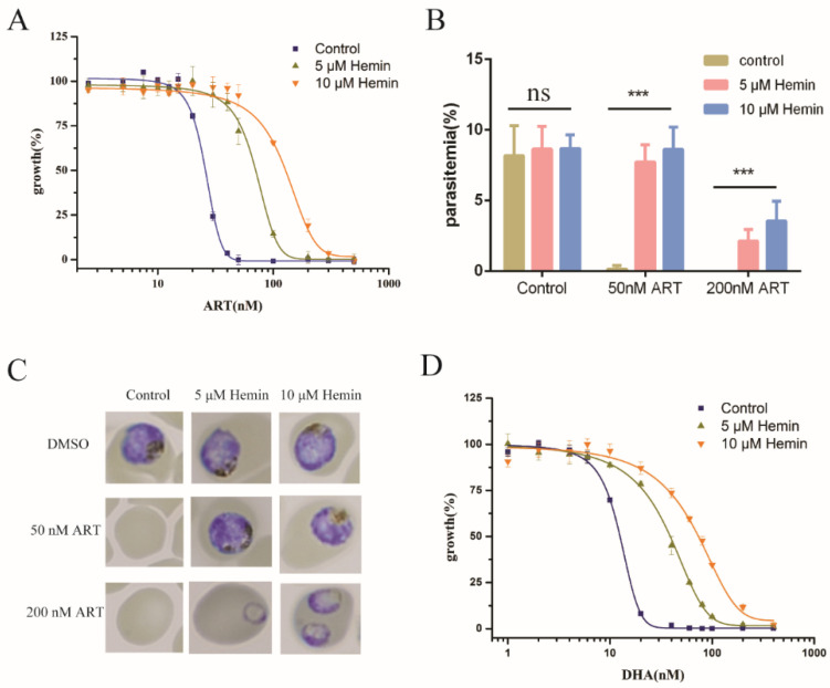
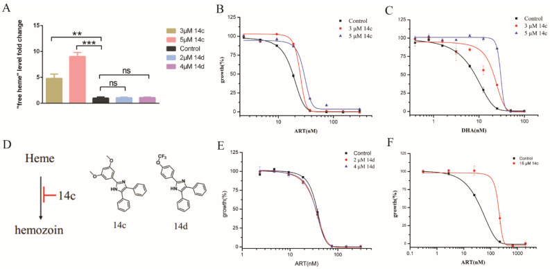
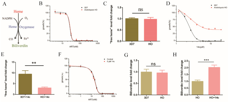
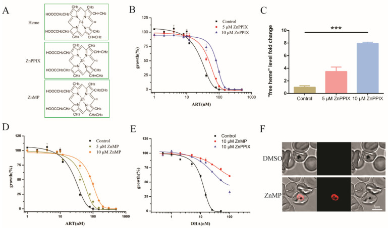
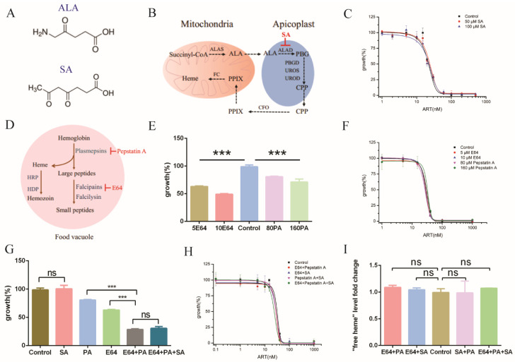
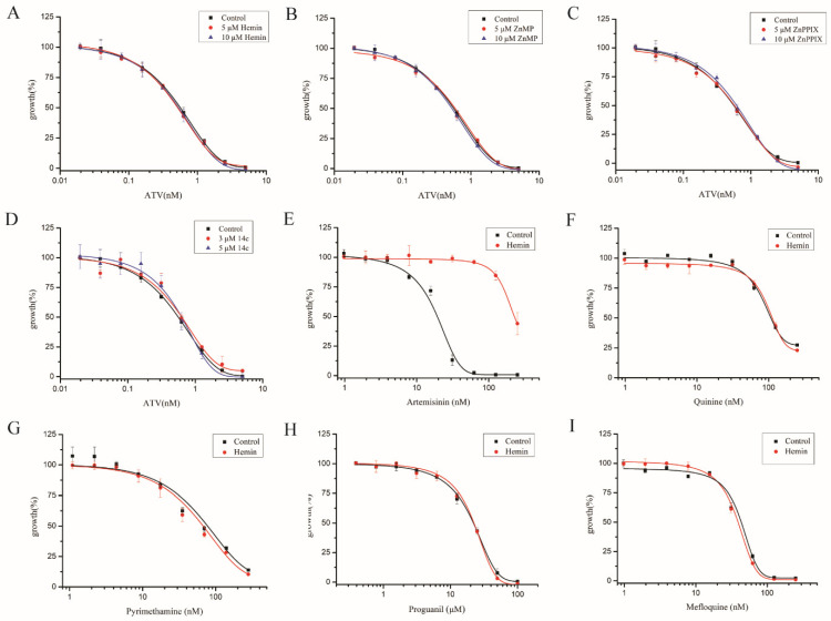
The rich source of heme within malarial parasites has been considered to underly the action specificity of artemisinin. We reasoned that increasing intraparasitic free heme levels might further sensitize the parasites to artemisinin. Various means, such as modulating heme synthesis, degradation, polymerization, or hemoglobin digestion, were tried to boost intracellular heme levels, and under several scenarios, free heme levels were significantly augmented. Interestingly, all results arrived at the same conclusion, i.e., elevating heme acted in a strongly negative way, impacting the antimalarial action of artemisinin, but exerted no effect on several other antimalarial drugs. Suppression of the elevated free heme level by introducing heme oxygenase expression effectively restored artemisinin potency. Consistently, zinc protoporphyrin IX/zinc mesoporphyrin, as analogues of heme, drastically increased free heme levels and, concomitantly, the EC50 values of artemisinin. We were unable to effectively mitigate free heme levels, possibly due to an unknown compensating heme uptake pathway, as evidenced by our observation of efficient uptake of a fluorescent heme homologue by the parasite. Our results thus indicate the existence of an effective and mutually compensating heme homeostasis network in the parasites, including an uncharacterized heme uptake pathway, to maintain a certain level of free heme and that augmentation of the free heme level negatively impacts the antimalarial action of artemisinin. Importance: It is commonly believed that heme is critical in activating the antimalarial action of artemisinins. In this work, we show that elevating free heme levels in the malarial parasites surprisingly negatively impacts the action of artemisinin. We tried to boost free heme levels with various means, such as by modulating heme synthesis, heme polymerization, hemoglobin degradation and using heme analogues. Whenever we saw elevation of free heme levels, reduction in artemisinin potency was also observed. The homeostasis of heme appears to be complex, as there exists an unidentified heme uptake pathway in the parasites, nullifying our attempts to effectively reduce intraparasitic free heme levels. Our results thus indicate that too much heme is not good for the antimalarial action of artemisinins. This research can help us better understand the biological properties of this mysterious drug.

Keywords: artemisinins; heme; heme oxygenase; hemozoin; triarylimidazole 14c.


Conflict of interest statement

The authors declare no conflict of interest.

Figures

Figure 1
Figure 1
Antimalarial effectiveness of artemisinin was reduced by exogenous hemin. (A) Inhibitory effect of artemisinin was decreased with the addition of exogenous hemin. (B) Hemin antagonized with artemisinin in inhibiting parasite multiplication, suppressing the parasitemia of P. falciparum. (C) Hemin released the inhibition of artemisinin on the development of the parasites after 72 h incubation. (D) Similarly to artemisinin, augmenting heme levels through hemin decreased sensitivity of the parasite to DHA. ART: artemisinin, DHA: dihydroartemisinin. Data are mean ± SD of three independent experiments. The data were analyzed by GraphPad Prism. *** p-value < 0.0005, and ns means no significant change (p-value > 0.05).
Figure 2
Figure 2
Hemozoin inhibition enhanced free heme levels and decreased sensitivity of artemisinin. (A) 14c but not 14d increased “free heme” levels in P. falciparum. Data are mean ± SD of three experiments. (B) EC50 of artemisinin was increased with 14c. (C) 14c obviously decreased sensitivity of parasites to dihydroartemisinin. (D) Chemical structure of 14c and 14d; (E) 14c analogue 14d had little effect on EC50 of artemisinin. (F) EC50 of artemisinin could also be significantly changed with higher dosages of 14c in the short-pulse assay. Early ring-stage parasites were used. Data are mean ± SD of three experiments. The data were analyzed by GraphPad Prism. ** p-value < 0.005, *** p-value < 0.0005, and ns = no significant change (p-value > 0.05).
Figure 3
Figure 3
HO neutralized 14c’s effect on heme and artemisinin. (A) HO executes the heme breakdown function. (B) The introduction of HO did not affect the EC50 value of artemisinin in transgenic parasites. (C) “Free heme” level in HO transgenic parasites had no significant change with 3D7 parasites. (D) HO transgenic parasites were more resistant to 14c than the wild-type parasite. (E) Arabidopsis HO1 mitigated the increase in free heme by 5 μM 14c. (F) HO expression in P. falciparum neutralized the antagonizing action of 14c on artemisinin. Data are mean ± SD of three experiments. (G) HO expression-strain-infected RBC had a similar level of biliverdin compared with 3D7-infected RBC. Data are mean ± SD of four experiments. (H) Treatment with 5 μM 14c enhanced biliverdin levels in HO expression-strain-infected RBC. The data were analyzed by GraphPad Prism. ** p-value < 0.005, *** p-value < 0.0005, and ns = no significant change (p-value > 0.05).
Figure 4
Figure 4
Heme analogues disrupted heme homeostasis and increased EC50 of artemisinin in P. falciparum. (A) Chemical structures of heme, ZnPPIX and ZnMP. (B) EC50 values of artemisinin in P. falciparum were increased by more than twofold in the presence of 10 μM ZnPPIX. (C) ZnPPIX elevated the free heme content by more than sixfold versus the control. (D) EC50 values of artemisinin were increased in the presence of ZnMP. (E) ZnPPIX and ZnMP decreased sensitivity of parasites to DHA. Data are mean ± SD of three experiments. (F) An amount of 20 μM ZnMP could efficiently reach into the parasite within an hour. The data were analyzed by GraphPad Prism. *** p-value < 0.0005.
Figure 5
Figure 5
Inhibiting heme biosynthesis and/or hemoglobin degradation had little effect on the effect of artemisinin. (A) Chemical structure of ALA and SA. (B) Heme de novo biosynthesis pathway in Plasmodium falciparum. (C) EC50 of artemisinin in P. falciparum was not changed by the addition of SA. (D) Hemoglobin degradation and hemozoin formation pathway in P. falciparum. (E) Amounts of 5 μM/10 μM E64 and 80 μM/160 μM pepstatin A inhibited the growth of parasites after 72 h incubation. (F) The EC50 value of artemisinin remained unaltered in the presence of cysteine protease inhibitor E64 or aspartic protease inhibitor pepstatin A. (G) An amount of 5 μM E64 was synergistic or additive with 80 μM pepstatin A on malaria growth inhibition. (H) Combinatory use of inhibitors (5 μM E64, 80 μM pepstatin A and 50 μM SA) still produced little effect on artemisinin after 72 h incubation. When three inhibitors were used together, a very mild or marginal effect on the sensitivity of artemisinin was seen. (I) Free heme levels in P. falciparum were not significantly changed with inhibitors (5 μM E64, 80 μM Pepstatin A and 50 μM SA) after 20 h incubation. Data are mean ± SD of three experiments. The data were analyzed by GraphPad Prism. *** p-value < 0.0005, and ns = no significant change (p-value > 0.05).
Figure 6
Figure 6
Heme had no effect on other antimalarial drugs. Parasites’ sensitivity to atovaquone had no significant change with 10 μM hemin (A), ZnPPIX (B), ZnMP (C) and 14c (D). Hemin decreased the sensitivity of artemisinin (E) but not antimalarial drugs quinine (F), pyrimethamine (G), mefloquine (H) and proguanil (I). ATV: atovaquone. Data are mean ± SD of three experiments.

References

    1. WHO . World Malaria Report 2018. WHO; Geneva, Switzerland: 2018.
    1. Björkman A., Phillips-Howard P.A. The epidemiology of drug-resistant malaria. Trans. R. Soc. Trop. Med. Hyg. 1990;84:177–180. doi: 10.1016/0035-9203(90)90246-B. - DOI - PubMed
    1. Wernsdorfer W.H., Payne D. The Dynamics of Drug Resistance in Plasmodium Falciparum. Pharmacol. Ther. 1991;50:95–121. doi: 10.1016/0163-7258(91)90074-V. - DOI - PubMed
    1. Wongsrichanalai C., Pickard A.L., Wernsdorfer W.H., Meshnick S.R. Epidemiology of drug-resistant malaria. Lancet Infect. Dis. 2002;2:209–218. doi: 10.1016/S1473-3099(02)00239-6. - DOI - PubMed
    1. Klayman D.L. Qinghaosu (artemisinin): An antimalarial drug from China. Science. 1985;228:1049–1055. doi: 10.1126/science.3887571. - DOI - PubMed

MeSH terms

LinkOut - more resources