. 2022 Mar 8;27(6):1777.
doi: 10.3390/molecules27061777.

Apigenin Isolated from Carduus crispus Protects against H2O2-Induced Oxidative Damage and Spermatogenic Expression Changes in GC-2spd Sperm Cells

Affiliations

Apigenin Isolated from Carduus crispus Protects against H2O2-Induced Oxidative Damage and Spermatogenic Expression Changes in GC-2spd Sperm Cells

Spandana Rajendra Kopalli et al. Molecules. .

Abstract

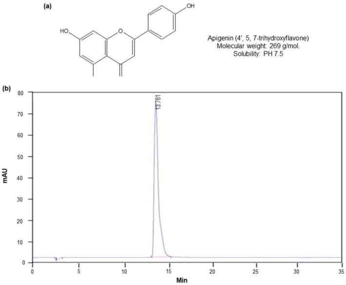
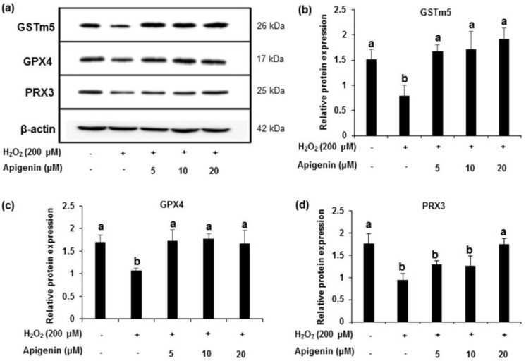
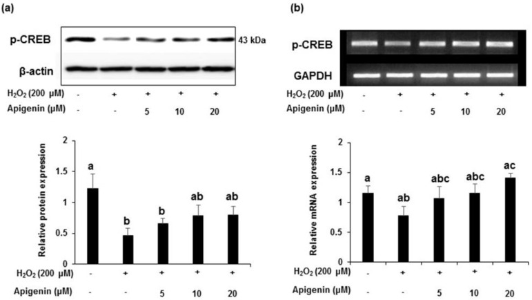
Testicular oxidative stress is one of the most common factors underlying male infertility. Welted thistle, Carduus crispus Linn., and its bioactive principles are attracting scientific interest in treating male reproductive dysfunctions. Here, the protective effects of apigenin isolated from C. crispus against oxidative damage induced by hydrogen peroxide (H2O2) and dysregulation in spermatogenesis associated parameters in testicular sperm cells was investigated. Cell viabilities, ROS scavenging effects, and spermatogenic associated molecular expressions were measured by MTT, DCF-DA, Western blotting and real-time RT-PCR, respectively. A single peak with 100% purity of apigenin was obtained in HPLC conditions. Apigenin treated alone (2.5, 5, 10 and 20 µM) did not exhibit cytotoxicity, but inhibited the H2O2-induced cellular damage and elevated ROS levels significantly (p < 0.05 at 5, 10 and 20 µM) and dose-dependently. Further, H2O2-induced down-regulation of antioxidant (glutathione S-transferases m5, glutathione peroxidase 4, and peroxiredoxin 3) and spermatogenesis-associated (nectin-2 and phosphorylated-cAMP response element-binding protein) molecular expression in GC-2spd cells were attenuated by apigenin at both protein and mRNA levels (p < 0.05). In conclusion, our study showed that apigenin isolated from C. crispus might be an effective agent that can protect ROS-induced testicular dysfunctions.

Keywords: CREB; apigenin; hydrogen peroxide; nectin; reactive oxygen species; testicular cells; welted thistle.


Conflict of interest statement

The authors declare no conflict of interest.

Figures

Figure 1
Figure 1
HPLC fingerprint of apigenin isolated from C. crispus. (a) Chemical name and structure of apigenin. (b) HPLC fingerprint of apigenin.
Figure 2
Figure 2
Effects of apigenin on cell viability in GC-2spd sperm cells. Cell viability was evaluated using the MTT assay. (a) The effect of apigenin (2.5, 5, 10, 20 and 40 μM) on the viability of GC-2spd cells. (b) The effect of apigenin (2.5, 5, 10 and 20 μM) in GC-2spd cells exposed to 200 μM hydrogen peroxide. Data are expressed as the mean ± standard deviation. The groups of control, H2O2, and different concentrations of apigenin were compared with each other, and letters on the top of the columns that do not share the same letters are statistically significant among the groups (p < 0.05) by one-way ANOVA.
Figure 3
Figure 3
Effect of apigenin on the inhibition of intracellular ROS formation. DCFH-DA fluorescence assay was performed and the fluorescence intensity (fold increase) in each treated group compared with control was shown. H2O2 (200 μM) treatment significantly increased the fluorescent intensity (fold increase) compared to control cells. Apigenin (2.5, 5, 10 and 20 μM) and positive control ascorbic acid (20 μM) treatment inhibited the H2O2-exposed increase in ROS generation by decreasing the florescence intensity in GC-2spd cells. Data are expressed as the mean ± standard deviation. The groups of control, H2O2, ascorbic acid, and different concentrations of apigenin were compared with each other and letters on the top of the columns that do not share the same letters are statistically significant among the groups (p < 0.05) by one-way ANOVA.
Figure 4
Figure 4
The effect of apigenin on the antioxidant enzyme protein expression level in H2O2-exposed cells. (a) The protein expression of GSTm5, GPX4 and PRX3 in H2O2-exposed GC-2spd cells was analyzed using Western blotting. Cell lysates from each groups were immunoblotted with specific antibodies. (bd) The protein band intensity of GSTm5, GPX4 and PRX3 in H2O2-exposed GC-2spd cells, respectively, normalized to that of β-actin is shown. The data represent the mean ± standard deviation. The groups of control, H2O2, and different concentrations of apigenin were compared with each other and letters on the top of the columns that do not share the same letters are statistically significant among the groups (p < 0.05) by one-way ANOVA. GSTm5, glutathione S-transferase m5; GPX4, glutathione peroxidase 4; PRX3, peroxiredoxin 3.
Figure 5
Figure 5
Effects of apigenin on the antioxidant enzyme mRNA expression level in hydrogen peroxide-exposed GC-2spd cells. (a) The mRNA expression level of the antioxidant enzymes GSTm5, GPX4 and PRX3 in H2O2-exposed GC-2spd cells. (bd) The polymerase chain reaction band intensity of GSTm5, GPX4 and PRX3 was analyzed using the ImageJ 1.41o software package and was normalized to that of glyceraldehyde 3-phosphate dehydrogenase. Data are expressed as the mean ± standard deviation. The groups of control, H2O2, and different concentrations of apigenin were compared with each other and letters on the top of the columns that do not share the same letters are statistically significant among the groups (p < 0.05) by one-way ANOVA. GSTm5, glutathione S-transferase m5; GPX4, glutathione peroxidase 4; PRX3, peroxiredoxin 3; GAPDH, glyceraldehyde 3-phosphate dehydrogenase.
Figure 6
Figure 6
The effect of apigenin on the expression level of nectin-2 in H2O2-exposed GC-2spd cells. (a) The protein expression level of nectin-2 was analyzed using Western blotting. Cell lysates were immunoblotted with specific antibodies with Beta-actin as the internal control (upper panel) and the band intensity of nectin-2 normalized to β-actin is shown in corresponding lower panel. (b) The mRNA expression of nectin-2 (upper panel) and corresponding band intensities normalized to GAPDH (lower panel). Data are expressed as the mean ± standard deviation. The groups of control, H2O2, and different concentrations of apigenin were compared with each other and letters on the top of the columns that do not share the same letters are statistically significant among the groups (p < 0.05) by one-way ANOVA.
Figure 7
Figure 7
The effect of apigenin on the expression level of p-CREB in H2O2-exposed GC-2spd cells. (a) The protein expression level of p-CREB was analyzed using Western blotting. Cell lysates were immunoblotted with specific antibodies with Beta-actin as the internal control (upper panel) and the band intensity of p-CREB normalized to β-actin is shown in corresponding lower panel. (b) The mRNA expression of p-CREB was shown in upper panel and corresponding band intensities normalized to GAPDH was shown in lower panel. Data are expressed as the mean ± standard deviation (n = 6). The groups of control, H2O2, and different concentrations of apigenin were compared with each other and letters on the top of the columns that do not share the same letters are statistically significant among the groups (p < 0.05) by one-way ANOVA.

References

    1. Vander Borght M., Wyns C. Fertility and infertility: Definition and epidemiology. Clin. Biochem. 2018;62:2–10. doi: 10.1016/j.clinbiochem.2018.03.012. - DOI - PubMed
    1. Agarwal A., Baskaran S., Parekh N., Cho C.-L., Henkel R., Vij S., Arafa M., Panner Selvam M.K., Shah R. Male infertility. Lancet. 2021;397:319–333. doi: 10.1016/S0140-6736(20)32667-2. - DOI - PubMed
    1. Chen L., Shi G., Huang D., Li Y., Ma C., Shi M., Su B., Shi G. Male sexual dysfunction: A review of literature on its pathological mechanisms, potential risk factors, and herbal drug intervention. Biomed. Pharmacother. 2019;112:108585. doi: 10.1016/j.biopha.2019.01.046. - DOI - PubMed
    1. Dutta S., Sengupta P., Slama P., Roychoudhury S. Oxidative Stress, Testicular Inflammatory Pathways, and Male Reproduction. Int. J. Mol. Sci. 2021;22:10043. doi: 10.3390/ijms221810043. - DOI - PMC - PubMed
    1. Fatima S. Novel Prospects in Oxidative and Nitrosative Stress. InTechOpen; London, UK: 2018. Role of Reactive Oxygen Species in Male Reproduction.

LinkOut - more resources