Review
. 2022 Mar 14;14(6):1153.
doi: 10.3390/polym14061153.

Poly-l-Lactic Acid (PLLA)-Based Biomaterials for Regenerative Medicine: A Review on Processing and Applications

Affiliations
Review

Poly-l-Lactic Acid (PLLA)-Based Biomaterials for Regenerative Medicine: A Review on Processing and Applications

Elisa Capuana et al. Polymers (Basel). .

Abstract

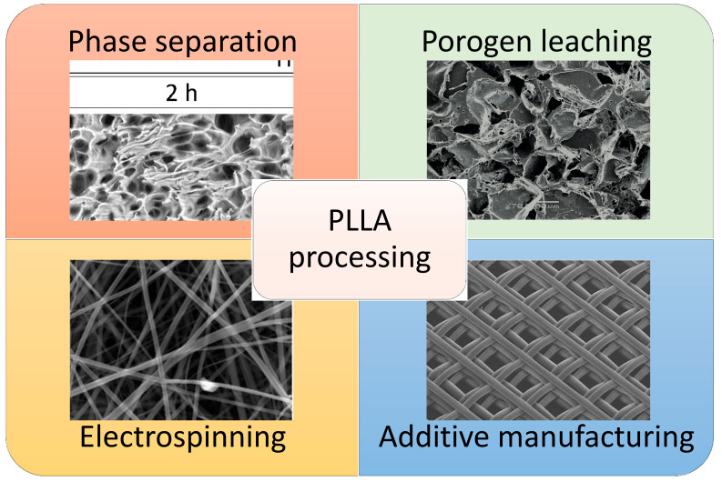
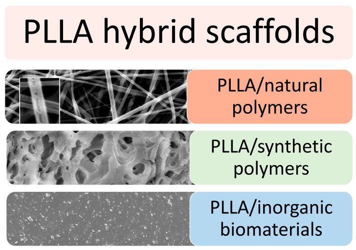
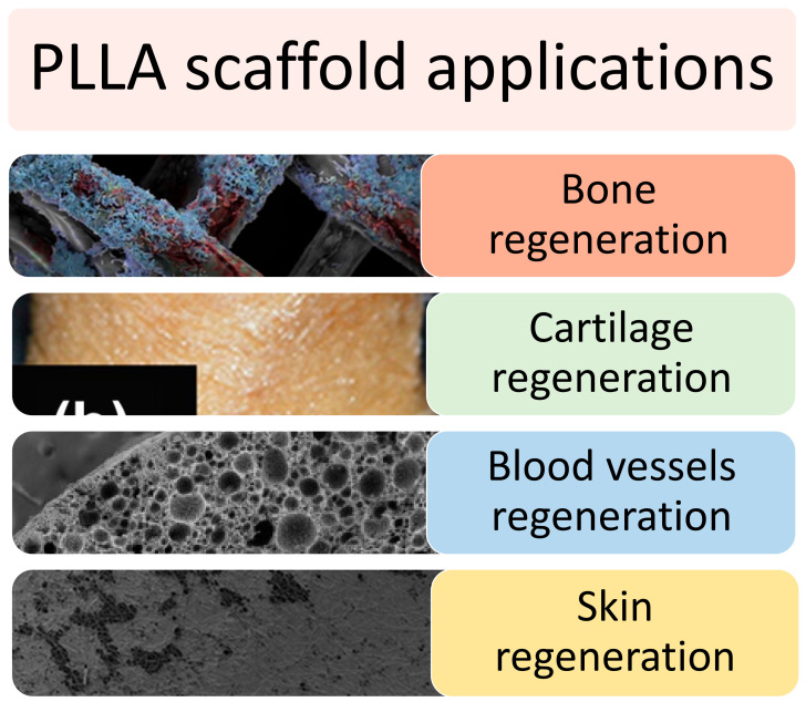
Synthetic biopolymers are effective cues to replace damaged tissue in the tissue engineering (TE) field, both for in vitro and in vivo application. Among them, poly-l-lactic acid (PLLA) has been highlighted as a biomaterial with tunable mechanical properties and biodegradability that allows for the fabrication of porous scaffolds with different micro/nanostructures via various approaches. In this review, we discuss the structure of PLLA, its main properties, and the most recent advances in overcoming its hydrophobic, synthetic nature, which limits biological signaling and protein absorption. With this aim, PLLA-based scaffolds can be exposed to surface modification or combined with other biomaterials, such as natural or synthetic polymers and bioceramics. Further, various fabrication technologies, such as phase separation, electrospinning, and 3D printing, of PLLA-based scaffolds are scrutinized along with the in vitro and in vivo applications employed in various tissue repair strategies. Overall, this review focuses on the properties and applications of PLLA in the TE field, finally affording an insight into future directions and challenges to address an effective improvement of scaffold properties.

Keywords: poly-l-lactic acid (PLLA); regenerative medicine; tissue engineering.


Conflict of interest statement

The authors declare no conflict of interest.

Figures

Figure 1
Figure 1
Enantiomeric forms of lactic acid [36]. Reprinted with permission from Frontiers.
Figure 2
Figure 2
(d) Obtained as-spun PLLA fiber reinforced with BaTiO3 particles by the pilot-scale melt-spinning, and (e) FE-SEM image of the as-spun PLLA/BaTiO3 fibers. Reprinted with permission from Nature Publishing Group [64].
Figure 3
Figure 3
SEM images of the surface and interior of PLLA/rifampicin blend particles prepared by the drop freeze-drying method. (a) PLLA = 2.0 wt%, surface, (b) PLLA = 2.0 wt%, interior, (c) PLLA = 4.9 wt%, surface, and (d) PLLA = 4.9 wt%, interior. Reprinted with permission from Hindawi Limited.
Figure 4
Figure 4
Confocal laser images of SMCs grown on SF/PLLA-CL/PRGF and SF/PLLA-CL for 1, 4, and 7 days. Reprinted with permission from Oxford Academic [73].
Figure 5
Figure 5
Pore morphologies of PLLA-based scaffolds prepared with some of the most used processing techniques. Reprinted with permission from MDPI [80,81], Hindawi [81], and Elsevier [82].
Figure 6
Figure 6
SEM images of different samples. Poly(L-lactide) (PLLA) (A), PLLA@PDA (B), PLLA@Ag1 (C), PLLA@Ag3 (D), PLLA@Ag6 (E), PLLA@Ag9 (F,G), and PLLA@Ag 24 (H,I). Reprinted with permission from Frontiers [130].
Figure 7
Figure 7
Microstructure of PLLA-based hybrid scaffold in combination with natural polymers, synthetic polymers or inorganic biomaterials. Reprinted with permission from RSC [135] and MDPI [136,137].
Figure 8
Figure 8
Three-dimensional electron micrograph images of (A) unseeded hybrid PLLA/PCL scaffolds, (B) hiPSCs-seeded scaffold on starting day, (C) induction cells to assembling, (D) induced cells to aggregate, and (E) islet-like clusters. Scale bars are (A): 10 mm; (BE): 100 mm [106]. Reprinted with permission from Taylor & Francis.
Figure 9
Figure 9
SEM micrographs of the scaffold prepared with a 90/10 PLLA/HA ratio at different demixing temperatures, keeping the demixing time constant. (A) 25 °C; (B) 30 °C; (C) 35 °C. Reprinted with permission from Elsevier [97].
Figure 10
Figure 10
Effect of guidance cues on the alignment of OEC. (A) Aligned SWCNT/PLLA nanofibers used as the substratum for OEC, scale bar = 10 µm; (B) OEC grown on culture plates showing random orientation, magnification 100×; (C) SEM micrographs of OEC aligned on nanofiber SWCNT/PLLA scaffolds, scale bar = 2 µm; (D) Fluorescence image of aligned OEC grown on SWCNT/PLLA nanofibrous scaffolds, magnification 100×. Reprinted with permission from EXCLI Journal [130].
Figure 11
Figure 11
Structure of PLLA-based scaffold designed for bone, cartilage, blood vessel, and skin regeneration. Reprinted with permission from Elsevier [82], Taylor & Francis [113], Dovepress [161], and RSC [162].
Figure 12
Figure 12
H&E histological analysis after 8 weeks of subcutaneous implantation at 100× magnification. Small pore scaffold (60–125 µm) (A) contained cartilage with typical morphology in the center of the scaffold. Small (125–250 µm), (B) medium (250–425 µm), (C) large-pore (425–600 µm), and (D) scaffold-supported bone formation on pore walls, shown by pink staining of bone matrix, with bone-marrow-like tissue within the pores. N = 3 for each group. Scale bars = 200 μm. Reprinted with permission from Acta Materialia Inc. [90].
Figure 13
Figure 13
The SEM images and pore diameter distributions of PLLA/PLGA/PCL composite scaffolds with various weight ratios. Reprinted with permission from Dove Press [161].

References

    1. Zhao P., Gu H., Mi H., Rao C., Fu J., Turng L.S. Fabrication of scaffolds in tissue engineering: A review. Front. Mech. Eng. 2018;13:107–119. doi: 10.1007/s11465-018-0496-8. - DOI
    1. Scaffaro R., Lopresti F., Botta L., Rigogliuso S., Ghersi G. Melt Processed PCL/PEG Scaffold With Discrete Pore Size Gradient for Selective Cellular Infiltration. Macromol. Mater. Eng. 2016;301:182–190. doi: 10.1002/mame.201500289. - DOI
    1. Scaffaro R., Lopresti F., Maio A., Botta L., Rigogliuso S., Ghersi G. Electrospun PCL/GO-g-PEG structures: Processing-morphology-properties relationships. Compos. Part A Appl. Sci. Manuf. 2017;92:97–107. doi: 10.1016/j.compositesa.2016.11.005. - DOI
    1. Scaffaro R., Lopresti F., Botta L. Preparation, characterization and hydrolytic degradation of PLA/PCL co-mingled nanofibrous mats prepared via dual-jet electrospinning. Eur. Polym. J. 2017;96:266–277. doi: 10.1016/j.eurpolymj.2017.09.016. - DOI
    1. Shafiee A., Atala A. Tissue Engineering: Toward a New Era of Medicine. Annu. Rev. Med. 2017;68:29–40. doi: 10.1146/annurev-med-102715-092331. - DOI - PubMed

LinkOut - more resources