. 2022 Mar 1;11(3):308.
doi: 10.3390/pathogens11030308.

Sexual Dimorphism of the Neuroimmunoendocrine Response in the Spleen during a Helminth Infection: A New Role for an Old Player?

Affiliations

Sexual Dimorphism of the Neuroimmunoendocrine Response in the Spleen during a Helminth Infection: A New Role for an Old Player?

Karen Elizabeth Nava-Castro et al. Pathogens. .

Abstract

The interaction of the nervous, immune, and endocrine systems is crucial in maintaining homeostasis in vertebrates, and vital in mammals. The spleen is a key organ that regulates the neuroimmunoendocrine system. The Taenia crassiceps mouse system is an excellent experimental model to study the complex host-parasite relationship, particularly sex-associated susceptibility to infection. The present study aimed to determine the changes in neurotransmitters, cytokines, sex steroids, and sex-steroid receptors in the spleen of cysticercus-infected male and female mice and whole parasite counts. We found that parasite load was higher in females in comparison to male mice. The levels of the neurotransmitter epinephrine were significantly decreased in infected male animals. The expression of IL-2 and IL-4 in the spleen was markedly increased in infected mice; however, the expression of Interleukin (IL)-10 and interferon (IFN)-γ decreased. We also observed sex-associated differences between non-infected and infected mice. Interestingly, the data show that estradiol levels increased in infected males but decreased in females. Our studies provide evidence that infection leads to changes in neuroimmunoendocrine molecules in the spleen, and these changes are dimorphic and impact the establishment, growth, and reproduction of T. crassiceps. Our findings support the critical role of the neuroimmunoendocrine network in determining sex-associated susceptibility to the helminth parasite.

Keywords: Helminths; Taenia crassiceps; cysticercosis; cytokines; immunity; infection; neuroimmunoendocrinology; neurotransmitters; parasite immunity; sexual dimorphism; spleen.


Conflict of interest statement

The authors declare no conflict of interest.

Figures
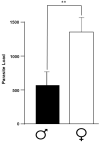
Figure 1
Figure 1
Parasite load in infected mice of both sexes. Data show the average of the number of parasites recovered from the peritoneal cavity of 10 female (white bar) and 10 male (black bar) BALB/c mice at 16 weeks post-infection. Each bar represents the average +/− SD in every sex at 16 weeks post-infection. **, p < 0.001 when comparing male and female infected mice.
Figure 2
Figure 2
Splenic levels of neurotransmitters in the control groups and in female and male mice infected with T. crassiceps. The levels of NE, EP, DOPA, and 5HT were measured in the spleen of the different experimental groups. Data show the levels of neurotransmitters corresponding to 20 female (10 controls and 10 infected) and 20 male (10 controls and 10 infected) BALB/c mice at 16 weeks post-infection The black lines show differences between gender, the red lines show differences between control and infected animals. p < 0.05 *, p < 0.01 **. Abbreviations: ND, Not Detected, NE, norepinephrine; EP, epinephrine; DOPA, dopamine; 5-HT, serotonin; C, uninfected control; I, infected. Significance in black indicates difference between the control groups of both sexes. Significance in red color indicates difference between the control group and the infected group of the same sex.
Figure 3
Figure 3
Levels of splenic IL-2, IL-4, and IL-10 in the control groups and in male and female mice infected with T. crassiseps. Data show the levels of different cytokines corresponding to 20 female (10 controls and 10 infected) and 20 male (10 controls and 10 infected) BALB/c mice at 16 weeks. The black lines show differences between gender; the red lines show differences between control and infected animals. p < 0.05 *, p < 0.01 **. Significance in black indicates difference between the control groups of both sexes. Significance in red color indicates difference between the control group and the infected group of the same sex.
Figure 4
Figure 4
Levels of splenic IFNγ, TNF-α, and IL-6 in the control groups and in male and female mice infected with T. crassiseps. Data show the levels of different cytokines corresponding to 20 female (10 controls and 10 infected) and 20 male (10 controls and 10 infected) BALB/c mice at 16 weeks. The black lines show differences between gender; the red lines show differences between control and infected animals. p < 0.05 *, p < 0.01 **. Significance in black indicates difference between the control groups of both sexes. Significance in red color indicates difference between the control group and the infected group of the same sex.
Figure 5
Figure 5
Splenic levels of steroid hormones, progesterone, estradiol, and testosterone, in the control groups and in male and female mice infected with T. crassiseps. Data show the levels of different hormones corresponding to 20 female (10 controls and 10 infected) and 20 male (10 controls and 10 infected) BALB/c mice at 16 weeks post-infection, p < 0.001 ** when comparing the control group and infected individuals. The black lines show differences between gender, the red lines show differences between control and infected animals. p < 0.01 **. Significance in black indicates difference between the control groups of both sexes. Significance in red color indicates difference between the control group and the infected group of the same sex.
Figure 6
Figure 6
Splenic levels of the different steroid receptors, ERα, ERβ, PR, and AR, evaluated in male and female mice non-infected and infected with T. crassiseps. A representative image (A) and the densitometry analysis of the different experiments (B) are shown. β-actin was used as the loading control. Normalization of values was carried out against the loading control. 20 female (10 controls and 10 infected) and 20 male (10 controls and 10 infected) BALB/c mice at 16 weeks were used. The black lines show differences between gender. p < 0.05 *.

References

    1. Larralde C., Sciutto E., Huerta L., Terrazas I., Fragoso G., Trueba L., Lemus D., Lomelí C., Tapia G., Montoya R.M. Experimental cysticercosis by Taenia crassiceps in mice: Factors involved in susceptibility. Acta Leiden. 1989;57:131–134. - PubMed
    1. Larralde C., Sotelo J., Montoya R.M., Palencia G., Padilla A., Govezensky T., Diaz M.L., Sciutto E. Immunodiagnosis of human cysticercosis in cerebrospinal fluid. Antigens from murine Taenia crassiceps cysticerci effectively substitute those from porcine Taenia solium. Arch. Pathol. Lab. Med. 1990;114:926–928. - PubMed
    1. Rishi A.K., McManus D.P. Molecular cloning of Taenia solium genomic DNA and characterization of taeniid cestodes by DNA analysis. Parasitology. 1988;97:161–176. doi: 10.1017/S003118200006683X. - DOI - PubMed
    1. Larralde C., Laclette J.P., Montoya R.M., Contreras L., Sandoval M., Bojalil R., Owen C.S., Arzate J., Goodsaid F., Diaz M.L., et al. Reliable Serology of Taenia solium Cysticercosis with Antigens from Cyst Vesicular Fluid: Elisa and Hemagglutination Tests. Am. J. Trop. Med. Hyg. 1986;35:965–973. doi: 10.4269/ajtmh.1986.35.965. - DOI - PubMed
    1. Toledo A., Fragoso G., Rosas G., Hernández M., Gevorkian G., López-Casillas F., Hernández B., Acero G., Huerta M., Larralde C., et al. Two Epitopes Shared by Taenia crassiceps and Taenia solium Confer Protection against Murine T. crassiceps Cysticercosis along with a Prominent T1 Response. Infect. Immun. 2001;69:1766–1773. doi: 10.1128/IAI.69.3.1766-1773.2001. - DOI - PMC - PubMed

LinkOut - more resources