. 2022 Feb 25;14(3):508.
doi: 10.3390/pharmaceutics14030508.

Iodine Nanoparticles (Niodx) for Radiotherapy Enhancement of Glioblastoma and Other Cancers: An NCI Nanotechnology Characterization Laboratory Study

Affiliations

Iodine Nanoparticles (Niodx) for Radiotherapy Enhancement of Glioblastoma and Other Cancers: An NCI Nanotechnology Characterization Laboratory Study

James F Hainfeld et al. Pharmaceutics. .

Abstract

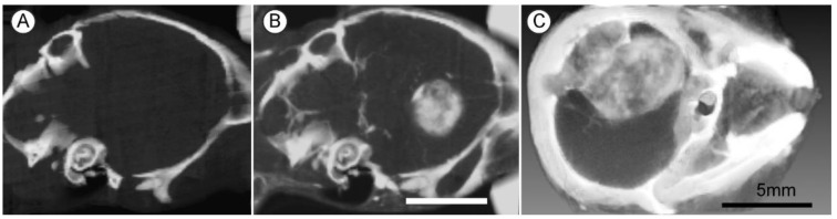
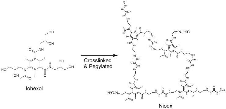
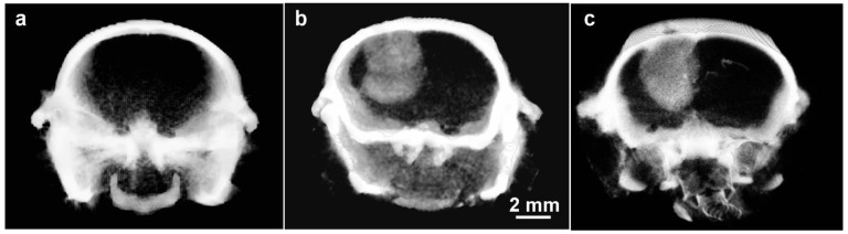
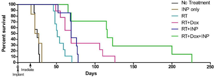
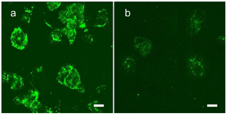
Effective and durable treatment of glioblastoma is an urgent unmet medical need. In this article, we summarize a novel approach of a physical method that enhances the effectiveness of radiotherapy. High atomic number nanoparticles that target brain tumors are intravenously administered. Upon irradiation, the nanoparticles absorb X-rays creating free radicals, increasing the tumor dose several fold. Radiotherapy of mice with orthotopic human gliomas and human triple negative breast cancers growing in the brain showed significant life extensions when the nanoparticles were included. An extensive study of the properties of the iodine-containing nanoparticle (Niodx) by the Nanotechnology Characterization Laboratory, including sterility, physicochemical characterization, in vitro cytotoxicity, in vivo immunological characterization, and in vivo toxicology, is presented. In summary, the iodine nanoparticle Niodx appears safe and effective for translational studies toward human use.

Keywords: brain metastases; brain tumors; breast cancer; cancer; dose enhancement; glioma; iodine; iodine nanoparticles; nanoparticles; radiotherapy.


Conflict of interest statement

James Hainfeld is a part owner of Nanoprobes, Inc. Other authors declare no conflicts of interest.

Figures

Figure 1
Figure 1
High-Z atoms absorb X-rays and eject electrons creating tissue-damaging free radicals.
Figure 2
Figure 2
X-rays that would normally pass through tissue are instead absorbed by high atomic number nanoparticles and the energy deposited locally, boosting local dose [11].
Figure 3
Figure 3
Iohexol (Omnipaque®). Molecular weight 821, 46.4% iodine by weight.
Figure 4
Figure 4
Survival graph showing 86% survival at 1 year after tumor loading with intravenous gold nanoparticles (AuNPs).
Figure 5
Figure 5
Live mouse microCT images of gliomas 9 days post implantation. (A,B) Same mouse before (A) and 15 h after (B) intravenous (IV) injection (4 g Au/kg); (C) Larger tumor imaged 15 h after IV injection of 1.7 g Au/kg. X-ray source was 45 kVp. Bars (B,C) = 5 mm.
Figure 6
Figure 6
Survival graph of mice bearing invasive orthotopic gliomas showing rapid demise with no treatment, no benefit from AuNPs only, some life extension with radiotherapy (RT) only, and 50% long-term survival (no tumor detectable after 1 year) when RT was combined with a prior IV injection of AuNPs. Durable responses seen here may have been due, in part, to a functional immune system.
Figure 7
Figure 7
(a) Normal nude mouse; (b) nude mouse 24 h after tail vein injection of 1 g/kg 15nm PEG coated AuNPs. Skin color change was almost immediate after AuNP injection and showed little change even after 1 year; (c) nude mouse 24 h after intravenous injection of 4 g iodine/kg iodine nanoparticles.
Figure 8
Figure 8
(a) Electron micrograph; (b) dynamic light scattering; and (c) schematic of the iodine nanoparticles (Niodx).
Figure 9
Figure 9
Niodx is made from crosslinked iohexol with a PEG coating.
Figure 10
Figure 10
MicroCT images of orthotopic gliomas growing in brains of athymic mice. (a) Before Niodx injection; (b) same mouse 24 h after intravenous injection (Niodx, 3.5 g iodine/kg); (c) Different mouse but taken 3 days after IV Niodx injection. X-ray source was 70 kVp.
Figure 11
Figure 11
Kaplan–Meier survival graph showing survival vs. days after implantation of orthotopic U87 tumors. RT only (15 Gy, n = 6) extended life on average (at the 50% level, small blue arrow) by 16 days (compared to no treatment, n = 8), but 7 g I/kg Niodx + 15 Gy (n = 8) extended life by 38 days (2.4-fold). Inset shows the Niodx without radiation had no effect on survival. X-ray source was 100 kVp.
Figure 12
Figure 12
Histological section of an orthotopic human U87 glioma growing in the mouse brain. (a) is a coronal section of the whole brain with tumor in upper left. Green is a fluorescent secondary antibody immunolabeling an anti-PEG antibody, specific for the PEGylated iodine nanoparticle. The Niodx targets the tumor and the surrounding edematous growing region. Blue is DAPI, staining nuclei. (b) is a higher magnification of the tumor region. Niodx highly stains endothelium surrounding all tumor capillaries (round-like “holes” in image). Bars: left image 1 mm, right image 50 μm.
Figure 13
Figure 13
Survival graph showing results with Doxil, Niodx (INP), and radiotherapy (RT, 15 Gy). Groups: no treatment (n = 7), INP only (n = 6), RT (n = 9), RT + INP (n = 7), RT+Dox (n = 9), and RT + Dox + INP (n = 7). X-ray source was 100 kVp.
Figure 14
Figure 14
Uptake of Niodx targeted with transferrin (a) and non-targeted Niodx (b) 16 h after in vitro incubation with U87 human glioma cells. The uptake of iodine is about 5 times with the transferrin targeting in this study. Bars = 20 μm.
Figure 15
Figure 15
Gliomas and metastatic tumors grow differently in the brain as shown by (a) MRI scan of patient with GBM, (b) MRI scan of patient with triple negative breast cancer that has metastasized to the brain, (c) U87 glioma growing in the brain of an athymic mouse, and (d) human triple negative breast cancer growing in the brain of an athymic mouse. (c,d) are microCT X-ray images (70 kVp) 24 h after IV Niodx injection.
Figure 16
Figure 16
Survival graph showing no treatment (blue), RT only (15 Gy, 100 kVp, green), and INP + RT (red). n denotes number of animals per group. Arrow indicates day of irradiation.
Figure 17
Figure 17
Confocal cryosection image of advanced human triple negative breast cancer tumor (MDA-MB-231) growing in athymic mouse brain 24 h after IV administration of Niodx. Tumor cells have been transduced with red fluorescent protein (td-tomato, red), and labeled for Niodx using an anti-PEG antibody (Alexa-488, green). No endothelial cells are present, and black spaces surrounded by tumor cells (red) are vascular channels formed by the tumor cells mimicking endothelial cells. Niodx shows strong binding to these channels [37]. Bar = 20 μm.
Figure 18
Figure 18
Absorption of iodine vs. gold and soft tissue. In a useful range, iodine absorbs 1.7 times more than gold and at iodine’s K-edge, it absorbs ~100 times more than tissue (on a weight basis). Absorption of high-Z elements compared to tissue is better in the kV range rather than the MV range.

References

    1. Karlsson J., Luly K.M., Tzeng S.Y., Green J.J. Nanoparticle designs for delivery of nucleic acid therapeutics as brain cancer therapies. Adv. Drug Deliv. Rev. 2021;179:113999. doi: 10.1016/j.addr.2021.113999. - DOI - PMC - PubMed
    1. Sevastre A.S., Costachi A., Tataranu L.G., Brandusa C., Artene S.A., Stovicek O., Alexandru O., Danoiu S., Sfredel V., Dricu A. Glioblastoma pharmacotherapy: A multifaceted perspective of conventional and emerging treatments (Review) Exp. Ther. Med. 2021;22:1408. doi: 10.3892/etm.2021.10844. - DOI - PMC - PubMed
    1. Boateng F., Ngwa W. Delivery of Nanoparticle-Based Radiosensitizers for Radiotherapy Applications. Int. J. Mol. Sci. 2019;21:273. doi: 10.3390/ijms21010273. - DOI - PMC - PubMed
    1. Butterworth K.T., McMahon S.J., Currell F.J., Prise K.M. Physical basis and biological mechanisms of gold nanoparticle radiosensitization. Nanoscale. 2012;4:4830–4838. doi: 10.1039/c2nr31227a. - DOI - PubMed
    1. Howard D., Sebastian S., Le Q.V., Thierry B., Kempson I. Chemical Mechanisms of Nanoparticle Radiosensitization and Radioprotection: A Review of Structure-Function Relationships Influencing Reactive Oxygen Species. Int. J. Mol. Sci. 2020;21:579. doi: 10.3390/ijms21020579. - DOI - PMC - PubMed

LinkOut - more resources