

Programmed Catalytic Therapy-Mediated ROS Generation and T-Cell Infiltration in Lung Metastasis by a Dual Metal-Organic Framework (MOF) Nanoagent
- PMID: 35335903
- PMCID: PMC8955711
- DOI: 10.3390/pharmaceutics14030527
Programmed Catalytic Therapy-Mediated ROS Generation and T-Cell Infiltration in Lung Metastasis by a Dual Metal-Organic Framework (MOF) Nanoagent
Abstract
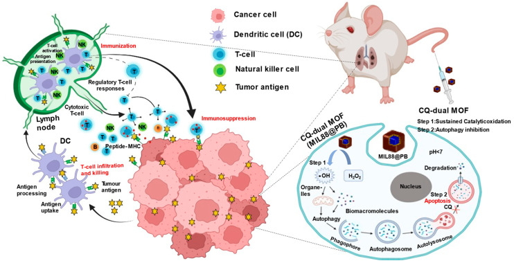
Nano-catalytic agents actuating Fenton-like reaction in cancer cells cause intratumoral generation of reactive oxygen species (ROS), allowing the potential for immune therapy of tumor metastasis via the recognition of tumor-associated antigens. However, the self-defense mechanism of cancer cells, known as autophagy, and unsustained ROS generation often restricts efficiency, lowering the immune attack, especially in invading metastatic clusters. Here, a functional core-shell metal-organic framework nanocube (dual MOF) doubling as a catalytic agent and T cell infiltration inducer that programs ROS and inhibits autophagy is reported. The dual MOF integrated a Prussian blue (PB)-coated iron (Fe2+)-containing metal-organic framework (MOF, MIL88) as a programmed peroxide mimic in the cancer cells, facilitating the sustained ROS generation. With the assistance of Chloroquine (CQ), the inhibition of autophagy through lysosomal deacidification breaks off the self-defense mechanism and further improves the cytotoxicity. The purpose of this material design was to inhibit autophagy and ROS efficacy of the tumor, and eventually improve T cell recruitment for immune therapy of lung metastasis. The margination and internalization-mediated cancer cell uptake improve the accumulation of dual MOF of metastatic tumors in vivo. The effective catalytic dual MOF integrated dysfunctional autophagy at the metastasis elicits the ~3-fold recruitment of T lymphocytes. Such synergy of T cell recruitment and ROS generation transported by dual MOF during the metastases successfully suppresses more than 90% of tumor foci in the lung.
Keywords: MOF; autophagy; drug delivery; immune response; lung metastasis; nano-catalytic medicine.
Conflict of interest statement
The authors declare no conflict of interest, financial or otherwise.
Figures

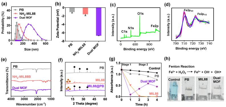
 ), MIL88(
 ), and dual MOF. Slight disappearances reflections of 2-Theta and intensities in the dual MOF were due to the shielding effect of MIL88 on PB. (g,h) Catalytic performance of control, PB, MIL88, and dual MOF.


Similar articles
-
Programmed T cells infiltration into lung metastases with harnessing dendritic cells in cancer immunotherapies by catalytic antigen-capture sponges.J Control Release. 2023 Aug;360:260-273. doi: 10.1016/j.jconrel.2023.06.033. Epub 2023 Jun 29. J Control Release. 2023. PMID: 37364798
-
A Metal-Organic Framework (MOF) Fenton Nanoagent-Enabled Nanocatalytic Cancer Therapy in Synergy with Autophagy Inhibition.Adv Mater. 2020 Mar;32(12):e1907152. doi: 10.1002/adma.201907152. Epub 2020 Feb 13. Adv Mater. 2020. PMID: 32053261
-
A Self-Cascading Catalytic Therapy and Antigen Capture Scaffold-Mediated T Cells Augments for Postoperative Brain Immunotherapy.Small. 2025 Feb;21(5):e2406178. doi: 10.1002/smll.202406178. Epub 2024 Dec 15. Small. 2025. PMID: 39676476
-
Dual Role of Reactive Oxygen Species and their Application in Cancer Therapy.J Cancer. 2021 Jul 25;12(18):5543-5561. doi: 10.7150/jca.54699. eCollection 2021. J Cancer. 2021. PMID: 34405016 Free PMC article. Review.
-
Considerations on the mechanism of action of artemisinin antimalarials: part 1--the 'carbon radical' and 'heme' hypotheses.Infect Disord Drug Targets. 2013 Aug;13(4):217-77. doi: 10.2174/1871526513666131129155708. Infect Disord Drug Targets. 2013. PMID: 24304352 Review.
Cited by
-
Unleashing the Potential of Metal Ions in cGAS-STING Activation: Advancing Nanomaterial-Based Tumor Immunotherapy.ACS Omega. 2025 Mar 17;10(12):11723-11742. doi: 10.1021/acsomega.4c10865. eCollection 2025 Apr 1. ACS Omega. 2025. PMID: 40191377 Free PMC article. Review.
-
Nanomedicine for cancer targeted therapy with autophagy regulation.Front Immunol. 2024 Jan 4;14:1238827. doi: 10.3389/fimmu.2023.1238827. eCollection 2023. Front Immunol. 2024. PMID: 38239356 Free PMC article. Review.
-
In Situ Forming of Nitric Oxide and Electric Stimulus for Nerve Therapy by Wireless Chargeable Gold Yarn-Dynamos.Adv Sci (Weinh). 2023 Nov;10(33):e2303566. doi: 10.1002/advs.202303566. Epub 2023 Oct 22. Adv Sci (Weinh). 2023. PMID: 37867218 Free PMC article.
-
Beyond borders: engineering organ-targeted immunotherapies to overcome site-specific barriers in cancer.Drug Deliv Transl Res. 2025 Aug 11. doi: 10.1007/s13346-025-01935-4. Online ahead of print. Drug Deliv Transl Res. 2025. PMID: 40788347
-
Advancing brain immunotherapy through functional nanomaterials.Drug Deliv Transl Res. 2025 Jan 9. doi: 10.1007/s13346-024-01778-5. Online ahead of print. Drug Deliv Transl Res. 2025. PMID: 39789307
References
-
- Hynes W.F., Pepona M., Robertson C., Alvarado J., Dubbin K., Triplett M., Adorno J.J., Randles A., Moya M.L. Examining metastatic behavior within 3D bioprinted vasculature for the validation of a 3D computational flow model. Sci. Adv. 2020;6:eabb3308. doi: 10.1126/sciadv.abb3308. - DOI - PMC - PubMed
Grants and funding
LinkOut - more resources
Full Text Sources
