. 2022 Feb 27;14(3):529.
doi: 10.3390/pharmaceutics14030529.

Acetylsalicylic Acid Suppresses Alcoholism-Induced Cognitive Impairment Associated with Atorvastatin Intake by Targeting Cerebral miRNA155 and NLRP3: In Vivo, and In Silico Study

Affiliations

Acetylsalicylic Acid Suppresses Alcoholism-Induced Cognitive Impairment Associated with Atorvastatin Intake by Targeting Cerebral miRNA155 and NLRP3: In Vivo, and In Silico Study

Doaa I Mohamed et al. Pharmaceutics. .

Abstract

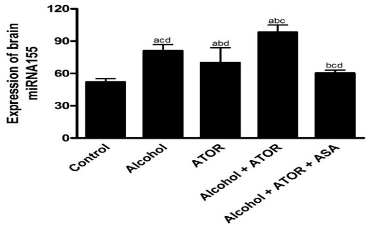
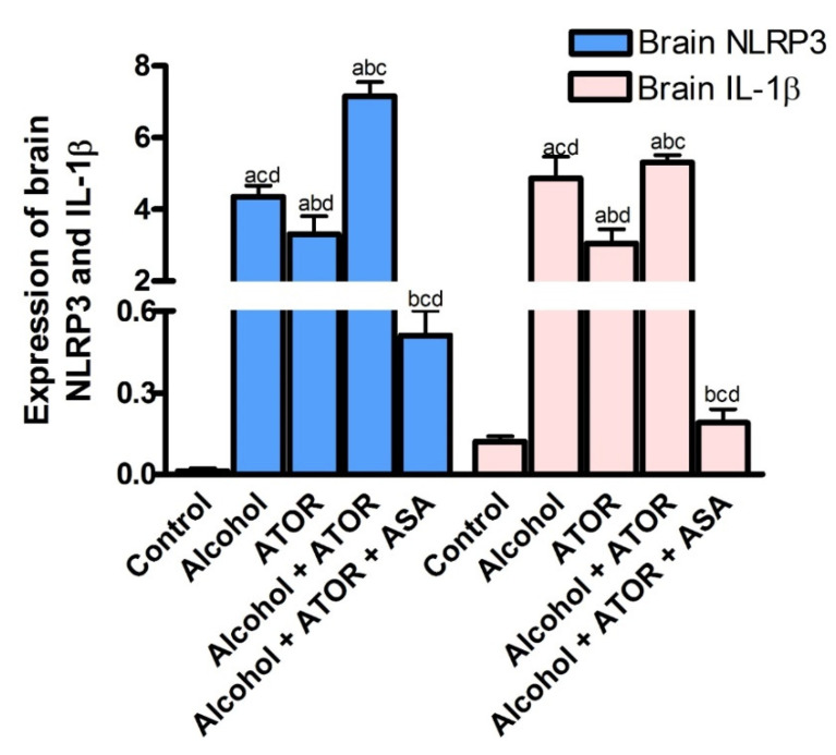
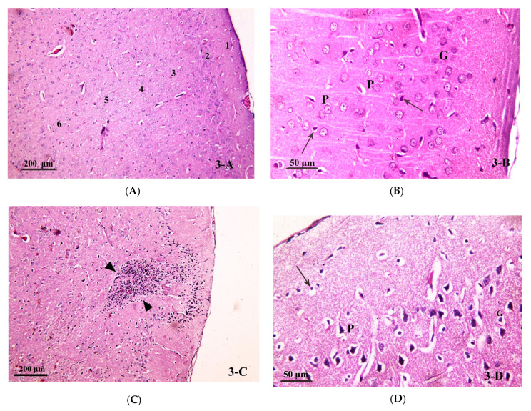
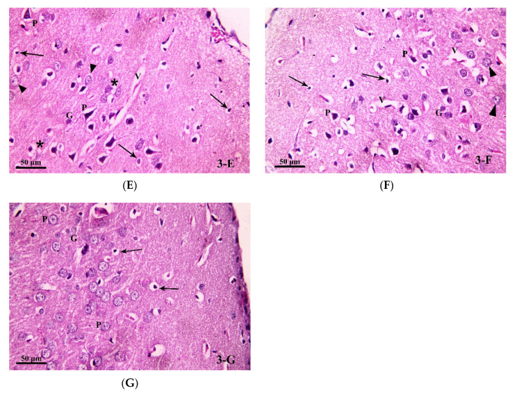
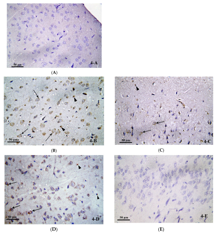
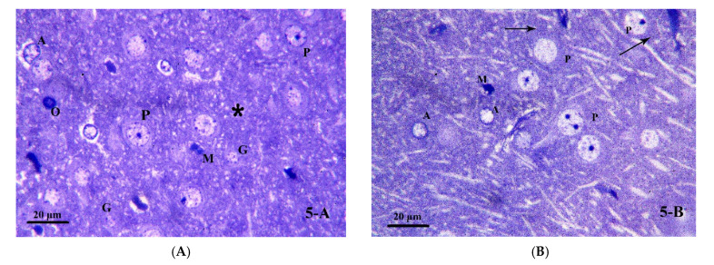
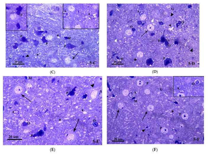
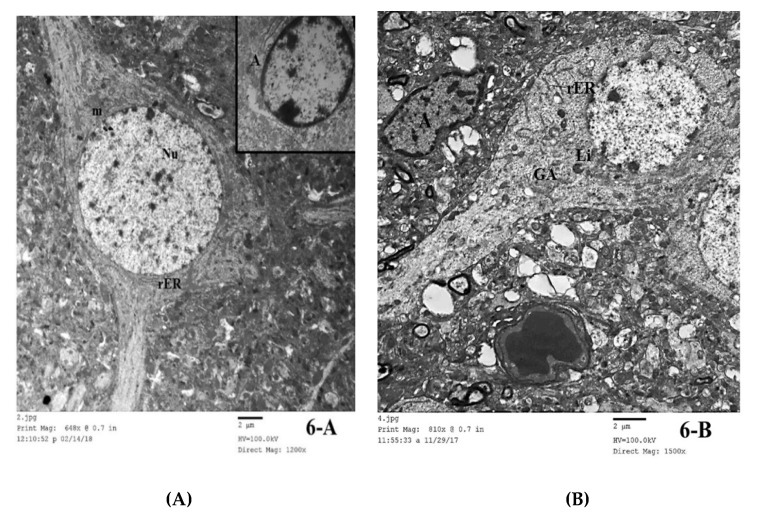
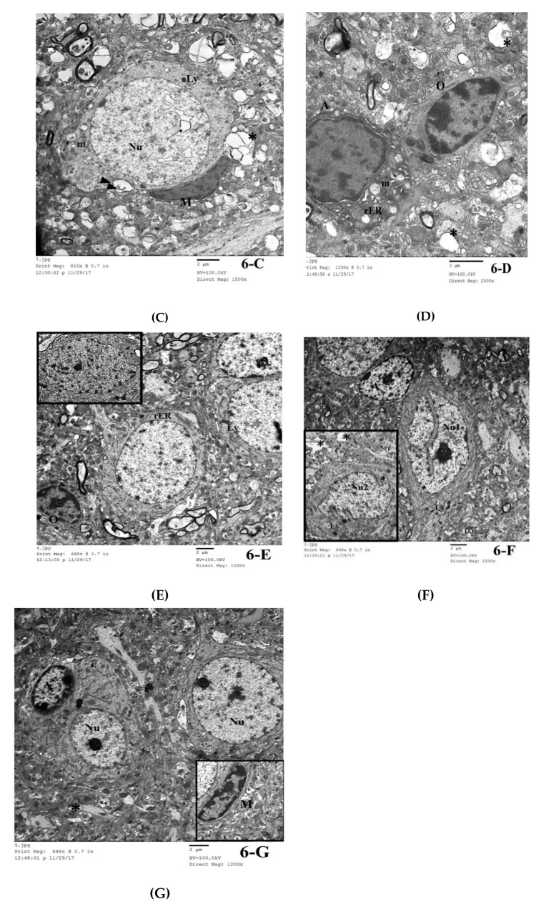
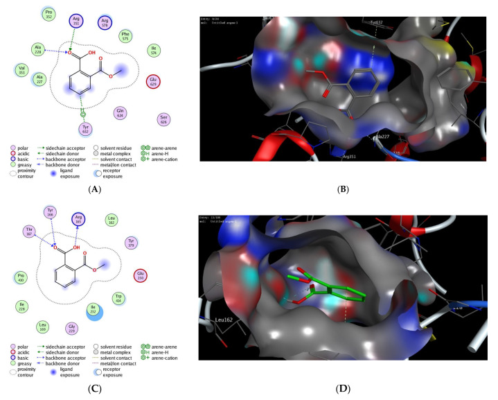
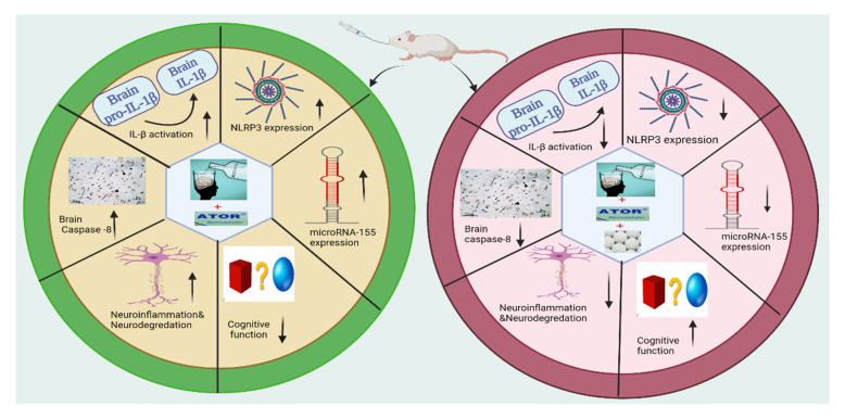
Alcoholism is one of the most common diseases that can lead to the development of several chronic diseases including steatosis, and cognitive dysfunction. Statins are lipid-lowering drugs that are commonly prescribed for patients with fatty liver diseases; however, the exact effect of statins on cognitive function is still not fully understood. In the present study, we have investigated the molecular and microscopic basis of cognitive impairment induced by alcohol and/or Atorvastatin (ATOR) administration to male Wistar albino rats and explored the possible protective effect of acetylsalicylic acid (ASA). The biochemical analysis indicated that either alcohol or ATOR or together in combination produced a significant increase in the nucleotide-binding domain-like receptor 3 (NLRP3), interleukin-1β (IL-1β) miRNA155 expression levels in the frontal cortex of the brain tissue. The histological and morphometric analysis showed signs of degeneration in the neurons and the glial cells with aggregations of inflammatory cells and a decrease in the mean thickness of the frontal cortex. Immunohistochemical analysis showed a significant increase in the caspase-8 immunoreaction in the neurons and glial cells of the frontal cortex. Interestingly, administration of ASA reversed the deleterious effect of the alcohol and ATOR intake and improved the cognitive function as indicated by biochemical and histological analysis. ASA significantly decreased the expression levels of miRNA155, NLRP3, and IL1B, and produced a significant decrease in caspase-8 immunoreaction in the neurons and glial cells of the frontal cortex with a reduction in the process of neuroinflammation and neuronal damage. To further investigate these findings, we have performed an extensive molecular docking study to investigate the binding affinity of ASA to the binding pockets of the NLRP3 protein. Our results indicated that ASA has high binding scores toward the active sites of the NLRP3 NACHT domain with the ability to bind to the NLRP3 pockets by a set of hydrophilic and hydrophobic interactions. Taken together, the present study highlights the protective pharmacological effect of ASA to attenuate the deleterious effect of alcohol intake and long term ATOR therapy on the cognitive function via targeting miRNA155 and NLRP3 proteins.

Keywords: NLRP3 inflammasomes; acetylsalicylic acid; alcoholism; atorvastatin; histopathology; miRNA155; molecular docking; statins.


Conflict of interest statement

The authors declare no conflict of interest.

Figures

Figure 1
Figure 1
Expression of brain miRNA155 in the different treated groups (mean ± SD). Data are mean ± SD of 6 rats per group. p < 0.05 is significant; a p versus control group, b p versus Alcohol group, c p versus ATOR group, d p versus Alcohol + ATOR + ASA group.
Figure 2
Figure 2
Effect of alcohol and/or ATOR administration on the expression of brain NLRP3, and IL-1β and assessment of ASA treatment (mean ± SD). Data are mean ± SD of 6 rats per group. p < 0.05 is significant; a p versus control group, b p versus Alcohol group, c p versus ATOR group, d p versus Alcohol + ATOR + ASA group.
Figure 3
Figure 3
Photomicrograph of brain tissue (H& E stain) from (A,B) Group I, showing the six layers of the cerebral cortex in the control group (H&E × 100). The nerve cells (pyramidal (P) and granule (G)) in the different layers of the cerebral cortex with large, rounded nuclei and prominent nucleoli. Nerve cells are separated by homogenous neuropil, containing small, deeply stained nuclei—most probably glial cells—(↑) (H&E × 400). (C,D) Group II showed distorted layers of the cerebral cortex in the alcoholic group. An area of aggregated mononuclear inflammatory cells (granuloma) can be seen (▲) (H&E × 100). Most of the pyramidal cells (P) and granule cells (G) in the different layers seen are shrunken, condensed, deeply stained and surrounded by a hallow. Small, deeply stained nuclei—most probably glial cells—are also surrounded by hallows (↑). Notice the pale, vacuolated neuropil (*) and the dilated blood vessel (V) (H&E × 400). (E) Group III, some pyramidal cells (P) and granule cells (G) are seen shrunken, condensed, deeply stained and surrounded by hallows. Notice some nerve cells have large, rounded nuclei and prominent nucleoli (▲). Small, deeply stained nuclei—most probably glial cells—are also surrounded by hallows (↑). The neuropil is pale and vacuolated (*). A dilated blood vessel (V) is seen (H&E × 400). (F) Group IV showed distorted layers of the cerebral cortex in group IV. Many pyramidal cells (P) and granule cells (G) are seen shrunken, condensed, deeply stained and surrounded by hallows. Notice few nerve cells have large, rounded nuclei and prominent nucleoli (▲). Small, deeply stained nuclei—most probably glial cells—are also surrounded by hallows (↑). Dilated blood vessels (V) are seen (H&E × 400). (G) Group V showed a more regular arrangement of layers of the cerebral cortex in group V. Most of the pyramidal(P) and granule (G) cells have large, rounded nuclei. Some nuclei have prominent nucleoli and others are vacuolated (∆). Some small, deeply stained nuclei—most probably glial cells—are surrounded by hallows (↑) (H&E × 400).
Figure 3
Figure 3
Photomicrograph of brain tissue (H& E stain) from (A,B) Group I, showing the six layers of the cerebral cortex in the control group (H&E × 100). The nerve cells (pyramidal (P) and granule (G)) in the different layers of the cerebral cortex with large, rounded nuclei and prominent nucleoli. Nerve cells are separated by homogenous neuropil, containing small, deeply stained nuclei—most probably glial cells—(↑) (H&E × 400). (C,D) Group II showed distorted layers of the cerebral cortex in the alcoholic group. An area of aggregated mononuclear inflammatory cells (granuloma) can be seen (▲) (H&E × 100). Most of the pyramidal cells (P) and granule cells (G) in the different layers seen are shrunken, condensed, deeply stained and surrounded by a hallow. Small, deeply stained nuclei—most probably glial cells—are also surrounded by hallows (↑). Notice the pale, vacuolated neuropil (*) and the dilated blood vessel (V) (H&E × 400). (E) Group III, some pyramidal cells (P) and granule cells (G) are seen shrunken, condensed, deeply stained and surrounded by hallows. Notice some nerve cells have large, rounded nuclei and prominent nucleoli (▲). Small, deeply stained nuclei—most probably glial cells—are also surrounded by hallows (↑). The neuropil is pale and vacuolated (*). A dilated blood vessel (V) is seen (H&E × 400). (F) Group IV showed distorted layers of the cerebral cortex in group IV. Many pyramidal cells (P) and granule cells (G) are seen shrunken, condensed, deeply stained and surrounded by hallows. Notice few nerve cells have large, rounded nuclei and prominent nucleoli (▲). Small, deeply stained nuclei—most probably glial cells—are also surrounded by hallows (↑). Dilated blood vessels (V) are seen (H&E × 400). (G) Group V showed a more regular arrangement of layers of the cerebral cortex in group V. Most of the pyramidal(P) and granule (G) cells have large, rounded nuclei. Some nuclei have prominent nucleoli and others are vacuolated (∆). Some small, deeply stained nuclei—most probably glial cells—are surrounded by hallows (↑) (H&E × 400).
Figure 4
Figure 4
Photomicrograph of brain tissue from (A) Group I, showing a negative caspase-8 immune-reaction in nerve cells and glial cells in the frontal cortex of the control group. (B) Group II showed positive immune-reaction to caspase-8 in the cytoplasm of some nerve cells (↑) and glial cells (▲). (C) Group III showed a positive immune-reaction to caspase-8 in the cytoplasm of some nerve cells (↑) and glial cells (▲). (D) Group IV, showing a positive immune-reaction to caspase-8 (↑) in the cytoplasm of some nerve cells and glial cells. (E) Group V showed a negative caspase-8 immune-reactions in nerve cells and glial cells in the frontal cortex of group V. (Avidin Biotin Peroxidase for Caspase-8 × 400).
Figure 5
Figure 5
Photomicrograph of brain tissue semi-thin sections (toluidine blue stained) for (A) Group I, showing pyramidal cells (P) with large, rounded nuclei and prominent nucleoli. Granule cells (G) have small rounded euchromatic nuclei. Notice the presence of astrocytes (A), oligodendrocyte (O) and microglia (M). The neuropil is homogenous (*). (B) Group I, showing pyramidal cells (P) with large, rounded nuclei and prominent nucleoli. Nissl granules (↑) are seen in their cytoplasm. Notice the presence of astrocytes (A) and microglia (M). (C) Group II, showing most of the pyramidal cells (P) are shrunken, condensed and deeply stained. Few pyramidal cells are seen with large, rounded nuclei but with absent Nissl granules (↑). Others have large rounded dark nuclei (▲). Notice the presence of astrocytes (A) with irregular nuclei and surrounded by vacuoles, oligodendrocyte (O) and microglia (M). The neuropil is vacuolated (*). (D) Group III, demonstrating some pyramidal cells are shrunken, condensed and deeply stained (P). Some are seen with dark nuclei (▲) while others have large, rounded nuclei but with absent Nissl granules (↑). Some granule cells (G) have small rounded euchromatic nuclei while others have darkly stained nuclei (∆). Notice the presence of astrocytes (A), oligodendrocyte (O) and microglia (M). The neuropil is vacuolated (*). (E) Group IV, showing some pyramidal cells are shrunken, condensed and deeply stained (P). Some are seen with dark nuclei (▲) Some others are seen with pale, irregular nuclei (∆) while others have large, rounded nuclei but with absent Nissl granules (↑). Notice the presence of astrocytes (A), oligodendrocyte (O) and microglia (M). Notice the large vacuoles in the neuropil (*). (F) Group V, showing most of the pyramidal cells have large, rounded nuclei and prominent nucleoli (P). Notice the presence of Nissl granules (↑). A pyramidal cell with dark irregular nucleus is present (▲). Granule cells (G) have small rounded euchromatic nuclei. Notice the presence of astrocytes (A), oligodendrocyte (O) and microglia (M). Small vacuoles are seen in the neuropil (*). (toluidine blue × 1000).
Figure 5
Figure 5
Photomicrograph of brain tissue semi-thin sections (toluidine blue stained) for (A) Group I, showing pyramidal cells (P) with large, rounded nuclei and prominent nucleoli. Granule cells (G) have small rounded euchromatic nuclei. Notice the presence of astrocytes (A), oligodendrocyte (O) and microglia (M). The neuropil is homogenous (*). (B) Group I, showing pyramidal cells (P) with large, rounded nuclei and prominent nucleoli. Nissl granules (↑) are seen in their cytoplasm. Notice the presence of astrocytes (A) and microglia (M). (C) Group II, showing most of the pyramidal cells (P) are shrunken, condensed and deeply stained. Few pyramidal cells are seen with large, rounded nuclei but with absent Nissl granules (↑). Others have large rounded dark nuclei (▲). Notice the presence of astrocytes (A) with irregular nuclei and surrounded by vacuoles, oligodendrocyte (O) and microglia (M). The neuropil is vacuolated (*). (D) Group III, demonstrating some pyramidal cells are shrunken, condensed and deeply stained (P). Some are seen with dark nuclei (▲) while others have large, rounded nuclei but with absent Nissl granules (↑). Some granule cells (G) have small rounded euchromatic nuclei while others have darkly stained nuclei (∆). Notice the presence of astrocytes (A), oligodendrocyte (O) and microglia (M). The neuropil is vacuolated (*). (E) Group IV, showing some pyramidal cells are shrunken, condensed and deeply stained (P). Some are seen with dark nuclei (▲) Some others are seen with pale, irregular nuclei (∆) while others have large, rounded nuclei but with absent Nissl granules (↑). Notice the presence of astrocytes (A), oligodendrocyte (O) and microglia (M). Notice the large vacuoles in the neuropil (*). (F) Group V, showing most of the pyramidal cells have large, rounded nuclei and prominent nucleoli (P). Notice the presence of Nissl granules (↑). A pyramidal cell with dark irregular nucleus is present (▲). Granule cells (G) have small rounded euchromatic nuclei. Notice the presence of astrocytes (A), oligodendrocyte (O) and microglia (M). Small vacuoles are seen in the neuropil (*). (toluidine blue × 1000).
Figure 6
Figure 6
Electron microscopic examination (TEM) of frontal cortex in brain tissue for (A) Group I, showing a pyramidal cell in cerebral cortex of Group I. A large, rounded, central and euchromatic nucleus (Nu) is seen surrounded by cytoplasm. Notice the rough endoplasmic reticulum (rER) and mitochondria (m). Inset showing an astrocyte (A) Inset (TEM × 2000). (B) Group I, demonstrating a nerve cell with dilated, irregularly arranged cisternae of rough endoplasmic reticulum (rER). Notice the presence of lysosomes (Ly), vacuoles (↑) and a Golgi apparatus (GA) in the nerve cell. Notice the presence of an astrocyte (A) with irregular dark nucleus. Large vacuoles (*) are seen in the neuropil (TEM × 1500). (C) Group II, showing a nerve cell in the frontal cortex of Group II with large, rounded, central and euchromatic nucleus (Nu). Notice the intranuclear vacuoles (↑). Lysosomes (Ly), elongated mitochondria (m) and cytoplasmic vacuoles are seen. Notice a microglial cell is present beside the nerve cell. Many large vacuoles are seen in the neuropil (*) (TEM × 1500). (D) Group II, showing two glial cells in the frontal cortex of Group II. An astrocyte (A) is seen with an irregular dark nucleus and a dense rim of peripheral heterochromatin. Notice the dilated and distorted cisternae of rough endoplasmic reticulum (rER), mitochondria (m) with destroyed cristae and cytoplasmic vacuoles (↑). An oligodendrocyte (O) is seen with its dark oval nucleus and large clumps of heterochromatin. Multiple dense bodies are seen in its cytoplasm (▲). Large vacuoles are seen in the neuropil (*) (TEM × 2500). (E) Group III, showing pyramidal cells in the frontal cortex of Group III with large, rounded, central and euchromatic nuclei (Nu). Notice mild dilatation of the rough endoplasmic reticulum (rER). Lysosomes are seen (Ly). An oligodendrocyte (O) is present beside a nerve cell. Inset: showing a pyramidal cell with darkly stained irregular nucleus (TEM × 1200). (F) Group IV, showing a pyramidal cell in frontal cortex with marked deep indentation of the nuclear membrane (Nu1). Many lysosomes are seen (Ly). An astrocyte (A) is present beside the nerve cells. Inset: showing a pyramidal cell with indented and irregular nucleus (Nu2). Large vacuoles are seen in the neuropil (*) (TEM × 1200). (G) Group V, showing pyramidal cells in the frontal cortex of Group V with large, rounded, central and euchromatic nuclei (Nu). Notice mild dilatation of a few rough endoplasmic reticulum cisternae (rER). Few lysosomes are seen (Ly). An astrocyte (A) is present beside a nerve cell. The neuropil is homogenous (*). Inset: showing a microglial cell (M) (TEM × 1200).
Figure 6
Figure 6
Electron microscopic examination (TEM) of frontal cortex in brain tissue for (A) Group I, showing a pyramidal cell in cerebral cortex of Group I. A large, rounded, central and euchromatic nucleus (Nu) is seen surrounded by cytoplasm. Notice the rough endoplasmic reticulum (rER) and mitochondria (m). Inset showing an astrocyte (A) Inset (TEM × 2000). (B) Group I, demonstrating a nerve cell with dilated, irregularly arranged cisternae of rough endoplasmic reticulum (rER). Notice the presence of lysosomes (Ly), vacuoles (↑) and a Golgi apparatus (GA) in the nerve cell. Notice the presence of an astrocyte (A) with irregular dark nucleus. Large vacuoles (*) are seen in the neuropil (TEM × 1500). (C) Group II, showing a nerve cell in the frontal cortex of Group II with large, rounded, central and euchromatic nucleus (Nu). Notice the intranuclear vacuoles (↑). Lysosomes (Ly), elongated mitochondria (m) and cytoplasmic vacuoles are seen. Notice a microglial cell is present beside the nerve cell. Many large vacuoles are seen in the neuropil (*) (TEM × 1500). (D) Group II, showing two glial cells in the frontal cortex of Group II. An astrocyte (A) is seen with an irregular dark nucleus and a dense rim of peripheral heterochromatin. Notice the dilated and distorted cisternae of rough endoplasmic reticulum (rER), mitochondria (m) with destroyed cristae and cytoplasmic vacuoles (↑). An oligodendrocyte (O) is seen with its dark oval nucleus and large clumps of heterochromatin. Multiple dense bodies are seen in its cytoplasm (▲). Large vacuoles are seen in the neuropil (*) (TEM × 2500). (E) Group III, showing pyramidal cells in the frontal cortex of Group III with large, rounded, central and euchromatic nuclei (Nu). Notice mild dilatation of the rough endoplasmic reticulum (rER). Lysosomes are seen (Ly). An oligodendrocyte (O) is present beside a nerve cell. Inset: showing a pyramidal cell with darkly stained irregular nucleus (TEM × 1200). (F) Group IV, showing a pyramidal cell in frontal cortex with marked deep indentation of the nuclear membrane (Nu1). Many lysosomes are seen (Ly). An astrocyte (A) is present beside the nerve cells. Inset: showing a pyramidal cell with indented and irregular nucleus (Nu2). Large vacuoles are seen in the neuropil (*) (TEM × 1200). (G) Group V, showing pyramidal cells in the frontal cortex of Group V with large, rounded, central and euchromatic nuclei (Nu). Notice mild dilatation of a few rough endoplasmic reticulum cisternae (rER). Few lysosomes are seen (Ly). An astrocyte (A) is present beside a nerve cell. The neuropil is homogenous (*). Inset: showing a microglial cell (M) (TEM × 1200).
Figure 7
Figure 7
The 2D and 3D molecular modelling interactions of ASA (green and grey in 3D interactions) with NLRP3 protein: 7alv (A,B), and 6npy (C,D). Dotted green arrows represent the hydrogen bonds; (C atoms are colored green, S yellow and O red).
Figure 8
Figure 8
Molecular and cellular effects of ASA on cognitive dysfunction induced by alcohol and/or ATOR intake.

Similar articles

Cited by

References

    1. Ishii T., Hashimoto E., Ukai W., Tateno M., Yoshinaga T., Ono T., Watanabe K., Saito S., Saito T. Epigenetic regulation in alcohol-related brain damage. Nihon Arukoru Yakubutsu Igakkai Zasshi. 2008;43:705–713. - PubMed
    1. Bajaj J.S. Alcohol, Liver Disease and the Gut Microbiota. Nat. Rev. Gastroenterol. Hepatol. 2019;16:235–246. doi: 10.1038/s41575-018-0099-1. - DOI - PubMed
    1. Piano M.R. Alcohol’s Effects on the Cardiovascular System. Alcohol. Res. 2017;38:219–241. - PMC - PubMed
    1. Moss M., Burnham E.L. Chronic Alcohol Abuse, Acute Respiratory Distress Syndrome, and Multiple Organ Dysfunction. Crit. Care Med. 2003;31:S207–S212. doi: 10.1097/01.CCM.0000057845.77458.25. - DOI - PubMed
    1. Osna N.A., Donohue T.M., Kharbanda K.K. Alcoholic Liver Disease: Pathogenesis and Current Management. Alcohol. Res. 2017;38:147–161. - PMC - PubMed

LinkOut - more resources