Review
. 2022 Mar 11;14(3):614.
doi: 10.3390/pharmaceutics14030614.

Challenges and Complications of Poly(lactic- co-glycolic acid)-Based Long-Acting Drug Product Development

Affiliations
Review

Challenges and Complications of Poly(lactic- co-glycolic acid)-Based Long-Acting Drug Product Development

Yi Wen Lim et al. Pharmaceutics. .

Abstract

Poly(lactic-co-glycolic acid) (PLGA) is one of the preferred polymeric inactive ingredients for long-acting parenteral drug products that are constituted of complex formulations. Despite over 30 years of use, there are still many challenges faced by researchers in formulation-related aspects pertaining to drug loading and release. Until now, PLGA-based complex generic drug products have not been successfully developed. The complexity in developing these generic drug products is not just due to their complex formulation, but also to the manufacturing process of the listed reference drugs that involve PLGA. The composition and product attributes of commercial PLGA formulations vary with the drugs and their intended applications. The lack of standard compendial methods for in vitro release studies hinders generic pharmaceutical companies in their efforts to develop PLGA-based complex generic drug products. In this review, we discuss the challenges faced in developing PLGA-based long-acting injectable/implantable (LAI) drug products; hurdles that are associated with drug loading and release that are dictated by the physicochemical properties of PLGA and product manufacturing processes. Approaches to overcome these challenges and hurdles are highlighted specifically with respect to drug encapsulation and release.

Keywords: PLGA microspheres; complex generic drug products; drug delivery; long acting injectable/implantable; poly(lactide-co-glycolide); sustained release.


Conflict of interest statement

The authors declare that they have no known competing financial interests or personal relationships that could have appeared to influence the work reported in this paper.

Figures

Figure 1
Figure 1
(A) The sample-and-separate method, and (B) the continuous flow method used in discrimination of the in vitro release profiles of risperidone from four PLGA microsphere formulations with equivalent compositions but different manufacturing processes. In comparison to the sample-and-separate method, the continuous flow method can better differentiate the release of risperidone ascribing to the porosity of microspheres (i.e., Formulations 3 and 4 vs. Formulations 1 and 2), as well as their particle sizes (i.e., Formulations 1 vs. 2). (Reprinted from Journal of Controlled Release, 218, Jie Shen, Stephanie Choi, Wen Qu, Yan Wang and Diane J. Burgess, In vitro-in vivo correlation of parenteral risperidone polymeric microspheres, 2–12, Copyright (2015), with permission from Elsevier [18]).
Figure 2
Figure 2
Cumulative release of risperidone from four microsphere formulations with similar drug loading but different porosities and glass transition temperatures. Formulation P, with a high porosity % but a low Tg, displayed the fastest drug release. (Adapted from International Journal of Pharmaceutics, 582, Moe Kohno, Janki V. Andhariya, Bo Wan, Quanying Bao, Sam Rothstein, Michael Hezel, Yan Wang and Diane J. Burgess, The effect of PLGA molecular weight differences on risperidone release from microspheres, 119339, Copyright (2020), with permission from Elsevier [43]).
Figure 3
Figure 3
Drug release profiles of PLGA microspheres prepared under varying temperatures using emulsification solvent extraction/evaporation technique. High processing temperature results in a longer lag phase and a more pronounced sigmoidal drug release profile. (Reprinted from European Journal of Pharmaceutics and Biopharmaceutics, 81, Kerstin Vay, Wolfgang Frieß and Stefan Scheler, A detailed view of microparticle formation by in-process monitoring of the glass transition temperature, 399–408, Copyright (2012), with permission from Elsevier [42]).
Figure 4
Figure 4
Pharmacokinetic profiles of Nutropin Depot (A) and Trelstar (B). The red arrows indicate the initial burst release region, and the green arrows indicate the therapeutically effective region. In both profiles, the serum drug concentrations in the initial burst release region are much greater than that of the therapeutically effective region (Reprinted from Journal of Controlled Release, 219, Yeon Hee Yun, Byung Kook Lee, Kinam Park, Controlled Drug Delivery: Historical perspective for the next generation, 2–7, Copyright (2015), with permission from Elsevier [60]).
Figure 5
Figure 5
(A) Dimensions of a cage implant. (B) Top and side view of the silicone rubber/stainless cage implant. (C,D) Cage that is subcutaneously implanted in a rat (E) Retrieved cage prior to analysis (Reprinted from Biomaterials, 109, Amy C. Doty, Keiji Hirota, Karl F. Olsen, Naoya Sakamoto, Rose Ackermann, Meihua R. Feng, Yan Wang, Stephanie Choi, Wen Qu, Anna Schwendeman, Steven P. Schwendeman, Validation of a cage implant system for assessing in vivo performance of long-acting release microspheres, 88–96. Copyright (2016) with permission from Elsevier [61]); (F,G) Release of triamcinolone acetonide and leuprolide, respectively in vivo and in vitro in pH 7.4. A faster-than-expected drug release was observed from both PLGA formulations in vivo (Reprinted from Journal of Controlled Release, 256, Amy C. Doty, David G. Weinstein, Keiji Hirota, Karl F. Olsen, Rose Ackermann, Yan Wang, Stephanie Choi, Steven P. Schwendeman, Mechanisms of in vivo release of triamcinolone acetonide from PLGA microspheres, 19–25, Copyright (2017), with permission from Elsevier [36]).
Figure 6
Figure 6
A two-compartment in vitro model of the eye (known as PK-Eye) used in study of the release profile of dexamethasone loaded PLGA microparticles. (Reprinted from Journal of Pharmaceutical Sciences, 104, Sahar Awwad, Alastair Lockwood, Steve Brocchini, Peng T. Khaw, The PK-Eye: A novel in vitro ocular flow model for use in preclinical drug development, 3330–3342, Copyright (2015), with permission from Elsevier [67]).
Figure 7
Figure 7
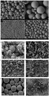
Scanning electron microscopic images of PLGA microparticles prepared using various processing techniques: (A,B) traditional W/O/W technique, (C,D) membrane emulsion method (Reproduced from Polymer Chemistry (2014), 5, Baoxia Liu, Xiao Zhou, Fei Yang, Hong Shen, Shenguo Wang, Bo Zhang, Guang Zhi, Decheng Wu, Fabrication of uniform sized polylactone microcapsules by premix membrane emulsification for ultrasound imaging, 1693–1701, with permission from the Royal Society of Chemistry [78]); (a,b) spray drying, (c,d) electrospray method (e,f) silicon microfluidic flow focusing device (MFFD). Among these techniques, MFFD produces the most uniform particles (Reprinted from International Journal of Pharmaceutics, 467, Kieran Keohane, Des Brennan, Paul Galvin, Brendan T. Griffin, Silicon microfluidic flow focusing devices for the production of size controlled PLGA-based drug loaded microparticles, 60–69, Copyright (2014), with permission from Elsevier [80]).
Figure 8
Figure 8
Schematic diagram of a spray drying process for manufacturing drug-loaded PLGA microspheres. Every aspect of the processing parameters, such as heat/mass transfer, inlet air temperature, and drying gas flow rate, requires optimisation in order to achieve the desired final products. (Reprinted from Journal of Controlled Release, 321, Nian-Qiu Shi, Jia Zhou, Jennifer Walker, Li Li, Justin K. Y. Hong, Karl F. Olsen, Jie Tang, Rose Ackermann, Yan Wang, Bin Qin, Anna Schwendeman, Steven P. Schwendeman, Microencapsulation of luteinizing hormone-releasing hormone agonist in poly (lactic-co-glycolic acid) microspheres by spray-drying, 756–772. Copyright (2020) with permission from Elsevier [87]).

Similar articles

Cited by

References

    1. Makadia H.K., Siegel S.J. Poly Lactic-co-Glycolic Acid (PLGA) as Biodegradable Controlled Drug Delivery Carrier. Polymers. 2011;3:1377–1397. doi: 10.3390/polym3031377. - DOI - PMC - PubMed
    1. Wang Y., Qu W., Choi S.H. FDA’s Regulatory Science Program for Generic PLA/PLGA-Based Drug Products|American Pharmaceutical Review—The Review of American Pharmaceutical Business & Technology. [(accessed on 22 October 2020)]. Available online: https://www.americanpharmaceuticalreview.com/Featured-Articles/188841-FD...
    1. Burgess D.J., Hussain A.S., Ingallinera T.S., Chen M.-L. Assuring Quality and Performance of Sustained and Controlled Release Parenterals: Workshop Report. AAPS PharmSci. 2002;4:13–23. doi: 10.1208/ps040205. - DOI - PMC - PubMed
    1. Martinez M.N., Rathbone M.J., Burgess D., Huynh M. Breakout Session Summary from AAPS/CRS Joint Workshop on Critical Variables in the In Vitro and In Vivo Performance of Parenteral Sustained Release Products. J. Control Release. 2009;142:2–7. doi: 10.1016/j.jconrel.2009.09.028. - DOI - PubMed
    1. Otte A., Sharifi F., Park K. Interfacial Tension Effects on the Properties of PLGA Microparticles. Colloids Surf. B Biointerfaces. 2020;196:111300. doi: 10.1016/j.colsurfb.2020.111300. - DOI - PMC - PubMed

LinkOut - more resources