. 2022 Mar 13;14(3):633.
doi: 10.3390/pharmaceutics14030633.

Liposomal siRNA Formulations for the Treatment of Herpes Simplex Virus-1: In Vitro Characterization of Physicochemical Properties and Activity, and In Vivo Biodistribution and Toxicity Studies

Affiliations

Liposomal siRNA Formulations for the Treatment of Herpes Simplex Virus-1: In Vitro Characterization of Physicochemical Properties and Activity, and In Vivo Biodistribution and Toxicity Studies

Doaa Jbara-Agbaria et al. Pharmaceutics. .

Abstract

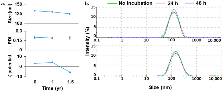
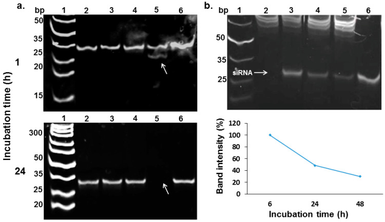
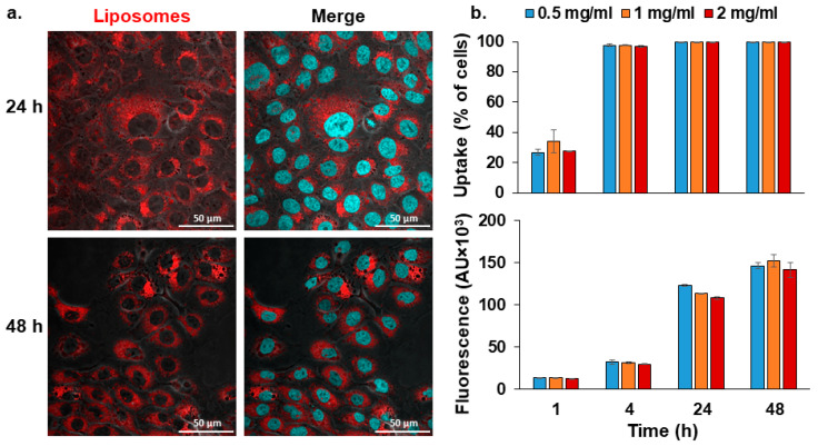
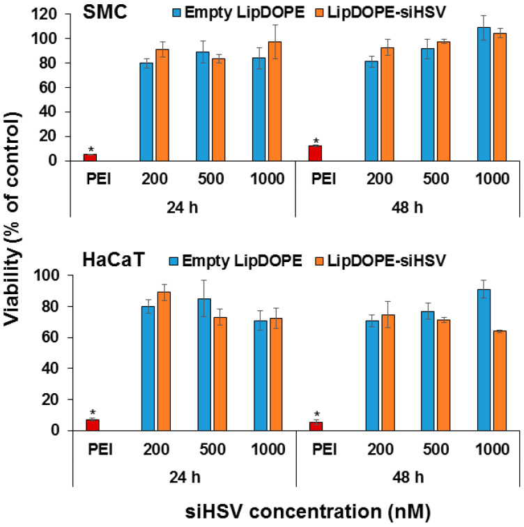
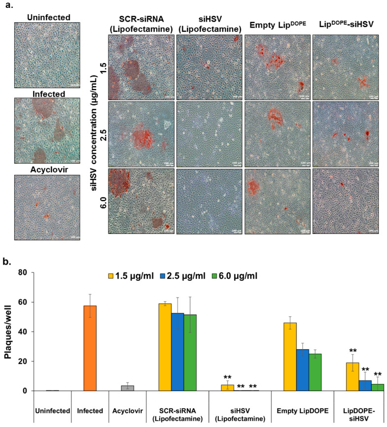
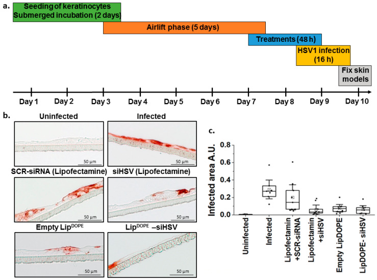
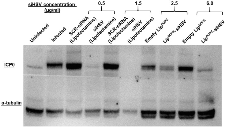
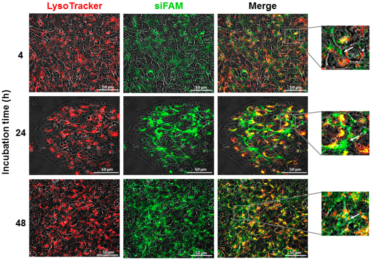
Herpes simplex virus-1 (HSV-1) is highly contagious, and there is a need for a therapeutic means to eradicate it. We have identified an siRNA (siHSV) that knocks down gene expression of the infected cell protein 0 (ICP0), which is important in the regulation of HSV infection. The selected siHSV was encapsulated in liposomes to overcome its poor stability, increase cell permeability, and prolonging siRNA circulation time. Several siRNAs against ICP0 have been designed and identified. We examined the role of various parameters, including formulation technique, lipids composition, and ratio. An optimal liposomal siHSV formulation (LipDOPE-siHSV) was characterized with desirable physiochemical properties, in terms of nano-size, low polydispersity index (PDI), neutral surface charge, high siHSV loading, spherical shape, high stability in physiologic conditions in vitro, and long-term shelf-life stability (>1 year, 4 °C). The liposomes exhibited profound internalization by human keratinocytes, no cytotoxicity in cell cultures, no detrimental effect on mice liver enzymes, and a gradual endo-lysosomal escape. Mice biodistribution studies in intact mice revealed accumulation, mainly in visceral organs but also in the trigeminal ganglion. The therapeutic potential of siHSV liposomes was demonstrated by significant antiviral activity both in the plaque reduction assay and in the 3D epidermis model, and the mechanism of action was validated by the reduction of ICP0 expression levels.

Keywords: 3D epidermis model; HSV-1; gene delivery; liposomes; nanomedicine.


Conflict of interest statement

The authors declare no conflict of interest.

Figures

Figure 1
Figure 1
(a) Identification of potential microRNAs in the latent state of HSV-1 infection against HSV-1 target protein, ICP0, by means of NGS-sequencing of differentiated, latently infected human neural progenitor cell line (ReNcell VM). A set of viral miRNAs highly expressed in differentiated ReNcell VM cells has been identified, and the miRNAs sequences, miR-H2 and miR-H6, have been further analyzed for antiviral activity; (b) the antiviral effect of the respective siRNAs in the plaque reduction assay (a confluent layer of HaCaT cells): free si-H2 and siH6 (upper panel) in comparison to siRNAs transfected by lipofectamine (lower panel). The formed syncytia, shown in brownish red, was visualized following immunohistochemistry staining of the cells (anti-HSV1 antibody). Brownish red depicts the envelope-protein of the virus, indicating proliferative infection (scale bar = 200 μm).
Figure 2
Figure 2
A representative cryo-TEM image depicting the structure of LipDOPE-siHSV (N:P ratio 5:1) (scale bar = 100 nm).
Figure 3
Figure 3
Stability and shelf-life of LipDOPE-siHSV. (a) The long-term shelf-life stability of LipDOPE-siHSV, suspended in TE buffer and stored at 4 °C, in terms of size, PDI, and surface charge changes. (b) The structural stability of LipDOPE-siHSV (bottom) over time in comparison to empty liposomes (top) following incubation in 10% w/v bovine serum albumin at 37 °C.
Figure 4
Figure 4
Evaluation of encapsulated siHSV stability in simulated physiological conditions. (a) The integrity of siHSV extracted from Lip-siHSV following incubation in 50% bovine serum albumin at 37 °C for 1 and 24 h: (1) Ladder; (2) extracted siHSV incubated in TE buffer; (3) extracted siHSV incubated in serum; (4) free siHSV incubated in TE buffer; (5) free siHSV incubated in serum; and (6) free siHSV without incubation. An aliquot of each sample (200 nM) was analyzed by native 24% acrylamide gel, stained with SYBER Gold. (b) The integrity of encapsulated siHSV in comparison to a spiked sample of empty liposomes with siHSV (200 nM) in keratinocytes (HaCaT) cell cultures: (1) ladder; (2) empty liposomes spiked with siHSV after 6 h; (3) Lip-siHSV after 6 h; (4) Lip-siHSV after 24 h; (5) Lip-siHSV after 48 h; and (6) free siHSV with no incubation. The semi-quantification (ImageJ analysis) of siHSV bands intensities is shown at the lower right-hand part of b. An aliquot from each sample of extracted RNA was analyzed (200 nM) by native 24% acrylamide gel, stained with ethidium bromide, and visualized. Free siHSV with no incubation (lane 6) was set as 100% recovery.
Figure 5
Figure 5
The internalization of LipDOPE by keratinocytes (HaCaT) cell culture. Qualitative (confocal microscopy; (a) quantitative (flow cytometry analysis; and (b) assessment of cellular uptake of LipDOPE (fluorescently labeled with Rhodamine-PE; 2 mg/mL lipids concentration). The liposomal membrane is depicted in red, and cell’s nuclei in cyan (scale bar = 50 μm). The fluorescence intensities were normalized to untreated cells (mean ± SD).
Figure 6
Figure 6
The dose- and time-dependent cytotoxicity of LipDOPE -siHSV in rat primary aortic smooth muscle cells (SMC) and keratinocytes (HaCaT) cell cultures. The cytotoxicity was determined by means of the MTT assay, and cell viability was normalized to untreated cells (n = 4; * p < 0.01).
Figure 7
Figure 7
The antiviral effect of LipDOPE-siHSV following treatment of HaCaT cells (keratinocytes) infected with HSV-1 (plaque reduction assay). (a) Treatment groups: LipDOPE-siHSV, empty LipDOPE and scrambled-siRNA (SCR-siRNA transfected by lipofectamine) served as negative controls, and acyclovir (125µM) and siHSV (transfected by lipofectamine) served as positive controls, in comparison to uninfected cells and untreated infected cells. Following 48 h of treatment, the cells were infected with HSV-1 (1 × 10⁴ PFU) for 16 h. The formed syncytia, shown in brownish red, was visualized following immunohistochemistry staining of the cells (anti-HSV1 antibody; n = 8). Brownish red staining depicts the envelope-protein of the virus, indicating proliferative infection, and no staining indicates full protection (magnification ×10; scale bar = 100 μm). (b) Semi-quantitative analysis of the antiviral efficacy (** p < 0.01).
Figure 8
Figure 8
The antiviral effect of LipDOPE-siHSV (DOTAP:siHSV N:P of 5:1) against HSV-1 infection in the 3D epidermis model of immortalized keratinocytes (HaCaT). (a) Illustration of the experimental setup. (b) Qualitative assessment of the treatments by immunohistochemistry of epidermis tissues treated with LipDOPE-siHSV (2.5 µg/mL), scrambled-siRNA (SCR) transfected by lipofectamine (1.5 µg/mL) and empty LipDOPE (negative controls), and siHSV transfected by lipofectamine (1.5 µg/mL; positive control), in comparison to uninfected and untreated infected cells. The epidermis tissues were infected with HSV-1 after 48 h and were analyzed after 16 h for tissue morphometry and integrity (scale bar = 50 μm). (c) Quantification of the immunohistochemistry staining of three independent experiments (each experiment consisting of two or three individual skin models) is shown in (b) by calculating the infected area in the epidermis tissue.
Figure 9
Figure 9
The expression levels of ICP0, following pre-treatment of HaCaT cells (keratinocytes) infected with HSV-1 (Western blot). Treatment groups: LipDOPE-siHSV, empty LipDOPE, and scrambled-siRNA (SCR-siRNA transfected by lipofectamine) served as negative controls, and siHSV (transfected by lipofectamine) served as positive control, in comparison to uninfected cells and untreated infected cells. Following 48 h of treatment, the cells were infected with HSV-1 (5 × 10⁴ PFU) for 16 h and lysed. Cell lysates were separated in a gel and transferred onto a membrane incubated with primary antibodies α-tubulin and HSV-1 ICP0.
Figure 10
Figure 10
The time-dependent internalization (confocal microscopy) and intracellular fate of LipDOPE-siRNA in keratinocytes (HaCaT) cell culture. Cells were treated with 6 µg/mL (siRNA concentration) and examined for siRNA endosomal/lysosomal escape. The endo-lysosomes are depicted in red (LysoTracker Red DND-99), fluorescently-labeled siRNA (siFAM) is depicted in green, and colocalization is depicted in yellow (white arrows). The fluorescence intensity was normalized to untreated cells; magnification ×60 (scale bar = 50 μm).

Similar articles

Cited by

References

    1. Lan K., Luo M.H. Herpesviruses: Epidemiology, pathogenesis, and interventions. Virol. Sin. 2017;32:347–348. doi: 10.1007/s12250-017-4108-2. - DOI - PMC - PubMed
    1. Chayavichitsilp P., Buckwalter J., Krakowski A.C., Friendlander S.F. Herpes simplex. Pediatr. Rev. 2009;30:119–129. doi: 10.1542/pir.30.4.119. - DOI - PubMed
    1. Simmons A. Clinical manifestations and treatment considerations of herpes simplex virus infection. J. Infect. Dis. 2002;186:S71–S77. doi: 10.1086/342967. - DOI - PubMed
    1. Klysik K., Pietraszek A., Karewicz A., Nowakowska M. Acyclovir in the treatment of herpes viruses—A review. Curr. Med. Chem. 2020;27:4118–4137. doi: 10.2174/0929867325666180309105519. - DOI - PubMed
    1. De Clercq E. In search of a selective therapy of viral infections. Antivir. Res. 2010;85:19–24. doi: 10.1016/j.antiviral.2009.10.005. - DOI - PubMed

LinkOut - more resources