Review
. 2022 Mar 17;14(3):660.
doi: 10.3390/pharmaceutics14030660.

PLGA-Gold Nanocomposite: Preparation and Biomedical Applications

Affiliations
Review

PLGA-Gold Nanocomposite: Preparation and Biomedical Applications

Alaaldin M Alkilany et al. Pharmaceutics. .

Abstract

A composite system consisting of both organic and inorganic nanoparticles is an approach to prepare a new material exhibiting "the best of both worlds". In this review, we highlight the recent advances in the preparation and applications of poly(lactic-co-glycolic acid)-gold nanoparticles (PLGA-GNP). With its current clinically use, PLGA-based nanocarriers have promising pharmaceutical applications and can "extract and utilize" the fascinating optical and photothermal properties of encapsulated GNP. The resulting "golden polymeric nanocarrier" can be tracked, analyzed, and visualized using the encapsulated gold nanoprobes which facilitate a better understanding of the hosting nanocarrier's pharmacokinetics and biological fate. In addition, the "golden polymeric nanocarrier" can reveal superior nanotherapeutics that combine both the photothermal effect of the encapsulated gold nanoparticles and co-loaded chemotherapeutics. To help stimulate more research on the development of nanomaterials with hybrid and exceptional properties, functionalities, and applications, this review provides recent examples with a focus on the available chemistries and the rationale behind encapsulating GNP into PLGA nanocarriers that has the potential to be translated into innovative, clinically applicable nanomedicine.

Keywords: PLGA; composite; gold nanoparticles; poly(lactic-co-glycolic acid).


Conflict of interest statement

The authors declare that they have no conflict of interest.

Figures

Figure 1
Figure 1
The collective rationale of encapsulating GNP into poly(lactic-co-glycolic acid) (PLGA) nanocarriers: (A) Utilizing the optical and electronic properties of GNP to enable the use of various tracking and visualization tools including electron and optical microscopies, ICP-MS and CT-scanning. The labeled PLGA nanocarrier can then be tracked and quantified with accuracy and precision to understand the nano-bio interaction of the PLGA host at the level of cell, tissue and whole organism. (B) The photothermal property of encapsulated GNP can be utilized in the development of a light-responsive drug delivery system as well as the synergetic photothermal-chemotherapeutic activity against cancer.
Figure 2
Figure 2
(A) Tunable optical properties of GNP. While suspension of spherical GNP (18 nm in diameter) exhibit a red color and plasmonic absorption peak around 520 nm, suspension of gold nanorods exhibit brown color with tunable optical extinction in the visible-infrared region of the spectrum. (B) Dark field image of GNP in solution (a) and inside cells (b). (C) Hyperspectral microscopy with adaptive detection analysis showing the location of gold nanorods tracked using their unique optical fingerprint (plasmonic optical extinction in the NIR) in orange and grayscale for tissue background. Images in B and C were reproduced from Refs. [18,19], respectively (Creative Commons Attribution 4.0 International Public License (CC-BY 4.0). (B) Reproduced from Zamora-Perez et al. [18] which is licensed under a Creative Commons Attribution CC BY 4.0 Internalional License. (C) Reproduced from SoRelle et al. [19] which is licensed under a Creative Commons Attribution CC BY 4.0 Internalional License.
Figure 3
Figure 3
Poly(lactic-co-glycolic acid) (PLGA) nanoparticles labeled with gold nanoprobes to track their nano-bio interaction: (A) TEM image showing single PLGA nanocarrier labeled with spherical GNP (15 nm) taken up by HeLa cancer cell, scale bar = 150 nm. (B) TEM image and (C) SEM backscattered electron image of one-to-one encapsulation of gold nanoprobes into PLGA carriers that allows for the tracking of the labeled PLGA carriers inside J774 cells (D) using SEM analysis with backscattered electron detection mode. (A) Reproduced with permission from [28], published by John Riley and Sons, 2019. (BD) Reproduced with permission from [29] published by Elsevier, 2017.
Figure 4
Figure 4
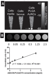
(A) In vitro CT image of macrophage cells (J774A) incubated with poly(lactic-co-glycolic acid) (PLGA) nanocarriers encapsulating GNP [arrow]. (B) In vitro X-ray CT images [upper panel] and the X-ray attenuation intensity (HU) of GNP loaded PLGA nanoparticles as a function of their concentrations. (A) Reproduced with permission from [32], published by Royal Society of Chemistry, 2012. (B) Reproduced with permission from [33], published by Elsevier, 2015.
Figure 5
Figure 5
Labeling poly(lactic-co-glycolic acid) (PLGA) particles with GNP. (A) o/w emulsion method in which pre-prepared hydrophobic GNP is suspended in the internal organic phase with PLGA polymers (Emulsion-evaporation method). (B) Co-precipitation of pre-prepared hydrophobic GNP and PLGA polymer from the use of a water-miscible organic solvent upon addition to aqueous system (nanoprecipitation method).
Figure 6
Figure 6
Labeling poly(lactic-co-glycolic acid) (PLGA) nanocarriers with GNP using the w/o/w double emulsion method. Left route: pre-prepared hydrophilic GNP are suspended in the internal aqueous phase. right route: both gold ions (precursor) and the reducing agent are dissolved in the internal aqueous phase. In-droplet reduction and solvent evaporation result in the formation and encapsulation of gold nanoparticles, respectively.
Figure 7
Figure 7
TEM images of poly(lactic-co-glycolic acid) (PLGA) nanoparticles encapsulating GNP prepared using various encapsulation methods. (A) Direct encapsulation of citrate-capped GNP in w/o/w emulsion (GNP are in the internal aqueous phase of the double emulsion). (B) Direct encapsulation of dodecanethiol-capped GNP in o/w emulsion (GNP are in the organic phase of the emulsion). (C) In situ reduction method in w/o/w emulsion (gold salt and reducing agent are in the internal aqueous phase of the double emulsion). Note that the different encapsulation routes resulted in different encapsulation outcomes; and the 1:1 encapsulation with minimal empty PLGA nanoparticles was obtained only when the in-situ reduction method was employed. Reproduced from Luque-Michel et al. [61] which is licensed under a Creative Commons Attribution CC BY 4.0 Internalional License.
Figure 8
Figure 8
Efficient encapsulation of GNP into poly(lactic-co-glycolic acid) (PLGA) nanoparticles according to the “like dissolves like” principle. (A) First, citrate-capped GNP are transferred from water to dichloromethane using PLGA-SH to prepare PLGA-capped GNP that can be re-dispersed in acetone. (B) Using the nanoprecipitation method, PEG- or PS- or PLGA-capped GNP are encapsulated into PLGA nanoparticles. (C) TEM images conforming the partitioning of PEG-GNP into the aqueous external phase, the poor encapsulation of PS-GNP, and the efficient encapsulation of PLGA-GNP into PLGA nanocarriers. Scale bars are 100 nm in all images. Reproduced from Alkilany et al. [28] which is licensed under a Creative Commons Attribution CC BY 4.0 Internalional License.

References

    1. Anselmo A.C., Mitragotri S. Nanoparticles in the clinic: An update. Bioeng. Transl. Med. 2019;4:e10143. doi: 10.1002/btm2.10143. - DOI - PMC - PubMed
    1. Page Faulk W., Malcolm Taylor G. Communication to the editors: An immunocolloid method for the electron microscope. Immunochemistry. 1971;8:1081–1083. doi: 10.1016/0019-2791(71)90496-4. - DOI - PubMed
    1. Montón H., Nogués C., Rossinyol E., Castell O., Roldán M. QDs versus Alexa: Reality of promising tools for immunocytochemistry. J. Nanobiotechnol. 2009;7:4. doi: 10.1186/1477-3155-7-4. - DOI - PMC - PubMed
    1. Chen F., Ehlerding E.B., Cai W. Theranostic nanoparticles. J. Nucl. Med. 2014;55:1919–1922. doi: 10.2967/jnumed.114.146019. - DOI - PMC - PubMed
    1. McCarthy J.R., Weissleder R. Multifunctional magnetic nanoparticles for targeted imaging and therapy. Adv. Drug Deliv. Rev. 2008;60:1241–1251. doi: 10.1016/j.addr.2008.03.014. - DOI - PMC - PubMed

LinkOut - more resources