. 2022 Mar 21;14(3):683.
doi: 10.3390/pharmaceutics14030683.

Study of Viral Photoinactivation by UV-C Light and Photosensitizer Using a Pseudotyped Model

Affiliations

Study of Viral Photoinactivation by UV-C Light and Photosensitizer Using a Pseudotyped Model

Mohammad Sadraeian et al. Pharmaceutics. .

Abstract

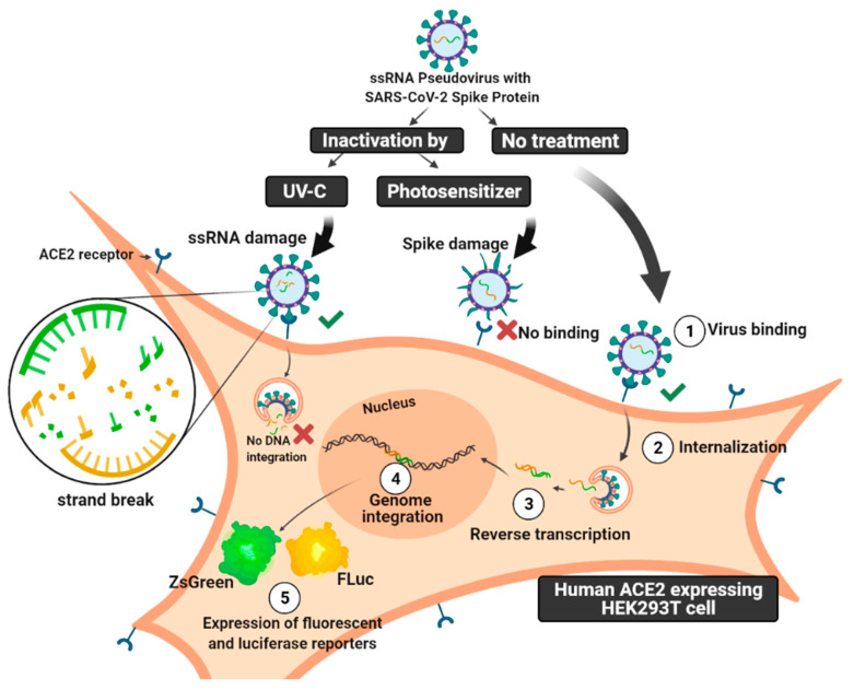
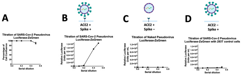
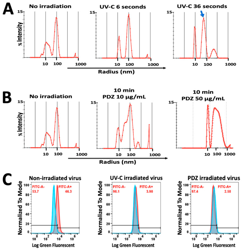
Different light-based strategies have been investigated to inactivate viruses. Herein, we developed an HIV-based pseudotyped model of SARS-CoV-2 (SC2) to study the mechanisms of virus inactivation by using two different strategies; photoinactivation (PI) by UV-C light and photodynamic inactivation (PDI) by Photodithazine photosensitizer (PDZ). We used two pseudoviral particles harboring the Luciferase-IRES-ZsGreen reporter gene with either a SC2 spike on the membrane or without a spike as a naked control pseudovirus. The mechanism of viral inactivation by UV-C and PDZ-based PDI were studied via biochemical characterizations and quantitative PCR on four levels; free-cell viral damage; viral cell entry; DNA integration; and expression of reporter genes. Both UV-C and PDZ treatments could destroy single stranded RNA (ssRNA) and the spike protein of the virus, with different ratios. However, the virus was still capable of binding and entering into the HEK 293T cells expressing angiotensin-converting enzyme 2 (ACE-2). A dose-dependent manner of UV-C irradiation mostly damages the ssRNA, while PDZ-based PDI mostly destroys the spike and viral membrane in concentration and dose-dependent manners. We observed that the cells infected by the virus and treated with either UV-C or PDZ-based PDI could not express the luciferase reporter gene, signifying the viral inactivation, despite the presence of RNA and DNA intact genes.

Keywords: SARS-CoV-2 pseudovirus; UV-C light; enveloped virus; photodynamic inactivation; photosensitizer; viral inactivation.


Conflict of interest statement

Authors declare no competing interests. Graphical figures were created with BioRender software ((https://biorender.com/ (accessed on 15 July 2021)).

Figures

Figure 1
Figure 1
Schematic picture of the mechanism of SARS-CoV-2 pseudovirus infectivity. Unlike SC2 ssRNA virus, which has viral reproduction independent of the host genome, this counterpart pseudovirus carries on reporter ssRNA with LTR, which causes integration into the genome. In this study, the pseudovirus has been treated with either UV-C irradiation or photodynamic inactivation (PDI) by Photodithazine photosensitizer. The mechanism of infectivity of photo-inactivated pseudovirus particles has been compared on four levels; free-cell viral damage; viral cell entry; DNA integration; and expression of reporter genes. The figure was created with BioRender software.
Figure 2
Figure 2
Titration of SARS-CoV-2 spike-pseudovirus particles in 293T cells expressing ACE-2. (A) Study of the percentage of cell viability during the viral infection respecting a serial dilution starting at 1:2 (0.5); (B,C) The graph shows the titers of the expression of Luciferase reporter as determined by measuring relative luciferase units (RLUs). The RLU data are the average of three-fold serial dilution of virus starting at 50 μL virus in a total volume of 100 μL (0.5) for the Spike-pseudovirus (B); naked pseudovirus without spike (C); or the Spike-pseudovirus with 293T cells without ACE2 receptor (D). After 8 h of pseudovirus incubation, the media were replaced with 150 µL fresh media.
Figure 3
Figure 3
Study of the effect of photo-inactivation of ssRNA pseudovirus considering the relative luciferase units with a time-dependent manner of UV-C irradiation at 1, 6, and 36 s corresponding to doses of 10, 60, and 360 mJ/cm2, respectively (A); and PDZ-based PDI in a serial dilution of 10, 50 and 250 µg/mL in a time-dependent manner of 1, 10 and 20 min which equal the light doses of 1.8, 18 and 36 J/cm2, respectively (B). Data are ± means S.E.M. (n = 3).
Figure 4
Figure 4
The pseudovirus has been treated with either 36 s UV-C irradiation or 10 min Photodynamic Inactivation in the presence of 50 µg/mL Photodithazine. The mechanism of infectivity of photo-inactivated pseudovirus particles has been compared on three levels; (A) viral RNA load referring to the free-cell virus; (B) proviral DNA load referring to the ability of the treated virus to complete the subsequent steps of cell internalization, reverse transcription and genome integration; (C) luciferase activity referring to the expression of luciferase reporter after DNA integration. nt/n0 represents the fraction of the targeted genome region that remained intact after treatment.
Figure 5
Figure 5
(A) Dynamic Light Scattering histograms of hydrodynamic radius (Rh) for pseudovirus showed optimal polydispersity with Rh of 100 nm. During 36 s of UV-C irradiation (360 mJ/cm2), a slight decrease in the size of pseudovirus was observed (Blue arrow) with no significant aggregation; (B) PDZ-based PDI significantly affected the size and polydispersity of the virus, resulting in major aggregated particles in a higher concentration of PDZ (50 µg/mL); (C) Flow cytometric diagram on the left demonstrated the percentage of pseudovirus-infected cells expressing ZsGreen Fluorescent protein. The middle and right diagrams represent the cells infected by UV irradiated-virus and PDZ-based PDI virus, which do not express ZsGreen protein. FITC rate indicates the green fluorescent emission from ZsGreen. Data are ± means S.E.M. (n = 2).
Figure 6
Figure 6
(A) Confocal microscopy images showing ZsGreen expression in 293T-ACE2 cells at 48 h after incubation with Spike-pseudotyped lentiviral particles with the ZsGreen backbone. The images are represented in spectral mode (panel (a)) and channel mode merged with a wide field transmission image (panel (c)). In panel (b), the green and red curves represent the regions of interest (ROI) of ZsGreen emission and the cell autofluorescence, respectively; (B) The positive control cells incubated with pseudovirus without treatment showed strong green fluorescent emission, indicating the expression of ZsGreen in comparison to negative control cells. The ZsGreen emission was detected between 492 and 532 nm (assigned the bright-blue false color), while the cellular autofluorescence can be differentiated by taking the emission (orange false color) in the spectral range from 585 to 695 nm; (C) The results of viruses with 36 s UV-C irradiation (360 mJ/cm2) did not show green fluorescent emission, while the cells with 1 s UV-C irradiation (10 mJ/cm2) were still showing somewhat ZsGreen expression; (D) The viruses were treated with Photoditazine (PDZ) (10 µg/mL) and irradiated in a time-dependent manner of 0 (so-called dark), 1 and 20 min. The cells infected by PDI virus after 20 min do not express ZsGreen protein. (A,B) scale bar: 20 µm.

References

    1. World Health Organization Coronavirus (COVID-19) Dashboard. [(accessed on 9 January 2022)]; Available online: https://covid19.who.int/
    1. Tran H.D.M., Boivin S., Kodamatani H., Ikehata K., Fujioka T. Potential of UV-B and UV-C irradiation in disinfecting microorganisms and removing N-nitrosodimethylamine and 1,4-dioxane for potable water reuse: A review. Chemosphere. 2021;286:131682. doi: 10.1016/j.chemosphere.2021.131682. - DOI - PubMed
    1. Simonet J., Gantzer C. Inactivation of poliovirus 1 and F-specific RNA phages and degradation of their genomes by UV irradiation at 254 nanometers. Appl. Environ. Microbiol. 2006;72:7671–7677. doi: 10.1128/AEM.01106-06. - DOI - PMC - PubMed
    1. Nogueira M.S. Ultraviolet-based biophotonic technologies for control and prevention of COVID-19, SARS and related disorders. Photodiagn. Photodyn. Ther. 2020;31:101890. doi: 10.1016/j.pdpdt.2020.101890. - DOI - PMC - PubMed
    1. Wallace S.S., Murphy D.L., Sweasy J.B. Base excision repair and cancer. Cancer Lett. 2012;327:73–89. doi: 10.1016/j.canlet.2011.12.038. - DOI - PMC - PubMed