Case Reports
. 2022 Mar 15;22(6):2263.
doi: 10.3390/s22062263.

A Novel Concept of Combined High-Level-Laser Treatment and Transcutaneous Photobiomodulation Therapy Utilisation in Orthodontic Periodontal Interface Management

Affiliations
Case Reports

A Novel Concept of Combined High-Level-Laser Treatment and Transcutaneous Photobiomodulation Therapy Utilisation in Orthodontic Periodontal Interface Management

Reem Hanna et al. Sensors (Basel). .

Abstract

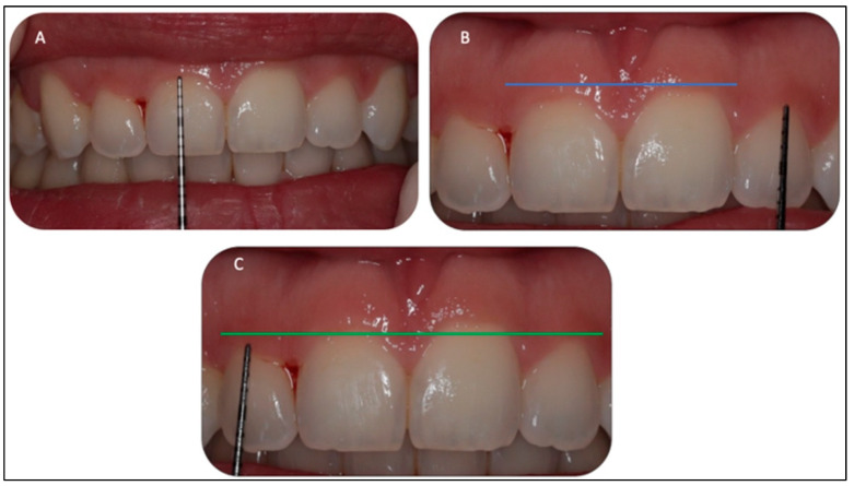
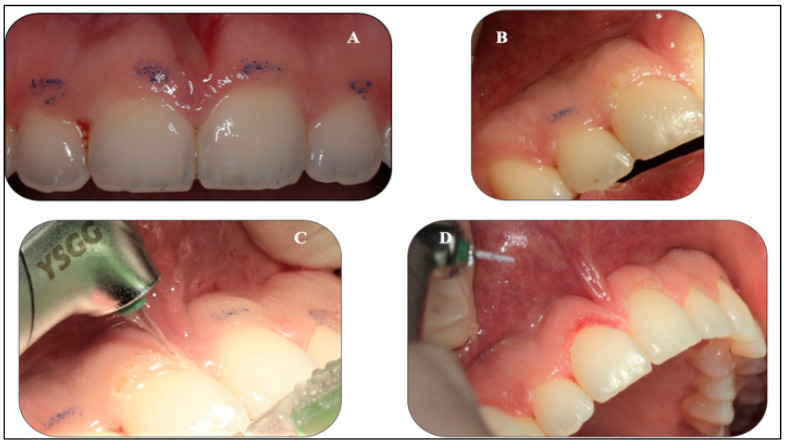
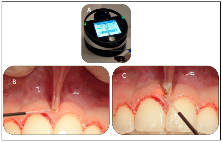
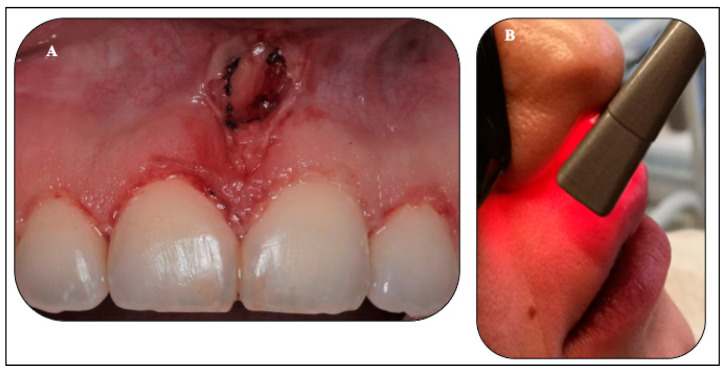
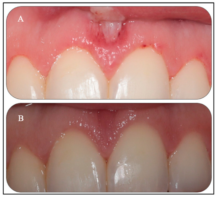
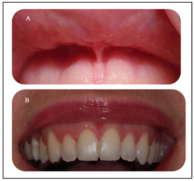
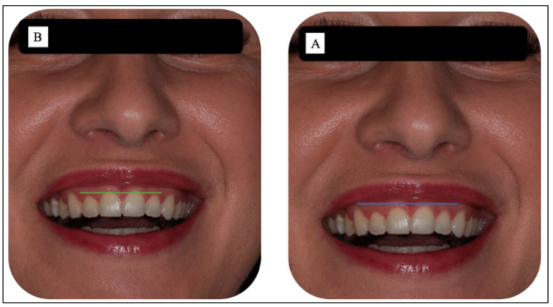
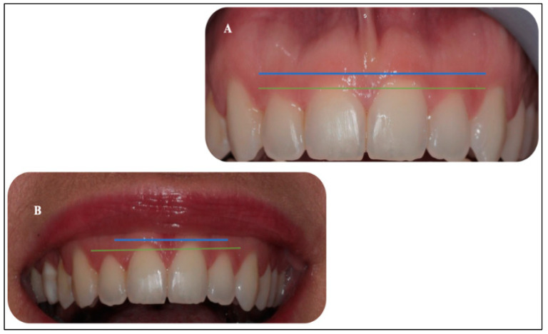
This case report is aimed to demonstrate the synergetic effects of λ940 nm laser photobiomodulation (PBM) therapy in augmenting the advantages of high-level-laser treatment (HLLT)-mediated reaction orthodontic periodontal interface management. Materials and Methods: A 32-year-old female who presented with a persistent gummy smile of upper incisors and low upper midline frenum attachment post-orthodontic treatment, was seeking a better smile appearance. She had a history of delayed wound healing without underlying medical conditions; otherwise, she was fit and healthy. She underwent laser ablation of the upper midline frenum and gingivoplasty of the upper incisors region with λ940 nm and λ2780, respectively, as well as transcutaneous PBM therapy (λ940 nm) to accelerate wound healing. The laser protocols were as follows: λ2780 nm: power output-2 W, pulse width-60 μs, free running pulse (FRP), spot area-0.0016 cm2, pulse repetition rate-25 pulses per second (s), 80 mJ/pulse, 90 s, λ940 nm: 1.2 W, continuous wave (CW) emission mode, 300 μm, 60 s; whereas the adjunctive λ940 nm induced-PBM parameters were as follows: power output-1.4 W, CW-120 s, single application, spot area-2.8 cm2. An acceleration of the wound healing was observed on the 4th day of treatment with no immediate or post-operative complications. The results showed no functional or aesthetic relapses at a long-term follow-up of 6 months. The authors concluded that λ940 nm laser-PBM can provide a synergetic effect to HLLT in accelerating wound healing and offering a precision smile with minimal to none post-operative complications. It is safe and justifiable to utilise dual therapy over the conventional methods, which serves our patients' needs in our daily practice and in various clinical indications. The concept and laser protocols of this clinical case report can pave the roadmap for future extensive studies.

Keywords: Er,Cr:YSGG; HLLT; LLLT; PBM; gingivoplasty; oral wound regeneration; photochemical; photothermal; surgical diode laser; upper midline frenectomy.


Conflict of interest statement

All authors have declared no conflict of interest.

Figures

Figure 1
Figure 1
An illustration of the concept of the surgical laser beam profile where the photonic energy λ940 nm, λ2970 nm of its first zone (HLLT) is absorbed by haemoglobin (Hb) and water, respectively, which is subsequently transformed into thermal energy and then causes tissue destruction (tissue ablation), whereas the last zone of laser beam is LLLT (Modified) [13,15]. The top right graph is an “Arndt–Schulz curve”, illustrating the biphasic dose response measured in the difference in the integrated area under the curve of the time course of wound size compared to a no-treatment control, with different modes of cell reaction at different levels of energy density [17].
Figure 2
Figure 2
Modified schematic description of the of mechanism of action for PBM, highlighting its four effects: the primary effects (photonic energy absorption by cytochrome C Oxidase (CCO)), secondary effects (mitochondrion of ATP, NO, and ROS), tertiary effects (downstream of intracellular responses effects (mitochondrion of ATP, NO, and ROS), and quaternary effect (indirect/distant effects such as gene transcription and cellular signaling) [21]. Abbreviations: PBM, photobiomodulation; ROS, reactive oxygen species; NO, nitric oxide; ATP, adenosine triphosphate.
Figure 3
Figure 3
(A–C). Clinical assessment of #11, #12, #21, and #22 revealed gingival overgrowth with no periodontal pocket or inflammation. The biological width was determined by measuring the proposed ablative gingival tissues with a periodontal probe and marking the proposed zones with an orthodontic marker. In addition, it shows the pocket depth determination with the periodontal probe. Biologic width requires the probe to pierce the attachments so that the clinician can accurately measure the distance to the osseous crest. Evidence of pre-operative discrepancy in the level of the gingival margin of the #11, compared to the #21, is highlighted in blue photo (B) as well for # 12 and #22, as highlighted in green photo (C).
Figure 4
Figure 4
(A,B). Pre-operative clinical and radiographic investigations. Photo (A) (top) is the periodontal charting, showing no evidence of any abnormality. Photo (B) (bottom) shows no evidence of any hard tissue abnormalities on the orthopantomogram.
Figure 5
Figure 5
(AD). Clinical photos (A,B) show the markings of the proposed gingival margin utilised by the orthodontic pencil based on the measurements, taking into consideration the biological width. The clinical photo (C) shows the angualtion of the Er,Cr:YSGG tip at 45° to follow the marked ginigval tissue. Clinical photo (D) shows an immediate gingivoplasty of #11.
Figure 6
Figure 6
(AC). Clinical photo (A) shows the tip initiation of the λ940 nm diode laser fibre using articulating paper, aiming to generate the maximum energy on its tip. Photo (B) is a peri-operative image, illustrating the upper midline frenum under stretch as well the light–tissue interaction, illustrating the initial ablation of the frenum attachments. Photo (C) is a peri-opearive image, showing the laser fiber direction at 90° to the frenum tissue and parallel to the alvoalr bone and away from the teeth positons.
Figure 7
Figure 7
(A,B). Clinical photo (A) was taken immediately following laser treatments, illustrating the rhomboid shape of the new position of the upper midline and the gingival re-contouring of the upper incisor teeth. Photo (B) is an extra-oral application of λ940 nm laser-PBM in the region of the treated sites.
Figure 8
Figure 8
(A,B). Clinical photo (A) reveals the healing status at the fourth day post-laser treatments, where the healing status of marginal gingival tissues of the #11, #12,# 21, and # 22 was grade IV, and the upper midline frenum shows re-epithelisation (grade IV), indicating an acceleration of the healing process. Clinical photo (B) reveals a complete healing of the upper midline frenum (Garde V) and great wound healing progress towards complete wound healing of the gingival margin of #11, #21, #12, and #22 (grade IV) at two weeks post-treatments.
Figure 9
Figure 9
(A,B) Clinical photos of both laser treatments at six-months post-laser treatments. Photo (A) shows a complete healing of the upper midline frenum with new frenum attachment without evidence of functional relapse. Photo (B) shows a complete healing of the gingival tissue with a great improvement in the gingival margins of the upper incisor teeth.
Figure 10
Figure 10
(A,B). At six-months post-laser treatment, photo (A) illustrates the level of the zeniths (blue line) of the gingival tissue of #11 and #21, whereas photo (B) illustrates the level of the zeniths (green line) of the gingival tissue of #12 and #22. The photos show an improvement in the clinical outcomes from an aesthetic standpoint.
Figure 11
Figure 11
(A,B) Illustration of the difference between the gingival overgrowth of the # 11, #12, #21, and #22 pre-operatively photo (A) and the (photo (B)) clinical outcome of the smile line at six-months post-laser gingivoplasty. Blue and green lines show the zeniths of the gingival tissue of #11 and #21, and #12 and #22, respectively. Photo (B) shows a great improvement in the gingival zeniths of upper incisors at six months, compared to those taken pre-operatively photo (A).
Figure 12
Figure 12
A periodontal chart at six-months post-laser treatments shows a healthy biological width and clinical attachment, as there is no evidence of gum recession and the pocket depth (PD) of the #11, #12, #21, and #22 ranged between 0–1 millimetres (mm). The PD was measured in mm at six points for each tooth (labial/buccal mesial, labial/buccal central, labial/buccal distal, palatal/lingual mesial, palatal/lingual central, and palatal/lingual distal). The empty circle represents a PD of 0 mm.

References

    1. Dym H., Pierre R. Diagnosis and Treatment Approaches to a “Gummy Smile”. Dent. Clin. N. Am. 2020;64:341–349. doi: 10.1016/j.cden.2019.12.003. - DOI - PubMed
    1. Nasr M.W., Jabbour S.F., Sidaoui J.A., Haber R.N., Kechichian E.G. Botulinum Toxin for the Treatment of Excessive Gingival Display: A Systematic Review. Aesthet. Surg. J. 2016;36:82–88. doi: 10.1093/asj/sjv082. - DOI - PubMed
    1. Amaral M.B., de Avila J.M., Abreu M.H., Mesquita R.A. Diode laser surgery versus scalpel surgery in the treatment of fibrous hyperplasia: A randomized clinical trial. Int. J. Oral Maxillofac. Surg. 2015;4:1383–1389. doi: 10.1016/j.ijom.2015.05.015. - DOI - PubMed
    1. Priyanka M., Sruthi R., Ramakrishnan T., Emmadi P., Ambalavanan N. An overview of frenal attachments. J. Indian Soc. Periodontol. 2013;17:12–15. doi: 10.4103/0972-124X.107467. - DOI - PMC - PubMed
    1. Bhatsange A., Meshram E.P., Waghamare A., Shiggaon L., Mehetre V.N., Shende A. A clinical and histological comparison of mucosal incisions produced by scalpel, electrocautery, and diode laser: A pilot study. J. Dent. Lasers. 2016;10:37–42. doi: 10.4103/2321-1385.196962. - DOI

Publication types

LinkOut - more resources