Review
. 2022 Mar 10;11(3):419.
doi: 10.3390/biology11030419.

Draining the Pleural Space: Lymphatic Vessels Facing the Most Challenging Task

Affiliations
Review

Draining the Pleural Space: Lymphatic Vessels Facing the Most Challenging Task

Eleonora Solari et al. Biology (Basel). .

Abstract

Lymphatic vessels exploit the mechanical stresses of their surroundings together with intrinsic rhythmic contractions to drain lymph from interstitial spaces and serosal cavities to eventually empty into the blood venous stream. This task is more difficult when the liquid to be drained has a very subatmospheric pressure, as it occurs in the pleural cavity. This peculiar space must maintain a very low fluid volume at negative hydraulic pressure in order to guarantee a proper mechanical coupling between the chest wall and lungs. To better understand the potential for liquid drainage, the key parameter to be considered is the difference in hydraulic pressure between the pleural space and the lymphatic lumen. In this review we collected old and new findings from in vivo direct measurements of hydraulic pressures in anaesthetized animals with the aim to better frame the complex physiology of diaphragmatic and intercostal lymphatics which drain liquid from the pleural cavity.

Keywords: breathing; chest wall; diaphragm; lung; lymphatic vessel; pleural cavity.


Conflict of interest statement

The authors declare no conflict of interest.

Figures

Figure 1
Figure 1
Representative image of lymphatic vessels organization on the pleural side of rat diaphragm, after the in vivo staining with a FITC-conjugated fluorescent tracer. Lymph enters lymphatic lacunae (asterisks) and then is propelled through vessels longitudinally (L) and/or perpendicularly (P) arranged with respect to the skeletal muscle fibers orientation. Lymphatic collectors located at the muscle periphery, next to the costal margin, are typically organized in complex loop structures (loop) and display intrinsic contractility. Scalebar 1 mm.
Figure 2
Figure 2
(A) Representative confocal image of a rat pleural diaphragmatic lymphatic vessel in vivo stained with FITC-dextrans (green signal) and whole mount stained for LMCs (red signal), highlighting the organization of the lymphatic muscle mesh surrounding the vessel (scalebar 100 µm). (B) Plot of rat diaphragmatic lymphatics intrinsic contraction amplitude (Δd) correlating to the density of lymphatic muscle in the vessels’ wall. (C) Plot of the dependence of rat diaphragmatic lymphatics intrinsic CF from temperature in the range 34–40 °C (green trace). Temperature-dependency is completely abolished by the selective TRPV4 channels antagonist HC067047 (2.5 µM, black dashed line). (D) Plot of osmolarity-induced modulation of rat diaphragmatic lymphatics intrinsic CF. The hyperosmolar environment (324 mOsm, red trace) induces a sigmoidal decease in CF. Hyposmolarity induces a two-phase response, as lymphatic vessels display an acute early CF increase (290 mOsm, blue dashed line) followed by a later CF decrease (blue solid line).
Figure 3
Figure 3
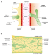
(A) Anatomical arrangement of parietal and visceral pleurae and the pleural space among them. Lymphatic stomata open directly into the pleural cavity. (B) Drawing of rat diaphragm cross section showing the reciprocal anatomical relationship among pleural and peritoneal submesothelial lymphatic lacunae, the transverse lymphatic network, and central lymphatic collectors (green). Skeletal muscle fiber bundles are shown in yellow and provide a strong extrinsic mechanical support to lymph propulsion during spontaneous breathing.

References

    1. Wiig H., Swartz M.A. Interstitial fluid and lymph formation and transport: Physiological regulation and roles in inflammation and cancer. Physiol. Rev. 2012;92:1005–1060. doi: 10.1152/physrev.00037.2011. - DOI - PubMed
    1. Leak L.V. Studies on the permeability of lymphatic capillaries. J. Cell Biol. 1971;50:300–323. doi: 10.1083/jcb.50.2.300. - DOI - PMC - PubMed
    1. Trzewik J., Mallipattu S.K., Artmann G.M., Delano F.A., Schmid-Schönbein G.W. Evidence for a second valve system in lymphatics: Endothelial microvalves. FASEB J. Off. Publ. Fed. Am. Soc. Exp. Biol. 2001;15:1711–1717. doi: 10.1096/fj.01-0067com. - DOI - PubMed
    1. Bazigou E., Wilson J.T., Moore J.E.J. Primary and secondary lymphatic valve development: Molecular, functional and mechanical insights. Microvasc. Res. 2014;96:38–45. doi: 10.1016/j.mvr.2014.07.008. - DOI - PMC - PubMed
    1. Baluk P., Fuxe J., Hashizume H., Romano T., Lashnits E., Butz S., Vestweber D., Corada M., Molendini C., Dejana E., et al. Functionally specialized junctions between endothelial cells of lymphatic vessels. J. Exp. Med. 2007;204:2349–2362. doi: 10.1084/jem.20062596. - DOI - PMC - PubMed

LinkOut - more resources