. 2022 Feb 22;14(3):450.
doi: 10.3390/v14030450.

Increased Heparanase Levels in Urine during Acute Puumala Orthohantavirus Infection Are Associated with Disease Severity

Affiliations

Increased Heparanase Levels in Urine during Acute Puumala Orthohantavirus Infection Are Associated with Disease Severity

Luz E Cabrera et al. Viruses. .

Abstract

Old-world orthohantaviruses cause hemorrhagic fever with renal syndrome (HFRS), characterized by acute kidney injury (AKI) with transient proteinuria. It seems plausible that proteinuria during acute HFRS is mediated by the disruption of the glomerular filtration barrier (GFB) due to vascular leakage, a hallmark of orthohantavirus-caused diseases. However, direct infection of endothelial cells by orthohantaviruses does not result in increased endothelial permeability, and alternative explanations for vascular leakage and diminished GFB function are necessary. Vascular integrity is partly dependent on an intact endothelial glycocalyx, which is susceptible to cleavage by heparanase (HPSE). To understand the role of glycocalyx degradation in HFRS-associated proteinuria, we investigated the levels of HPSE in urine and plasma during acute, convalescent and recovery stages of HFRS caused by Puumala orthohantavirus. HPSE levels in urine during acute HFRS were significantly increased and strongly associated with the severity of AKI and other markers of disease severity. Furthermore, increased expression of HPSE was detected in vitro in orthohantavirus-infected podocytes, which line the outer surfaces of glomerular capillaries. Taken together, these findings suggest the local activation of HPSE in the kidneys of orthohantavirus-infected patients with the potential to disrupt the endothelial glycocalyx, leading to increased protein leakage through the GFB, resulting in high amounts of proteinuria.

Keywords: Puumala hantavirus; acute kidney injury; glycocalyx; heparanase; podocytes; proteinuria; syndecan–1.


Conflict of interest statement

The authors declare no conflict of interest. The funders had no role in the design of the study; in the collection, analyses, or interpretation of data; in the writing of the manuscript, or in the decision to publish the results.

Figures
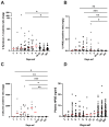
Figure 1
Figure 1
Heparanase and syndecan–1 levels during PUUV–HFRS. (A) Urinary syndecan–1:creatinine ratio (B), HPSE:creatinine ratio and (C) albumin:creatinine ratio. (D) HPSE concentration in plasma. Concentrations were measured from sequential samples of 49 (55 for plasma) patients hospitalized due to PUUV–HFRS (with a total of 159 urine and 262 plasma samples). Days after onset of fever (aof) 3–9 represent the acute stage, 20–30 the convalescent and 182–365 the controls. Differences between individual time points and the last time point (365 aof), which was considered to represent full recovery and used as controls, were assessed by GEE and significant differences indicated as *** <0.001, ** <0.01 and * < 0.05. Red lines represent mean ± standard deviation.
Figure 2
Figure 2
Spearman’s rank correlation coefficient matrix. The colors of the round circles indicate the level of the correlation coefficient and increasing size with a lower p–value. Statistical significance is specified as * = p < 0.05, **= p < 0.01, ***= p < 0.001, ****= p < 0.0001. AOF = after onset of fever, LOS = length of stay, eGFR = estimated glomerular filtration rate, P = plasma, U = urine, crea = creatinine, alb = albumin, HPSE = heparanase, Synd–1 = syndecan–1, IL = interleukin, IP–10 = interferon gamma–induced protein 10, MCP–1 = monocyte chemoattractant protein 1, min MAP = minimum mean arterial pressure, WBC = white blood cells, MPO = plasma myeloperoxidase, CRP = plasma C–reactive protein, tromb = blood thrombocytes.
Figure 3
Figure 3
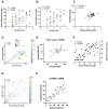
Simple and multiple linear regression models. Simple linear regression models of rank–transformed urinary albumin:creatinine ratio (A) and rank–transformed urinary HPSE:creatinine ratio (B) with severity score. (C) Simple linear regression model of plasma albumin and MAP. (D) Multiple linear regression model of plasma albumin, rank–transformed urinary HPSE:creatinine ratio and rank–transformed albumin on MAP. (E) Predicted vs. actual MAP of multiple linear regression model in (D). (F) Simple linear regression model of rank–transformed urinary HPSE:creatinine ratio and albumin:creatinine ratio. (G) Multiple linear regression model of eGFR and urinary HPSE on albuminuria. (H) Predicted vs. actual data of the multiple regression model in (G).
Figure 4
Figure 4
Increased HPSE concentration in HTNV–infected podocytes. Podocytes were infected with live HTNV (MOI 10 or 1), UV–inactivated HTNV (MOI 10) or remained uninfected (mock). Supernatants were collected and cells either fixed for immunofluorescence analysis or subjected to RNA extraction at 2 and 4 dpi. (A) Fixed and permeabilized podocytes were stained with rabbit serum against viral nucleocapsid N protein followed by AF488–conjugated secondary antibody (green) or Hoechst 33,420 to detect nuclei (blue). (B) HPSE levels measured from podocyte supernatants. (C) Isolated RNA was subjected to multiplex RT–qPCR with primers and probes detecting HPSE and GAPDH mRNA. The signal for HPSE mRNA was normalized based on GAPDH mRNA levels and fold change calculated in reference to mock–infected podocytes. Statistically significant differences were assessed with Tukey’s multiple comparisons test. ****, *** and * indicate p < 0.0001, p < 0.001 and p < 0.05, respectively.

References

    1. Jonsson C.B., Figueiredo L.T., Vapalahti O. A global perspective on hantavirus ecology, epidemiology, and disease. Clin. Microbiol. Rev. 2010;23:412–441. doi: 10.1128/CMR.00062-09. - DOI - PMC - PubMed
    1. Vaheri A., Strandin T., Hepojoki J., Sironen T., Henttonen H., Mäkelä S., Mustonen J. Uncovering the mysteries of hantavirus infections. Nat. Rev. Microbiol. 2013;11:539–550. doi: 10.1038/nrmicro3066. - DOI - PubMed
    1. Hepojoki J., Vaheri A., Strandin T. The fundamental role of endothelial cells in hantavirus pathogenesis. Front Microbiol. 2014;22:727. doi: 10.3389/fmicb.2014.00727. - DOI - PMC - PubMed
    1. Mustonen J., Outinen T., Laine O., Pörsti I., Vaheri A., Mäkelä S. Kidney disease in Puumala hantavirus infection. Infect. Dis. 2017;49:321–332. doi: 10.1080/23744235.2016.1274421. - DOI - PubMed
    1. Ala–Houhala I., Koskinen M., Ahola T., Harmoinen A., Kouri T., Laurila K., Mustonen J., Pasternack A. Increased glomerular permeability in patients with nephropathia epidemica caused by Puumala hantavirus. Nephrol. Dial. Transplant. 2002;17:246–252. doi: 10.1093/ndt/17.2.246. - DOI - PubMed

Publication types

LinkOut - more resources