Review
. 2022 Mar 12;14(3):585.
doi: 10.3390/v14030585.

Viral Shrimp Diseases Listed by the OIE: A Review

Affiliations
Review

Viral Shrimp Diseases Listed by the OIE: A Review

Dain Lee et al. Viruses. .

Abstract

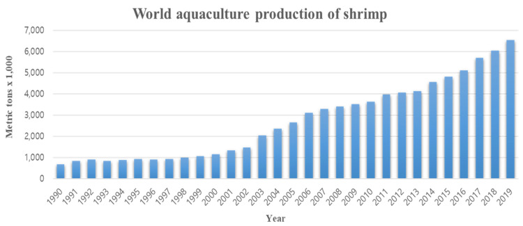
Shrimp is one of the most valuable aquaculture species globally, and the most internationally traded seafood product. Consequently, shrimp aquaculture practices have received increasing attention due to their high value and levels of demand, and this has contributed to economic growth in many developing countries. The global production of shrimp reached approximately 6.5 million t in 2019 and the shrimp aquaculture industry has consequently become a large-scale operation. However, the expansion of shrimp aquaculture has also been accompanied by various disease outbreaks, leading to large losses in shrimp production. Among the diseases, there are various viral diseases which can cause serious damage when compared to bacterial and fungi-based illness. In addition, new viral diseases occur rapidly, and existing diseases can evolve into new types. To address this, the review presented here will provide information on the DNA and RNA of shrimp viral diseases that have been designated by the World Organization for Animal Health and identify the latest shrimp disease trends.

Keywords: DNA and RNA virus; OIE; shrimp disease; viral disease.


Conflict of interest statement

The funders had no role in the design of the study; in the collection, analyses, or interpretation of data; in the writing of the manuscript, or in the decision to publish the results.

Figures

Figure 1
Figure 1
World aquaculture production of shrimp from 1990 to 2019 (Source: FAO yearbook of Fishery and Aquaculture Statistics).
Figure 2
Figure 2
Proportion of the major shrimp species Penaeus monodon and Penaeus vannamei in aquaculture production from 1990 to 2019 (Source: FAO yearbook of Fishery and Aquaculture Statistics).
Figure 3
Figure 3
Total shrimp aquaculture production for Penaeus monodon and Penaeus vannamei in Thailand from 1980 to 2019 (Source: FAO Global Aquaculture Production Statistics from FishstatJ Software for Fishery and Aquaculture Statistical Time Series).
Figure 4
Figure 4
Total shrimp aquaculture production for Penaeus chinensis and Penaeus vannamei in the Republic of Korea from 1980 to 2019 (Source: FAO Global Aquaculture Production Statistics from FishstatJ Software for Fishery and Aquaculture Statistical Time Series).
Figure 5
Figure 5
Distribution map showing the geographical occurrence of white spot syndrome disease (WSSD) (Reprinted from CABI, 2019, White spot syndrome virus. In: Invasive Species Compendium. Wallingford, UK: CAB International, with permission from CABI).
Figure 6
Figure 6
External white spot symptoms indicating white spot syndrome virus (WSSV) infection. (A) Penaeus monodon and (BD) Penaeus vannamei infected with WSSV. (A) Reprinted from Letter in Applied Microbiology, Vol. 60 (2), Hossain, A., Nandi, S.P., Siddique, M.A., Sanyal, S.K., Sultana, M., Hossain, M.A., Prevalence and distribution of White Spot Syndrome Virus in cultured shrimp, p. 7, Copyright (2014), with permission from John Wiley and Sons; (B) Reprinted from Elsevier Books, Dashtiannasab, A., Emerging and Reemerging Viral Pathogens, p. 12, Copyright (2020), with permission from Elsevier; (C,D) Reprinted from Journal of Fish Diseases, Vol. 36 (12), Cheng, L., Lin, W.H., Wang, P.C., Tsai, M.A., Hsu, J.P., Chen, S.C., White spot syndrome virus epizootic in cultured Pacific white shrimp Litopenaeus vannamei (Boone) in Taiwan, p. 9, Copyright (2013), with permission from John Wiley and Sons).
Figure 7
Figure 7
Penaeusvannamei infected with white spot syndrome virus (WSSV). The infection progresses through different stages that can be seen in the nucleus via histology. (A) Early-stage infected cells display enlarged nuclei with marginalized chromatin and a homogenous eosinophilic central region. These then develop an intranuclear eosinophilic Cowdry A-type inclusion (*); this can be surrounded by a clear halo beneath the nuclear membrane (white arrow). Scale bar = 25 µm; (B) The eosinophilic inclusion usually expands to fill the nucleus (*). This inclusion becomes basophilic when staining and denser in color as the infection progresses (white arrow). Nuclei then disintegrate so that the content fuses with the cytoplasm (black arrow). Scale bar = 10 µm. H & E stain; (C) WSSV virions appear ovoid in shape and contain an electron-dense nucleocapsid (white arrow) within a trilaminar envelope (black arrow). Scale bar = 0.2 µm. Inset. Negatively stained WSSV nucleocapsid, showing the presence of cross-hatched or striated material that is structured as a series of stacked rings of subunits and is a key diagnostic feature of WSSV. Scale bar = 20 nm; (D) Presumptive nucleocapsid material within the nucleus prior to envelopment. This material is cross-hatched or striated in appearance and linear prior to its incorporation in the formation of mature WSSV particles. This linear nucleocapsid material is observed sporadically in the manufacture of the WSSV particles. Scale bar = 100 nm. Transmission electron microscopy images (Source: Verbruggen et al., 2016, https://doi.org/10.3390/v8010023 accessed on 11 May 2018).
Figure 8
Figure 8
Distribution maps showing the geographical occurrence of infectious hypodermal and hematopoietic necrosis virus (Reprinted from CABI, 2019, Infectious hypodermal and hematopoietic necrosis. In: Invasive Species Compendium. Wallingford, UK: CAB International, with permission from CABI).
Figure 9
Figure 9
External symptoms of infectious hypodermal and hematopoietic necrosis virus (IHHNV) on shrimp. (A,B) subadult Penaeus vannamei with bent (to the left) rostrums, a classic sign of ‘runt deformity syndrome’ (RDS); (C) a juvenile P. vannamei with RDS. In this specimen the rostrum is bent to the right and the antennal flagella are wrinkled, brittle and mostly broken-off; (D) juvenile P. vannamei with RDS from a nursery population at approximately 60 days post stocking (Reprinted from Journal of Invertebrate Pathology, Vol. 106 (1), Lightner D.V., Virus diseases of farmed shrimp in the Western Hemisphere (the Americas) A rieview, p. 21, Copyright (2011), with permission from Elsevier).
Figure 10
Figure 10
Size variations observed in 50-day-old Penaeus monodon with infectious hypodermal and hematopoietic necrosis virus (IHHNV) (A,B) (Reprinted from Aquaculture, Vol. 289 (3–4), Rai, P., Pradeep, B., Karunasagar, I., Karunasagar, I., Detection of viruses in Penaeus monodon from India showing signs of slow growth syndrome, p. 5, Copyright (2009), with permission from Elsevier).
Figure 11
Figure 11
Electron microscopy and histological analysis of the changes in shrimp with infectious hypodermal and hematopoietic necrosis virus (IHHNV). (A) Electron microscopy of negatively stained IHHNV VLPs under self-assembly and disassembly conditions in Penaeus vannamei; (B) Cowdry type A eosinophilic inclusion of IHHNV in a nucleus of subcuticular epithelial cells of the pleopod of P. monodon (H & E, 1000×); (C) Histological detection of Procambarus clarkii gills negative to IHHNV detected by PCR. The gill cells were normal, no hypertrophied nucleus was observed; (D) Histological detection of P. clarkii gills positive to IHHNV detected by PCR. Several hypertrophied nuclei (arrow) were observed. ((A) Reprinted from Journal of Invertebrate Pathology, Vol. 166, Zhu, Y.P., Li, C., Wan, X.Y., Yang, Q., Xie, G.S., Huang, J., Delivery of plasmid DNA to shrimp hemocytes by infectious hypodermal and hematopoietic necrosis virus (IHHNV) nanoparticles expressed from a baculovirus insect cell system, p. 1, Copyright (2019), with permission from Elsevier; (B) Reprinted from Aquaculture, Vol. 289 (3–4), Rai, P., Pradeep, B., Karunasagar, I., Karunasagar, I., Detection of viruses in Penaeus monodon from India showing signs of slow growth syndrome, p. 5, Copyright (2009), with permission from Elsevier; (C,D) Reprinted from Aquaculture, Vol. 477, Chen, B.K., Dong, Z., Liu, D.P., Yan, Y.B., Pang, N.Y., Nian, Y.Y., Yan, D.C., Infectious hypodermal and hematopoietic necrosis virus (IHHNV) infection in freshwater crayfish Procambarus clarkii, p. 4, Copyright (2017), with permission from Elsevier).
Figure 12
Figure 12
Distribution map showing the geographical occurrence of infectious myonecrosis virus (IMNV) (Reprinted from CABI, 2019, Infectious myonecrosis virus. In: Invasive Species Compendium. Wallingford, UK: CAB International, with permission from CABI).
Figure 13
Figure 13
External symptoms of infectious myonecrosis virus (IMNV) on shrimp. (A) IMNV-infected Penaeus vannamei with reddish opaque muscles at the distal abdominal segments; (B) P. vannamei injected with IMNV propagated in a C6/36 cell line with reddish opaque muscle at the distal abdominal segments as observed in the natural infection; (C,D) P. vannamei infected with IMNV and displaying focal to extensive white necrotic areas in the striated muscle, especially of the distal abdominal segments and tail fan, and exposure of the paired lymphoid organs (LO) by simple dissection will show that the paired LO are hypertrophic to twice or more their normal size. ((A) Reprinted from Journal of Fish Diseases, Vol. 40 (12), Sahul Hameed, A.S., Abdul Majeed, S., Vimal, S., Madan, N., Rajkumar, T., Santhoshkumar, S., Sivakumar, S., Studies on the occurrence of infectious myonecrosis virus in pond-reared Litopenaeus vannamei (Boone, 1931) in India, p. 8, Copyright (2017), with permission from John Wiley and Sons; (B) Reprinted from Journal of Fish Diseases, Vol. 44 (7), Santhosh Kumar, S., Sivakumar, S., Abdul Majeed, S., Vimal, S., Taju, G., Sahul Hameed, A.S., In vitro propagation of infectious myonecrosis virus in C6/36 mosquito cell line, p. 6, Copyright (2021), with permission from John Wiley and Sons; (C,D) Reprinted from Journal of Invertebrate Pathology, Vol. 106(1), Lightner, D.V., Virus diseases of farmed shrimp in the Western Hemisphere (the Americas) a review, p. 21, Copyright (2011), with permission from Elsevier).
Figure 14
Figure 14
Electron microscopy and histological changes in shrimp with infectious myonecrosis virus (IMNV). (A) TEM of a purified preparation of IMNV from naturally infected Penaeus vannamei from Brazil. Photomicrographs of tissue sections from P. vannamei examined for IMNV lesions (BD) (Scale bar = 50 μm); (B) Focal hemocytic infiltration in muscle tissue; (C) Muscle coagulation necrosis accompanied by infiltration of hemocytes; (D) Muscle liquefactive necrosis and fibrosis. ((A) Reprinted from Journal of Invertebrate Pathology, Vol. 106 (1), Lightner, D.V., Virus diseases of farmed shrimp in the Western Hemisphere (the Americas) a review, p. 21, Copyright (2011), with permission from Elsevier; (BD) Reprinted from Aquaculture, Vol. 380, Feijó, R.G., Kamimura, M.T., Oliveira-Neto, J.M., Vila-Nova, C.M., Gomes, A.C., Maria das Graças, L.C., Maggioni, R., Infectious myonecrosis virus and white spot syndrome virus co-infection in Pacific white shrimp (Litopenaeus vannamei) farmed in Brazil, p. 5, Copyright (2013), with permission from Elsevier).
Figure 15
Figure 15
Distribution map showing the geographical occurrence of yellow head virus genotype 1 (YHV genotype 1) (Reprinted from CABI, 2019, Yellow head virus. In: Invasive Species Compendium. Wallingford, UK: CAB International, with permission from CABI).
Figure 16
Figure 16
External symptoms on yellow head virus genotype 1 (YHV genotype 1)-infected shrimp. (A) P. monodon showing signs of yellow head disease (YHD) Yellow (light gray in print version) to yellow-brown (dark gray in print version) discoloration of the cephalothorax. Three shrimp with (left) and without (right) YHD; (B) discoloration of the gill region. ((A,B) Reprinted from Elsevier Books, Samocha, Sustainable biofloc systems for marine shrimp, p. 23, Copyright (2019), with permission from Elsevier).
Figure 17
Figure 17
Electron microscopy and histological changes in shrimp infected with yellow head virus genotype 1 (YHV). (A) TEM of negative-strained YHV virions (Scale bars = 100 nm); (B) LO tissue of moribund shrimp from YHV immersion challenged P. vannamei at day 5 showing numerous pyknotic nuclei (arrows), karyorrhectic nucleic and cytoplasmic inclusion (arrow heads); (C) Hemolymph from normal and YHV infected shrimp identified by staining hemolymph smears; (D) Gills of YHV infected shrimp stained with H&E in rapidly fixed and stained (3 h) whole mounts. ((A) Reprinted from Advances in virus research, Vol. 63, Dhar, A.K., Cowley, J.A., Hasson, K.W., Walker, P.J., Genomic organization, biology, and diagnosis of Taura syndrome virus and yellow head virus of penaeid shrimp, p. 69, Copyright (2004), with permission from Elsevier; (B) Reprinted from Developmental & Comparative Immunology, Vol. 32 (6), Anantasomboon, G., Poonkhum, R., Sittidilokratna, N., Flegel, T.W., Withyachumnarnkul, B., Low viral loads and lymphoid organ spheroids are associated with yellow head virus (YHV) tolerance in whiteleg shrimp Penaeus vannamei, p. 14, Copyright (2008), with permission from Elsevier; (C,D) Reprinted from Aquaculture, Vol. 258 (1–4), Flegel, T.W., Detection of major penaeid shrimp viruses in Asia, a historical perspective with emphasis on Thailand, p. 33, Copyright (2006), with permission from Elsevier).
Figure 18
Figure 18
Distribution map showing the geographical occurrence of Taura syndrome virus (TSV) (Reprinted from CABI, 2019, Taura syndrome virus. In: Invasive Species Compendium. Wallingford, UK: CAB International, with permission from CABI).
Figure 19
Figure 19
External symptoms of Taura syndrome virus (TSV) on infected shrimp. (A,B) Penaeus vannamei showing typical signs of TSV at the end of the acute phase: Multifocal and melanized lesions on the thorax and tail (indicated by arrow); (C,D) P. vannamei showing signs of TSV: red tail fan with rough edges on the cuticular epithelium of uropods (indicated by arrow) and multiple melanized cuticular lesions. ((A) Reprinted from Elsevier Books, Dhar, A.K., Allnutt, F.T., Taura Syndrome Virus. In Encyclopedia of virology, p. 8, Copyright (2008), with permission from Elsevier; (B) Reprinted from Aquaculture, Vol. 260 (1–4), Phalitakul, S., Wongtawatchai, J., Sarikaputi, M., Viseshakul, N., The molecular detection of Taura syndrome virus emerging with White spot syndrome virus in penaeid shrimps of Thailand, p. 9, Copyright (2006), with permission from Elsevier; (C,D) Reprinted from Elsevier Books, Samocha, Sustainable biofloc system for marine shrimp, p. 23, Copyright (2019), with permission from Elsevier).
Figure 20
Figure 20
Electron microscopy and histological changes in shrimp infected with Taura syndrome virus (TSV). (A) TEM of CsCl gradient-purified and negative-strained (with 2% PTA) TSV particle isolated from Penaeus vannamei in Ecuador; (B) the section of intestine with 400 × magnification has cytoplasmic inclusion bodies in the lymphoid organ of Penaeus monodon (arrow); (C,D) spheroids (LOS) in the lymphoid organ tissue and ectopic spheroids in the connective tissue of P. vannamei from Venezuela, when stained with H&E, respectively (Scale bar = 25 μm). ((A) Reprinted from Advances in virus research, Vol. 63, Dhar, A.K., Cowley, J.A., Hasson, K.W., Walker, P.J., Genomic organization, biology, and diagnosis of Taura syndrome virus and yellowhead virus of penaeid shrimp, p. 69, Copyright (2004), with permission from Elsevier; (B) Reprinted from Aquaculture, Vol. 260 (1–4), Phalitakul, S., Wongtawatchai, J., Sarikaputi, M., Viseshakul, N., The molecular detection of Taura syndrome virus emerging with White spot syndrome virus in penaeid shrimps of Thailand, p. 9, Copyright (2006), with permission from Elsevier; (C,D) Reprinted from Aquaculture, Vol. 480, Tang, K.F., Aranguren, L.F., Piamsomboon, P., Han, J.E., Maskaykina, I.Y., Schmidt, M.M., Detection of the microsporidian Enterocytozoon hepatopenaei (EHP) and Taura syndrome virus in Penaeus vannamei cultured in Venezuela, p. 5, Copyright (2017), with permission from Elsevier).
Figure 21
Figure 21
Distribution map of the geographical occurrence of White tail disease (WTD). (Reprinted from CABI, 2019, Macrobrachium rosenbergii nodavirus. In: Invasive Species Compendium. Wallingford, UK: CAB International, with permission from CABI).
Figure 22
Figure 22
External symptoms of shrimps with White tail disease (WTD). (A) MrNV-infected Penaeus vannamei showing signs of whitish muscle in the tail (arrows); (B) Cherax quadricarinatus showing signs of WTD with necrosis and myositis (arrows); (C,D) Clinical signs, whitish abdominal muscles (arrows), in the infected post-larvae of Penaeus indicus ((A) Reprinted from Aquaculture, Vol. 483, Jariyapong, P., Pudgerd, A., Weerachatyanukul, W., Hirono, I., Senapin, S., Dhar, A.K., Chotwiwatthanakun, C., Construction of an infectious Macrobrachium rosenbergii nodavirus from cDNA clones in Sf9 cells and improved recovery of viral RNA with AZT treatment, p. 9, Copyright (2018), with permission from Elsevier; (B) Reprinted from Aquaculture, Vol. 319 (1–2), Hayakijkosol, O., La Fauce, K., Owens, L., Experimental infection of redclaw crayfish (Cherax quadricarinatus) with Macrobrachium rosenbergii nodavirus, the aetiological agent of white tail disease, p. 5, Copyright (2011), with permission from Elsevier; (C,D) Reprinted from Aquaculture, Vol. 292(1–2), Ravi, M., Basha, A.N., Sarathi, M., Idalia, H.R., Widada, J.S., Bonami, J.R., Hameed, A.S., Studies on the occurrence of white tail disease (WTD) caused by MrNV and XSV in hatchery-reared post-larvae of Penaeus indicus and P. monodon, p. 4, Copyright (2009), with permission from Elsevier).
Figure 23
Figure 23
Histological changes in shrimp tissues when infected with White tail disease (WTD) and stained with H&E. (A) Uninfected shrimp; (B) Histological detection included the aggregation of cells into clumps of various sizes and coagulative necrosis in P. vannamei skeletal muscle (72 h post-infection); (C,D) Muscle degeneration and necrotic muscle tissues in MrNV-infected C. quadricarinatus (arrow). ((A,B) Reprinted from Aquaculture, Vol. 483, Jariyapong, P., Pudgerd, A., Weerachatyanukul, W., Hirono, I., Senapin, S., Dhar, A.K., Chotwiwatthanakun, C., Construction of an infectious Macrobrachium rosenbergii nodavirus from cDNA clones in Sf9 cells and improved recovery of viral RNA with AZT treatment, p. 9, Copyright (2018), with permission from Elsevier; (C,D) Reprinted from Aquaculture, Vol. 319 (1–2), Hayakijkosol, O., La Fauce, K., Owens, L., Experimental infection of redclaw crayfish (Cherax quadricarinatus) with Macrobrachium rosenbergii nodavirus, the aetiological agent of white tail disease, p. 5, Copyright (2011), with permission from Elsevier).

References

    1. Walker P.J., Mohan C.V. Viral disease emergence in shrimp aquaculture: Origins, impact and the effectiveness of health management strategies. Rev. Aquac. 2009;1:125–154. doi: 10.1111/j.1753-5131.2009.01007.x. - DOI - PMC - PubMed
    1. Lightner D.V., Redman R.M., Pantoja C.R., Tang K.F.J., Noble B.L., Schofield P., Navarro S.A. Historic emergence, impact and current status of shrimp pathogens in the Americas. J. Invertebr. Pathol. 2012;110:174–183. doi: 10.1016/j.jip.2012.03.006. - DOI - PubMed
    1. Roy S., Bossier P., Norouzitallab P., Vanrompay D. Trained immunity and perspectives for shrimp aquaculture. Rev. Aquac. 2020;12:2351–2370. doi: 10.1111/raq.12438. - DOI
    1. Manan H., Ikhwanuddin M. Triploid induction in penaeid shrimps aquaculture: A review. Rev. Aquac. 2021;13:619–631. doi: 10.1111/raq.12489. - DOI
    1. Morshed M., Islam M.S., Lohano H.D., Shyamsundar P. Production externalities of shrimp aquaculture on paddy farming in coastal Bangladesh. Agric. Water. Manag. 2020;238:106213. doi: 10.1016/j.agwat.2020.106213. - DOI

Publication types