. 2022 Mar 15;14(3):606.
doi: 10.3390/v14030606.

RNA Structural Requirements for Nucleocapsid Protein-Mediated Extended Dimer Formation

Affiliations

RNA Structural Requirements for Nucleocapsid Protein-Mediated Extended Dimer Formation

Françoise Chaminade et al. Viruses. .

Abstract

Retroviruses package two copies of their genomic RNA (gRNA) as non-covalently linked dimers. Many studies suggest that the retroviral nucleocapsid protein (NC) plays an important role in gRNA dimerization. The upper part of the L3 RNA stem-loop in the 5' leader of the avian leukosis virus (ALV) is converted to the extended dimer by ALV NC. The L3 hairpin contains three stems and two internal loops. To investigate the roles of internal loops and stems in the NC-mediated extended dimer formation, we performed site-directed mutagenesis, gel electrophoresis, and analysis of thermostability of dimeric RNAs. We showed that the internal loops are necessary for efficient extended dimer formation. Destabilization of the lower stem of L3 is necessary for RNA dimerization, although it is not involved in the linkage structure of the extended dimer. We found that NCs from ALV, human immunodeficiency virus type 1 (HIV-1), and Moloney murine leukemia virus (M-MuLV) cannot promote the formation of the extended dimer when the apical stem contains ten consecutive base pairs. Five base pairs correspond to the maximum length for efficient L3 dimerization induced by the three NCs. L3 dimerization was less efficient with M-MuLV NC than with ALV NC and HIV-1 NC.

Keywords: HIV-1; RNA dimerization; RNA secondary structure; Rous sarcoma virus (RSV); nucleocapsid protein; retrovirus.


Conflict of interest statement

The authors declare no conflict of interest.

Figures

Figure 1
Figure 1
Predicted secondary structures for the ALV 5′-leader RNA [30]. Numbering is relative to the genomic RNA cap site (+1). R, repeated sequence; U5, unique sequence in 5′; PBS, primer binding site; SL-A, B, and C, stem-loop structures of the minimal packaging signal [31]; L3 in blue font, stem-loop structure involved in ALV gRNA dimerization; the structural elements of L3 are indicated in blue; AUG in bold, gag initiation codon; SD, splice donor site.
Figure 2
Figure 2
Secondary structures for the L3 stem-loop in the monomer and the extended dimer.
Figure 3
Figure 3
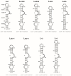
Predicted secondary structures for the L3 sequence of LRNAs containing mutations or deletions. The mfold program [40] predicted the most stable secondary structure for each RNA. Numbering is relative to the genomic RNA cap site (+1). The lower-case letters in the boxes indicate the mutations.
Figure 4
Figure 4
Dimerization of L RNAs harboring mutations in loop B. The 5′-end-labeled L RNAs in the absence (lanes C) or the presence of NCp12 (lanes 3–6) were incubated at 37 °C as described in Materials and Methods. After phenol–chloroform extraction, the samples were analyzed by electrophoresis on 12% polyacrylamide gels at 25 °C in the TBE buffer. Heat-denatured L RNAs (lanes D) were used to identify the bands corresponding to monomeric L RNAs. Lanes 3–6, protein to nucleotide molar ratios were 1:8, 1:4, 1:2, and 1:1. The monomeric and dimeric forms of L RNAs are indicated by m and d, respectively. The graph was derived from the gels shown in this figure. Filled circles, Lwt RNA; open circles, Lm1LB RNA; open squares, Lm2LB RNA; open triangles, LdLB RNA.
Figure 5
Figure 5
Thermal stability of the dimeric LdLB RNA. The melting curve was determined as described in Materials and Methods. Data are normalized according to the percentage of dimer at 30 °C and result from three experiments. Error bars show standard deviations.
Figure 6
Figure 6
Deletion of loop A and the G bulge prevents NCp12-mediated L3 RNA dimerization. The 5′-end-labeled LdLA RNA in the absence (lane C) or the presence of NCp12 (lanes 3–6) was incubated at 37 °C as described in Materials and Methods. After phenol–chloroform extraction, the samples were analyzed by electrophoresis on a 12% polyacrylamide gel at 25 °C in the TBE buffer. Heat-denatured LdLA RNA (lane D) was used to identify the band corresponding to monomeric LdLA RNA. Lanes 3–6, protein to nucleotide molar ratios were 1:8, 1:4, 1:2, and 1:1. The monomeric and dimeric forms of LdLA RNA are indicated by m and d, respectively. The graph was derived from the gels shown in this figure and Figure 4. Filled circles, Lwt; open circles, LdLA.
Figure 7
Figure 7
Influence of stem C extensions on NCp12-mediated L3 RNA dimerization. The 5′-end-labeled L RNAs in the absence (lanes C) or the presence of NCp12 (lanes 3–6) were incubated at 37 °C as described in Materials and Methods. After phenol–chloroform extraction, the samples were analyzed by electrophoresis on 12% polyacrylamide gels at 25 °C in the TBE buffer. Heat-denatured L RNAs (lanes D) were used to identify the bands corresponding to monomeric L RNAs. Lanes 3–6, protein to nucleotide molar ratios were 1:8, 1:4, 1:2, and 1:1. The monomeric and dimeric forms of L RNAs are indicated by m and d, respectively. The graph was derived from the gels shown in this figure and Figure 4. Filled circles, Lwt RNA; open circles, LAU+1 RNA; open squares, LGC+1 RNA; open triangles, L+2 RNA; open diamonds, L+5 RNA.
Figure 8
Figure 8
Influence of stem C extensions on L3 RNA dimerization induced by NCp7 and NCp10. (A) NCp7-mediated L3 RNA dimerization. (B) NCp10-mediated L3 RNA dimerization. The 5′-end-labeled L RNAs in the absence (lanes C) or the presence of NCp7 or NCp10 (lanes 3–6) were incubated at 37 °C as described in Materials and Methods. After phenol–chloroform extraction, the samples were analyzed by electrophoresis on 12% polyacrylamide gels at 25 °C in the TBE buffer. Heat-denatured L RNAs (lanes D) were used to identify the bands corresponding to monomeric L RNAs. Lanes 3–6, protein to nucleotide molar ratios were 1:8, 1:4, 1:2, and 1:1. The monomeric and dimeric forms of L RNAs are indicated by m and d, respectively. The graphs were derived from the gels shown in this figure. Filled circles, Lwt RNA; open circles, LAU+1 RNA; open squares, LGC+1 RNA; open triangles, L+2 RNA; open diamonds, L+5 RNA.

Similar articles

References

    1. Dubois N., Marquet R., Paillart J.-C., Bernacchi S. Retroviral RNA Dimerization: From Structure to Functions. Front. Microbiol. 2018;9:527. doi: 10.3389/fmicb.2018.00527. - DOI - PMC - PubMed
    1. Onafuwa-Nuga A., Telesnitsky A. The Remarkable Frequency of Human Immunodeficiency Virus Type 1 Genetic Recombination. Microbiol. Mol. Biol. Rev. 2009;73:451–480. doi: 10.1128/MMBR.00012-09. - DOI - PMC - PubMed
    1. Pornillos O., Ganser-Pornillos B.K. Maturation of Retroviruses. Curr. Opin. Virol. 2019;36:47–55. doi: 10.1016/j.coviro.2019.05.004. - DOI - PMC - PubMed
    1. Stoltzfus C.M., Snyder P.N. Structure of B77 Sarcoma Virus RNA: Stabilization of RNA after Packaging. J. Virol. 1975;16:1161–1170. doi: 10.1128/jvi.16.5.1161-1170.1975. - DOI - PMC - PubMed
    1. Fu W., Rein A. Maturation of Dimeric Viral RNA of Moloney Murine Leukemia Virus. J. Virol. 1993;67:5443–5449. doi: 10.1128/jvi.67.9.5443-5449.1993. - DOI - PMC - PubMed

LinkOut - more resources