. 2022 Mar 18;14(3):632.
doi: 10.3390/v14030632.

Investigation of the Low-Populated Excited States of the HIV-1 Nucleocapsid Domain

Affiliations

Investigation of the Low-Populated Excited States of the HIV-1 Nucleocapsid Domain

Assia Mouhand et al. Viruses. .

Abstract

The nucleocapsid domain (NCd), located at the C-terminus of the HIV-1 Gag protein, is involved in numerous stages of the replication cycle, such as the packaging of the viral genome and reverse transcription. It exists under different forms through the viral life cycle, depending on the processing of Gag by the HIV-1 protease. NCd is constituted of two adjacent zinc knuckles (ZK1 and ZK2), separated by a flexible linker and flanked by disordered regions. Here, conformational equilibria between a major and two minor states were highlighted exclusively in ZK2, by using CPMG and CEST NMR experiments. These minor states appear to be temperature dependent, and their populations are highest at physiological temperature. These minor states are present both in NCp7, the mature form of NCd, and in NCp9 and NCp15, the precursor forms of NCd, with increased populations. The role of these minor states in the targeting of NCd by drugs and its binding properties is discussed.

Keywords: CEST; CPMG; HIV-1; NCp15; NCp7; NCp9; NMR; dark-state; dynamic; low-populated state; nucleocapsid.


Conflict of interest statement

The authors declare no conflict of interest.

Figures
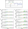
Figure 1
Figure 1
15N-CPMG relaxation dispersion experiments measured on NCp7 at 700 MHz and different conditions of temperature (10 °C and 35 °C), salt concentration (25 or 50 mM) and pH (5.5, 6.0, or 6.5). (A) Sequence of the HIV-1 C-terminal domain of Gag (NCp15). The first cleavage by the HIV-1 protease first liberates the NCp15 protein, then NCp9. and finally, the mature form of NCd called NCp7. The dashed line represents the two sites of protease cleavage present in NCp15. Residues are colored in grey for residues in the N-terminal part of NCd, green for those in the ZKs. orange for residues in the linker between the two ZKs of NCd, and purple for the C-terminal domain of NCd; (B) Rex measured for NCp7 at 25 mM NaCl and 10 °C for three pH (5.5, 6.0, and 6.5); (C) Rex measured for NCp7 at 25 mM NaCl and 35 °C for three pH (5.5, 6, and 6.5); (D) Rex measured for NCp7 at 50 mM NaCl and 10 °C for three pH (5.5, 6, and 6.5); (E) Rex measured for NCp7 at 50 mM NaCl and 35 °C for three pH (5.5, 6, and 6.5). In panels (B)–(E), the bars are colored using the same color code as the amino acid sequence in panel (A).
Figure 2
Figure 2
15N-CPMG relaxation dispersion experiments measured for NCp9 (upper panel), and NCp15 (lower panel) at 700 MHz, pH 6.5 and at (A) 10 °C or (B) 35 °C. (C) 15N-CPMG relaxation dispersion profiles of residues in NCp15 (in red) and NCp9 (in purple) that coordinate the zinc atom in ZK2 (C36, C39, H44, and C49). The experimental data are represented with dots and the fit curve of the data by lines. The fit was done with ChemEx for a three-state model, with one major and two minor states that interconvert.
Figure 3
Figure 3
15N-chemical shift differences for ZK2 residues. (A) ΔωAB and (B) ΔωAc between major and minor species obtained from the fit of 15N-CPMG relaxation dispersion experiment for NCp7 (black), NCp9 (purple), and NCp15 (red). The signs of ΔωAB and ΔωAC were obtained from the CEST data of NCp7 and the same signs were used to represent ΔωAB and ΔωAC of NCp9 and NCp15.
Figure 4
Figure 4
15N-CEST intensity profiles for C39, G40, Q45, and W37 residues obtained at 600 MHz and 25 °C with a B1 field of 25 Hz and Tex = 0.4 s. The main dip is at the resonance frequency of the major state and minor dips were observed at the frequency of the minor states. For the figure, the ChemEx software was used to fit the data, using an individual fit for each residue.
Figure 5
Figure 5
15N-CPMG relaxation dispersion profiles measured at 950 MHz and 35 °C at two different protein concentrations, 100 μM (blue) and 400 μM (green), for six residues of NCp7.

References

    1. Darlix J.-L., de Rocquigny H., Mauffret O., Mély Y. Retrospective on the All-in-One Retroviral Nucleocapsid Protein. Virus Res. 2014;193:2–15. doi: 10.1016/j.virusres.2014.05.011. - DOI - PMC - PubMed
    1. Levin J.G., Mitra M., Mascarenhas A., Musier-Forsyth K. Role of HIV-1 Nucleocapsid Protein in HIV-1 Reverse Transcription. RNA Biol. 2010;7:754–774. doi: 10.4161/rna.7.6.14115. - DOI - PMC - PubMed
    1. Thomas J.A., Gorelick R.J. Nucleocapsid Protein Function in Early Infection Processes. Virus Res. 2008;134:39–63. doi: 10.1016/j.virusres.2007.12.006. - DOI - PMC - PubMed
    1. Darlix J.-L., Godet J., Ivanyi-Nagy R., Fossé P., Mauffret O., Mély Y. Flexible Nature and Specific Functions of the HIV-1 Nucleocapsid Protein. J. Mol. Biol. 2011;410:565–581. doi: 10.1016/j.jmb.2011.03.037. - DOI - PubMed
    1. Godet J., Mély Y. Biophysical Studies of the Nucleic Acid Chaperone Properties of the HIV-1 Nucleocapsid Protein. RNA Biol. 2010;7:687–699. doi: 10.4161/rna.7.6.13616. - DOI - PubMed

Publication types

MeSH terms

Substances

LinkOut - more resources