. 2022 Mar 13;15(3):347.
doi: 10.3390/ph15030347.

ApoA-I Nanoparticles as Curcumin Carriers for Cerebral Endothelial Cells: Improved Cytoprotective Effects against Methylglyoxal

Affiliations

ApoA-I Nanoparticles as Curcumin Carriers for Cerebral Endothelial Cells: Improved Cytoprotective Effects against Methylglyoxal

Sai Sandhya Narra et al. Pharmaceuticals (Basel). .

Abstract

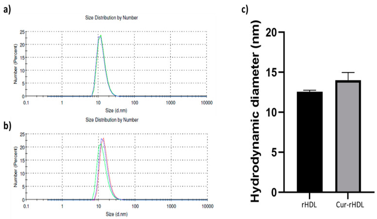
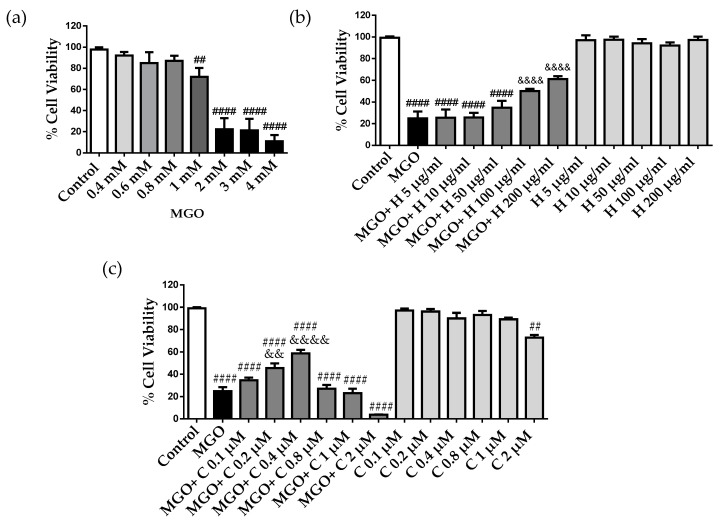
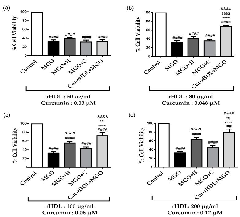
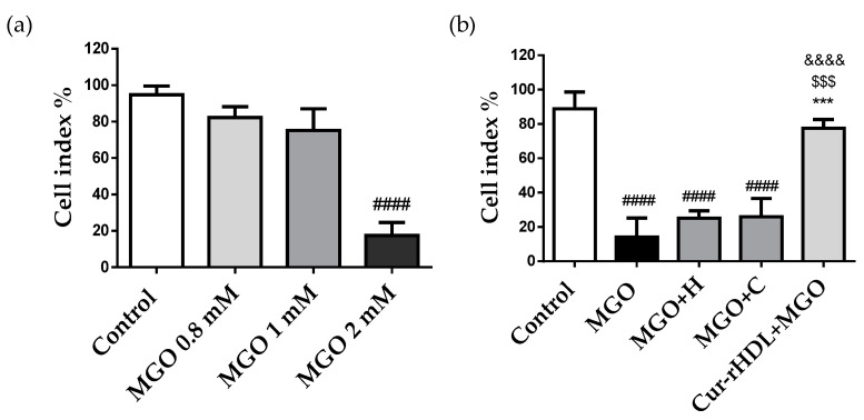
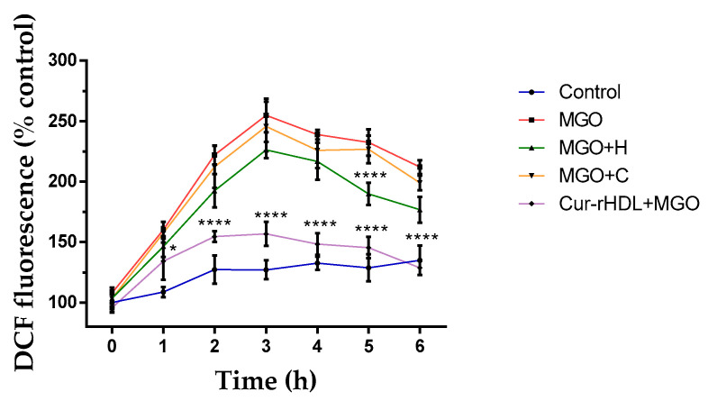
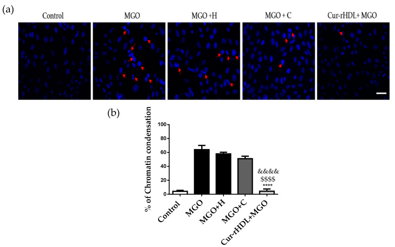
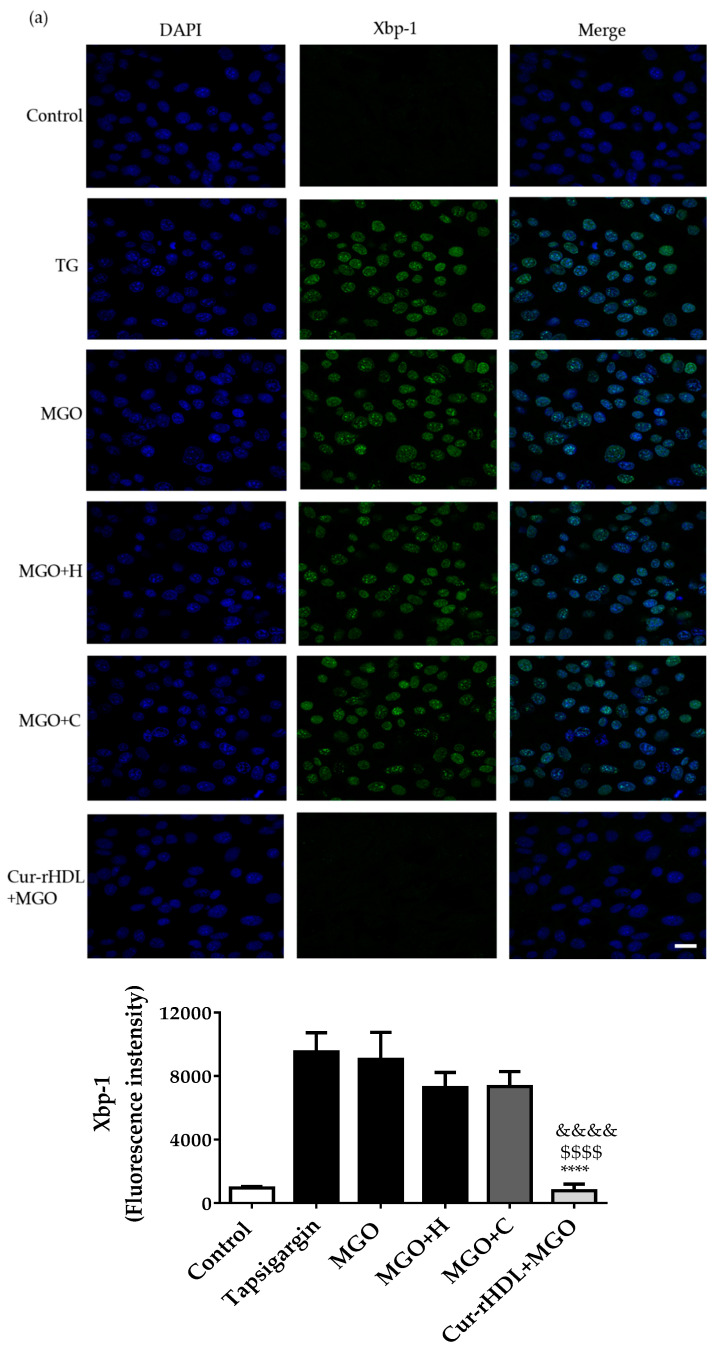
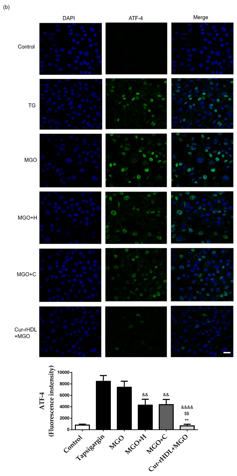
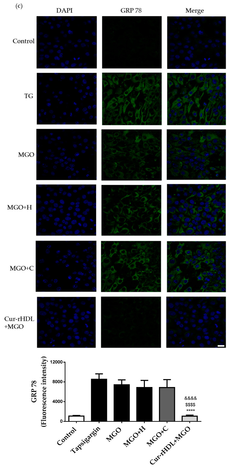
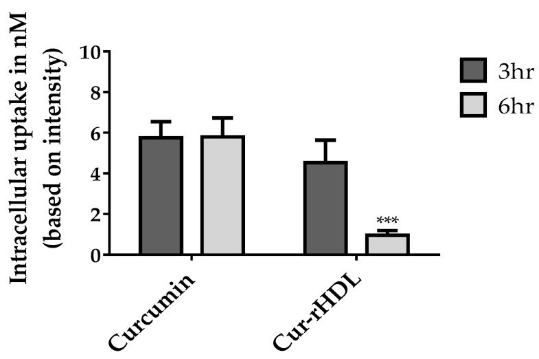
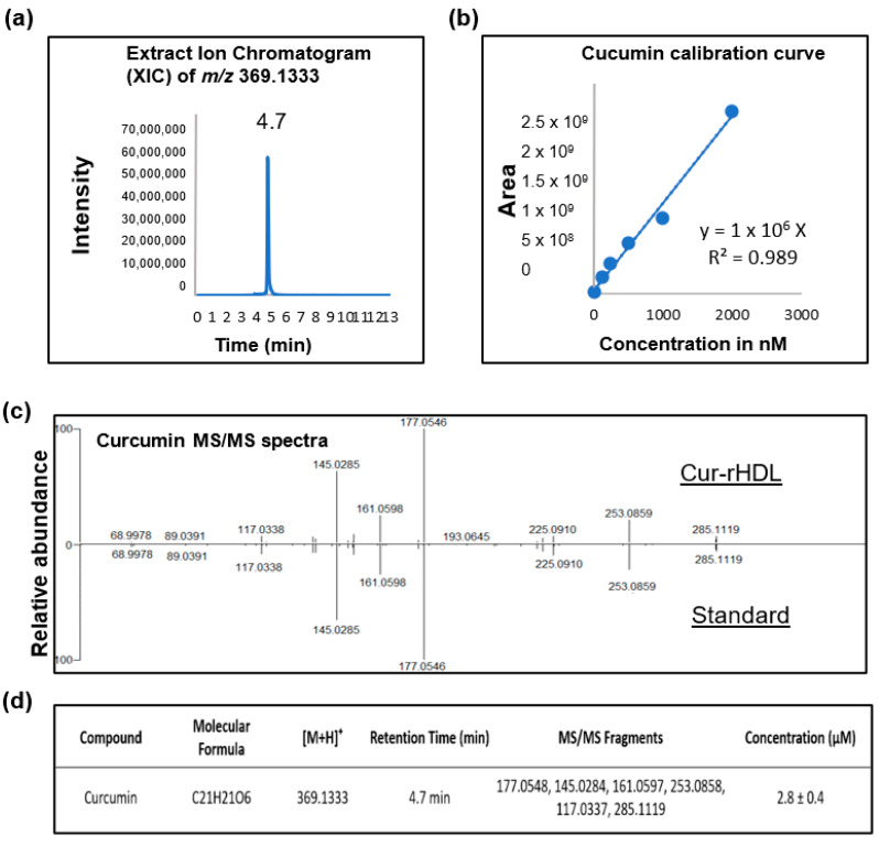
Methylglyoxal (MGO) is a highly reactive metabolite of glucose present at elevated levels in diabetic patients. Its cytotoxicity is associated with endothelial dysfunction, which plays a role in cardiovascular and cerebrovascular complications. Although curcumin has many therapeutic benefits, these are limited due to its low bioavailability. We aimed to improve the bioavailability of curcumin and evaluate a potential synergistic effect of curcumin and reconstituted high-density lipoprotein (rHDL) nanoparticles (Cur-rHDLs) on MGO-induced cytotoxicity and oxidative stress in murine cerebrovascular endothelial cells (bEnd.3). Cur-rHDL nanoparticles (14.02 ± 0.95 nm) prepared by ultracentrifugation and containing curcumin were quantified by LC-MS/MS. The synergistic effect of cur-rHDL nanoparticles was tested on bEnd.3 cytotoxicity, reactive oxygen species (ROS) production, chromatin condensation, endoplasmic reticulum (ER) stress, and endothelial barrier integrity by impedancemetry. The uptake of curcumin, alone or associated with HDLs, was also assessed by mass spectrometry. Pretreatment with Cur-rHDLs followed by incubation with MGO showed a protective effect on MGO-induced cytotoxicity and chromatin condensation, as well as a strong protective effect on ROS production, endothelial cell barrier integrity, and ER stress. These results suggest that Cur-rHDLs could be used as a potential therapeutic agent to limit MGO-induced dysfunction in cerebrovascular endothelial cells by enhancing the bioavailability and protective effects of curcumin.

Keywords: HDL; cerebral endothelial cells; curcumin; endothelial dysfunction; methylglyoxal; nanoparticle.


Conflict of interest statement

The authors declare no conflict of interest.

Figures

Figure 1
Figure 1
Particle size distribution of rHDLs and Cur-rHDLs determined by DLS analysis. The samples were prepared in PBS at 1 mg/mL. (a) Size distribution of rHDLs and (b) Cur-rHDLs and (c) Histograms representing the hygrodynamic particle diameter of rHDLs and Cur-rHDLs. Data are presented as the mean ± SD of three independent measurements for each sample.
Figure 2
Figure 2
MGO cytotoxicity in bEnd3 cerebral endothelial cells, effect of rHDLs (H) and curcumin (C). (a) The cells were treated with different MGO concentrations (0.4–4 mM) for 24 h. (b) The cells were pre-treated with different concentrations of rHDLs, 5–200 μg/mL, (H) and (c) curcumin (C), 0.1–2 μM, for 1 h, followed by the addition of 2 mM MGO for 24 h. Cell viability was assessed by the MTT assay. Data are presented as mean ± SD of three independent experiments (n = 3). ## p ˂ 0.01, #### p ˂ 0.0001 as compared to control. && p ˂ 0.01, &&&& p ˂ 0.0001 as compared to MGO.
Figure 3
Figure 3
Effect of curcumin-enriched rHDLs on MGO cytotoxicity in cerebral endothelial cells. The cells were pre-treated with different concentrations of rHDLs (H), curcumin (C), and curcumin-enriched rHDLs (Cur-rHDLs) for 1 h before the addition of 2 mM MGO for 24 h. (a) H: 50μg/mL, C: 0.03 μM and Cur-rHDL: 50 μg/mL + 0.03 μM. (b) H: 80 μg/mL, C: 0.048 μM and Cur-rHDLs: 80 μg/mL + 0.048 μM. (c) H: 100 μg/mL, C: 0.06 μM and Cur-rHDLs: 100 μg/mL + 0.06 μM (d) H: 200 μg/mL, C: 0.12 μM and Cur-rHDLs: 200 μg/mL + 0.12 μM. Data are presented as mean ± SD of three independent experiments (n = 3). #### p < 0.0001, ## p < 0.01 as compared to control, $$ p < 0.01, $$$$ p < 0.0001 as compared to MGO+H, &&&& p < 0.0001 as compared to MGO and **** p ˂ 0.0001 as compared to MGO+C.
Figure 4
Figure 4
Effect of curcumin-enriched rHDLs on the cerebral endothelial cell monolayer integrity assessed by the measurement of electrical impedance. (a) The cells were treated with different MGO concentrations (0.8–2 mM). (b) The cells were pre-treated with HDL (80 μg/mL), curcumin (0.048 μM), curcumin-enriched rHDLs (Cur-rHDLs-80 μg/mL + 0.048 μM) for 1 h before the addition of 2 mM MGO. The cell index was measured as electrical impedance generated by cell attachment and proliferation detected by electrodes present at the bottom of the plate. Data are presented as mean ± SD of three independent experiments (n = 3). #### p ˂ 0.0001 as compared to control, $$$ p ˂ 0.001 as compared to MGO+H, *** p ˂ 0.001 as compared to MGO+C and &&&& p ˂ 0.0001 as compared to MGO.
Figure 5
Figure 5
Effect of curcumin-enriched rHDLs on intracellular ROS production induced by MGO in cerebral endothelial cells evaluated by the DCFH-DA assay. The cells were pretreated with rHDLs (H-80 μg/mL), curcumin (C-0.048 μM), and curcumin-enriched rHDLs (H+C-80 μg/mL + 0.048 μM) for 1 h before the addition of 2 mM MGO for 1–6 h. Intracellular ROS were quantified by the measurement of DCFH-DA fluorescence. Data are rpresented as mean ± SD of three independent experiments (n = 3). * p ˂ 0.05, **** p ˂ 0.0001 as compared to MGO.
Figure 6
Figure 6
Effect of curcumin-enriched rHDLs on chromatin condensation induced by MGO in cerebral endothelial cells by DAPI staining. (a) DAPI staining images; the cells were treated with rHDLs (H-80 μg/mL), curcumin (C-0.048 μM), and curcumin-enriched rHDLs (Cur-rHDL-80 μg/mL + 0.048 μM) before the addition of 2 mM MGO for 24 h. Red arrows indicate a typical example of chromatin condensation. (b) The results were obtained by counting the number of cells with condensed chromatin. Scale bar: 60 μm. Data are presented as mean ± SD of three independent experiments (n = 3). $$$$ p ˂ 0.0001 as compared to MGO+H, **** p ˂ 0.0001 as compared to MGO+C and &&&& p ˂ 0.0001 as compared to MGO.
Figure 7
Figure 7
Effect of curcumin-enriched rHDLs on ER stress induced by MGO in cerebral endothelial cells assessed by immunohistofluorescence analysis. (a) Confocal microscopy images of the nuclear translocation of Xbp-1, (b) ATF-4, and (c) GRP 78 markers. The cells were treated with 1 μg/mL of thapsigargin, used as positive control for ER stress (TG), 2 mM methylglyoxal (MGO), pretreated with rHDLs (H, 80 μg/mL), curcumin (C, 0.048 μM), or curcumin-enriched rHDLs (Cur-rHDL, 80 μg/mL + 0.048 μM) before the addition of 2 mM MGO for 6 h. Scale bar: 24 μm. Data are presented as mean ± SD of three independent experiments (n = 3). $$ p ˂ 0.01, $$$$ p < 0.0001 as compared to MGO+H, ** p < 0.01, **** p < 0.0001 as compared to MGO+C and && p < 0.01, &&&& p < 0.0001 as compared to MGO.
Figure 7
Figure 7
Effect of curcumin-enriched rHDLs on ER stress induced by MGO in cerebral endothelial cells assessed by immunohistofluorescence analysis. (a) Confocal microscopy images of the nuclear translocation of Xbp-1, (b) ATF-4, and (c) GRP 78 markers. The cells were treated with 1 μg/mL of thapsigargin, used as positive control for ER stress (TG), 2 mM methylglyoxal (MGO), pretreated with rHDLs (H, 80 μg/mL), curcumin (C, 0.048 μM), or curcumin-enriched rHDLs (Cur-rHDL, 80 μg/mL + 0.048 μM) before the addition of 2 mM MGO for 6 h. Scale bar: 24 μm. Data are presented as mean ± SD of three independent experiments (n = 3). $$ p ˂ 0.01, $$$$ p < 0.0001 as compared to MGO+H, ** p < 0.01, **** p < 0.0001 as compared to MGO+C and && p < 0.01, &&&& p < 0.0001 as compared to MGO.
Figure 8
Figure 8
LC–MS/MS analysis of curcumin uptake in cerebral endothelial cells after incubation (3–6 h) with curcumin-enriched rHDLs or curcumin alone. The samples were prepared and analyzed by LC–MS/MS as described in the materials and methods. Data are presented as ±SD of three independent experiments (n = 3). *** p ˂ 0.001 as compared to curcumin.
Figure 9
Figure 9
Quantification of curcumin in curcumin-enriched rHDLs by LC–MS/MS analysis. (a) Representative ion chromatogram of curcumin, (b) Calibration curve used to quantify curcumin, (c) LC–MS/MS analysis spectrum of curcumin in the Cur-rHDL sample vs. the curcumin standard and (d) Combined LC–MS/MS analysis details of curcumin quantification.

Similar articles

Cited by

References

    1. McLellan A.C., Thornalley P.J., Benn J., Sonksen P.H. Glyoxalase system in clinical diabetes mellitus and correlation with diabetic complications. Clin. Sci. 1994;87:21–29. doi: 10.1042/cs0870021. - DOI - PubMed
    1. Van Eupen M.G.A., Schram M.T., Colhoun H.M., Hanssen N.M.J., Niessen H.W.M., Tarnow L., Parving H.H., Rossing P., Stehouwer C.D.A., Schalkwijk C.G. The methylglyoxal-derived AGE tetrahydropyrimidine is increased in plasma of individuals with type 1 diabetes mellitus and in atherosclerotic lesions and is associated with sVCAM-1. Diabetologia. 2013;56:1845–1855. doi: 10.1007/s00125-013-2919-8. - DOI - PubMed
    1. Kilhovd B.K., Giardino I., Torjesen P., Birkeland K., Berg T., Thornalley P., Brownlee M., Hanssen K. Increased serum levels of the specific AGE-compound methylglyoxal-derived hydroimidazolone in patients with type 2 diabetes. Metabolism. 2003;52:163–167. doi: 10.1053/meta.2003.50035. - DOI - PubMed
    1. Hanssen N.M.J., Westerink J., Scheijen J.L., van der Graaf Y., Stehouwer C.D., Schalkwijk C.G., Algra A., Grobbee R.D., Rutten G.E., Visseren F.L., et al. Higher Plasma Methylglyoxal Levels Are Associated With Incident Cardiovascular Disease and Mortality in Individuals with Type 2 Diabetes. Diabetes Care. 2018;41:1689–1695. doi: 10.2337/dc18-0159. - DOI - PubMed
    1. Bourajjaj M., Stehouwer C., Van Hinsbergh V., Schalkwijk C. Role of methylglyoxal adducts in the development of vascular complications in diabetes mellitus. Biochem. Soc. Trans. 2003;31:1400–1402. doi: 10.1042/bst0311400. - DOI - PubMed

LinkOut - more resources